RESEARCH ARTICLE
How does a slender tibia resist buckling? Effect of material,
structural and geometric characteristics on buckling behaviour of
the hindleg tibia in stick insect postembryonic development
Maximilian Schmitt1,2, Thies H. Büscher1, Stanislav N. Gorb1and Hamed Rajabi1,*
ABSTRACT
During the lifespan of the stick insectCarausius morosus, their long and narrow tibiae experience substantial compressive loads. The mechanical load on the tibiae increases as the weight of the insect rises. The increase in body weight is accompanied by a notable increase in the insect’s body size and, accordingly, by an increase in the length of the tibiae. Both of these changes can raise the risk of buckling of the tibiae. In this study, we tracked changes in the material and geometric properties of the hindleg tibia ofC. morosusduring growth. The results show that although buckling (either by Euler buckling or local buckling) is the dominant failure mode under compression, the tibia is very capable of maintaining its buckling resistance in each postembryonic developmental stage. This is essentially the result of a compromise between the increasing slenderness of the tibia and its increasing material stiffness. The use of an optimal radius to thickness ratio, a soft resilin-dominated core, and chitin fibres oriented in both longitudinal and circumferential directions are presumably additional strategies preventing buckling of the tibia. This study, providing the first quantitative data on changes in the biomechanical properties of cuticle during the entire life of an insect, is expected to shed more light on the structure–property– function relationship in this complex biological composite.
KEY WORDS:Carausius morosus, Biomechanics, Material gradient,
Cuticle, Stiffness, Slenderness
INTRODUCTION
The term ‘compression member’ is employed in engineering to describe structures that are designed to resist compressive forces (Budynas and Nisbett, 2015). Such structures are widely used in civil and mechanical engineering for load-carrying and supporting purposes. Although structurally very efficient, compression members have a major disadvantage: when slender, they are susceptible to failure by buckling.
Buckling is a complex mechanical phenomenon arising from the instability of a structure subjected to compressive stresses. In slender compression members, buckling takes place spontaneously and at stresses far below the strength of the material of which the structure is made (Beer et al., 2006). Hence, it is known to be one of
the most catastrophic failure modes (Levy and Salvadori, 1992). This is somewhat reflected in a relatively large factor of safety of engineering structures subjected to axial compression, considered to be about four times higher than that of a similar structure under tension (Grote and Feldhusen, 2011). This higher factor of safety is often achieved by three key strategies: (i) the use of materials with higher Young’s moduli, (ii) adding extra material to the cross-section and (iii) adjusting the geometric parameters of the member (Clausen, 1851; Keller, 1960; Galambos, 1998).
Buckling, however, is not only limited to metals and traditional engineering materials. Many biological systems have evolved strategies to overcome the problem of failure by buckling (Sakes et al., 2016). An example is the hollow stem of the bamboo plant, known as bamboo culm (Harries et al., 2017). The culm is subdivided into sections of regular size by so-called nodes, recognizable by the presence of external circular ridges. While the hollow structure increases the structural stiffness of the culm for a given weight, the nodes improve its resistance to local buckling (Kappel et al., 2004). The latter is achieved by providing extra reinforcement and reducing the effective length of the culm.
A foam-like infill is another strategy utilized by some biological systems. The foam core, which provides internal support against kinking, can be found in plant stems, echidna spines and porcupine quills, all of which have closed-cell foam-filled cores (Karam and Gibson, 1994). The foam may be additionally stiffened by thin, solid, radially extended ribs (Vincent and Owers, 1986). In some cases, such as in hedgehog spines, the foam may form a honeycomb structure, resulting in a remarkable increase in buckling resistance compared with an equivalent hollow cylinder (Karam and Gibson, 1994).
In comparison to the above-mentioned structural examples, some members of parasitic hymenopteran families (e.g. Aulacidae) employ an adaptive behaviour as a buckling-prevention mechanism in their long ovipositor (Sakes et al., 2016). Although the ovipositor in this group shows several morphological adaptations (Vilhelmsen et al., 2001), it is additionally supported by the insects’hindlegs (Sakes et al., 2016). During penetration into a substrate, the ovipositor is held between the legs, reducing its unsupported length.
Similar to many other body parts, insect legs, and in particular their tibiae, experience excessive bending and compressive stresses during their lifespan (Parle et al., 2016a). Taking into account their thin-walled hollow structure, such stresses increase the risk of buckling failure in tibiae, and thus potentially reduce insect survival. Therefore, it is reasonable to expect that insect tibiae are adapted to better resist buckling. Our knowledge about the buckling behaviour of insect tibiae, however, is limited to only a few recent reports (Parle et al., 2016a,b; Parle and Taylor, 2017). Although these studies suggest that tibiae in some insects have undergone adaptation to reduce the likelihood of buckling, we are not fully
Received 26 October 2017; Accepted 14 December 2017
1Functional Morphology and Biomechanics, Institute of Zoology,
Kiel University, Am Botanischen Garten 1-9, D-24098 Kiel, Germany. 2Westphalian Institute for Biomimetics, Department of Mechanical Engineering,
Westphalian University of Applied Sciences, Münsterstrasse 265, 46397 Bocholt, Germany.
*Author for correspondence ([email protected]; [email protected])
H.R., 0000-0002-1792-3325
Journal
of
Experimental
aware of the mechanisms underlying this ability (Parle et al., 2016b). Here, therefore, we utilized a combination of mechanical testing and modern imaging techniques to address these questions experimentally. The well-established model organism in insect locomotion (Graham, 1972; Schütz and Dürr, 2011), sensory physiology (Tichy, 1979) and control (Dean, 1991), the stick insect Carausius morosus (Sinéty 1901), was used in this study. Stick insects (Phasmatodea) in general possess considerably elongated extremities, usually with very long and slender tibiae. It is currently unknown whether, during daily activities, the tibiae are in danger of buckling. But, especially in case of a possible drop down from elevated vegetation in which most species dwell (Büscher and Gorb, 2017), they seem to be notably prone to buckling.
As these stick insects are hemimetabolous, their tibiae scale up over time from the first nymphal stage to the adult. We tracked changes in the geometric characteristics, material properties and mechanical behaviour of the hindleg tibia, which is assumed to experience highest forces during the insect locomotion, compared with the other legs (Pfeiffer et al., 1991). We aimed to answer the following questions. (i) How do geometric parameters of the tibia, such as length, radius, thickness, slenderness ratio and cross-sectional shape, change during development? (ii) Does the Young’s modulus of the tibia cuticle change during the insect’s lifetime? (iii) How do the geometry of the tibia and the Young’s modulus of its material influence the buckling resistance of the tibia at each postembryonic developmental stage? (iv) Which biomechanical strategies influence the buckling behaviour of the tibia?
MATERIALS AND METHODS Species
The stick insectC. morosushas six nymphal stages followed by the adult stage. Stick insects were taken from our laboratory culture and raised separately, in order to precisely track the nymphal stage and observe moulting events. They were fed with fresh blackberry leaves and water ad libitum, and kept under a natural day/night cycle. We always used hindleg tibiae of those specimens which were in the second week after moulting. For this purpose, the insects were
first anaesthetized with CO2, and then either the right or the left
hindleg tibia was cut using a sharp razor blade. Afterwards, the insects were quarantined until the cut legs had been autotomized thoroughly by the insects, and then returned to the laboratory culture after recovery. All procedures comply with ethical guidelines at Kiel University.
Mechanical testing
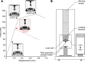
Compression tests were performed using a ZwickiLine uniaxial compression testing machine (Zwick Roell, Ulm, Germany) equipped with a 5 N load cell (Xforce P load cell, Zwick Roell) (Fig. 1B). The distal end of the freshly cut hindleg tibiae was placed on a 3D-printed sample holder which had a 1 mm-deep hole in the middle. The specimens were fixed in the hole using a small drop of hot wax. The proximal end of the specimens was placed in contact with a piece of rough sandpaper adhered to the load cell. This enabled us to more precisely control the straightness of specimens prior to the experiment. Compression tests were always performed at an increasing displacement of 5 mm min−1. Force and displacement
were continuously recorded by the software testXpert (v3.5, Zwick Roell) throughout the experiment. We finished each experiment within a maximum of 15 min after removal of the tibiae from the insects’body. The whole testing process was filmed using a Nikon D5300 digital camera (Nikon Corp., Tokyo, Japan) equipped with a macro lens (Canon Macro Lens EF 100 mm, Canon Inc., Tokyo, Japan). Mechanical tests were conducted on 10–17 hindleg tibiae at each postembryonic developmental stage (Table 1).
We estimated the effective Young’s modulus (E) of the tibia cuticle using the following equation:
E¼FL
Ad; ð1Þ
whereFis the measured force at any point of the linear portion of the force–displacement curve andδis the corresponding displacement. Aand Lare the average cross-sectional area and length of tested tibiae, respectively.
0 0.2 0.4 0.6 0.8 1.0
160
140
120
100
80
60
40
20
0
Load cell
Tibia specimen
Displacement (mm)
Force (mN)
sample holder
Elastic deformation Onset of Euler buckling
Formation of a plastic hinge
Loading direction Moving stroke
Onset of local buckling
A
B
Fig. 1. Uniaxial compression tests on thehindleg tibia of the stick insectCarausius
morosus.(A) Representative force–
displacement curves of specimens at the first five nymphal stages (L1–L5, red dashed line) and those at the last nymphal (L6) and adult stage (black dashed line). (B) Schematic diagram of the compression testing machine. Tibia specimens were fixed on a 3D-printed sample holder and subjected to a displacement-controlled compressive load.
Journal
of
Experimental
[image:2.612.50.400.481.738.2]The stress at buckling was calculated by dividing the force at buckling (Fb) by the cross-sectional area of specimens at the
buckling site:
sb¼F
b
A: ð2Þ
The cross-sectional area at the buckling site was taken as the average cross-sectional area of at least three specimens at that region (see ‘Micro-computed tomography scanning’, below). The buckling sites were identified from the recorded videos.
When loaded in axial compression, a column-like structure could fail at either a material or a structural level. Failure at the material level is essentially due to fracture or plasticity. In such cases, failure occurs when the stress at any point of the structure exceeds the ultimate compressive strength (σuc) or the yield strength (σy) of the
material of which it is made. Failure at a structural level, however, could occur by either Euler or local buckling. The stress required to cause failure by Euler buckling can be theoretically predicted using the following formula (Timoshenko, 1953):
sEuler¼p 2
EI AL2
e
; ð3Þ
where I and Le are the minimum second moment of area and
effective length of the column, respectively. The stress needed to induce local buckling can be estimated by (Timoshenko and Gere, 1961; Rees, 1997):
slocal¼ E
2pffiffiffiffiffiffiffiffiffiffiffiffiffiffiffiffiffiffiffi3ð1n2Þ
t
r; ð4Þ
where t and r are the thickness and outer radius of the column, respectively.νis Poisson’s ratio of the column material, and here it is assumed to be 0.3.
Micro-computed tomography scanning
Micro-computed tomography (µCT) scanning was used to measure the geometric parameters of the hindleg tibia of the stick insect at all seven postembryonic developmental stages. The tibiae were dried in an ascending ethanol series prior to scanning. All tibiae were scanned using a Skyscan 1172 µCT scanner (Bruker, Kontich, Belgium) with a resolution of 1–3 µm at a peak voltage of 40 kV and a current of 250 µA. The scans were performed on at least three tibiae at each developmental stage.
The obtained data were used to calculate the second moment of area (I), cross-sectional area (A), thickness (t) and radius (r) of the specimens. Measurements were performed using BoneJ (Doube et al., 2010) and ImageJ software (v1.5i, National Institutes of Health, Bethesda, MD, USA).
The slenderness ratio (SR) of specimens was calculated by dividing the length (L) to minimum radius of gyration (rg):
SR¼ L
rg; ð
5Þ
wherergis obtained by:
rg¼
ffiffiffi I A r
: ð6Þ
We used the data from µCT scans to measure the area and second moment of area for more than 2000 cross-sections of each scanned specimen. Eqn 6 was then used to calculate the radius of gyration of each cross-section. The minimum obtained value was taken as the minimum radius of gyration of that specimen. The minimum radius of gyration of specimens at each developmental stage was assumed to be equal to the average of those of at least three tibiae at that stage.
Scanning electron microscopy
For scanning electron microscopy (SEM), three tibiae at each developmental stage were first air dried and then broken into smaller pieces. The fractures were mounted on SEM stubs using carbon Leit-tabs (Plano GmbH, Wetzlar, Germany). They were sputter coated with a ∼10 nm-thick gold–palladium layer. The broken sections of the tibiae were imaged using a Hitachi S-4800 scanning electron microscope (Hitachi High-Technologies, Tokyo, Japan) at 3 kV accelerating voltage.
Confocal laser scanning microscopy
Confocal laser scanning microscopy (CLSM) was utilized to obtain an understanding of the material composition of the tibia cuticle. We used the method described by Michels and Gorb (2012) and previously employed by us to demonstrate the presence of the soft and hard cuticle in insect exoskeletons (Rajabi et al., 2015, 2017a, 2018). For this purpose, three freshly cut tibiae at each developmental stage were first washed in distilled water. They were then carefully cut into pieces of∼0.5 mm thickness using a sharp razor blade. The obtained sections were mounted in glycerine between a glass slide and a coverslip. We used a Zeiss LSM 700 microscope (Carl Zeiss Microscopy, Jena, Germany) to visualize the cross-section of the specimens. The microscope was equipped with four stable solid-state lasers (405, 488, 555 and 639 nm) and four emission filters (BP420–480, LP490, LP560 and LP640 nm).
Statistics
[image:3.612.48.569.70.165.2]Statistical analyses were performed according to the protocol suggested by Nayak and Hazra (2011). Because the obtained data were unpaired and non-parametric, the Kruskal–WallisHtest was utilized to compare datasets. The analysis was followed by Dunn’s
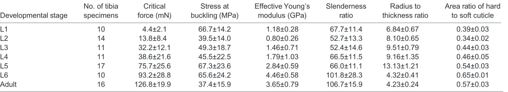
Table 1. Results of mechanical and geometric measurements at each developmental stage
Developmental stage
No. of tibia specimens
Critical force (mN)
Stress at buckling (MPa)
Effective Young’s modulus (GPa)
Slenderness ratio
Radius to thickness ratio
Area ratio of hard to soft cuticle
L1 10 4.4±2.1 66.7±14.2 1.18±0.28 67.7±11.4 6.84±0.67 0.39±0.03
L2 14 13.8±8.4 39.5±14.0 0.80±0.26 52.7±13.3 8.10±0.65 0.34±0.02
L3 11 32.2±12.1 49.3±18.7 1.46±0.71 52.4±14.6 9.51±0.79 0.44±0.03
L4 11 38.6±21.6 45.5±22.5 1.79±1.03 66.5±11.5 9.16±1.35 0.46±0.05
L5 17 75.7±25.6 67.3±23.6 2.84±0.59 66.0±11.1 13.13±1.21 0.54±0.03
L6 10 93.2±28.8 65.6±24.2 4.46±0.58 101.8±28.3 4.32±0.41 0.65±0.01
Adult 16 126.8±19.9 37.4±15.9 3.65±0.79 106.7±15.9 4.23±0.24 0.57±0.03
Data are means±s.d.
Journal
of
Experimental
post hoctest when appropriate. The values in the text are means± s.d., unless stated otherwise.
RESULTS
Representative force–displacement curves obtained from uniaxial compression tests on the hindleg tibiae of the stick insect C. morosus are shown in Fig. 1A. Prior to buckling, the tibiae exhibited a linear elastic behaviour. Once buckling began, the slope of the force–displacement curve decreased (onset of buckling). The buckling of the tibiae at different postembryonic developmental stages appeared to occur in two different modes (shown by black and red dashed lines in Fig. 1A). The first buckling mode was associated with the sideways bending of the whole sample (Fig. 1A, onset of Euler buckling). This buckling mode, known as Euler buckling, resulted in instability of the tibia. In this state, any further loading caused the formation of a plastic hinge and, finally, the collapse of the specimen. This buckling mode was characteristic of specimens at the last nymphal (L6) and adult stages. The hindleg tibiae of individuals in the first five nymphal stages (L1–L5), however, buckled in a different way. In these specimens, the instability was initiated by formation of a localized kink (Fig. 1A, onset of local buckling). The initial local buckling was subsequently followed by out-of-straightness of the tibia. In contrast to Euler buckling, localized buckling resulted in a sudden reduction of the resistance of the tibia to the applied displacement. Movies 1 and 2 show the onset and evolution of buckling in two tibia specimens at the fifth nymphal stage (L5) and adult stage, respectively.
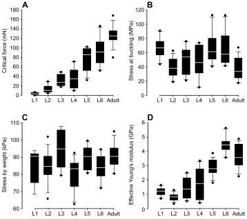
Fig. 2A shows the critical buckling force of the hindleg tibia at each postembryonic developmental stage. The specimens at the first nymphal stage (L1) had the lowest critical force, 4±2 mN, and this
increased to a value of 39±21 mN at the fourth nymphal stage (L4). From this stage, critical force increased again, but at a slightly higher rate until it reached a maximum value of 127±20 mN at the adult stage.
The stress at buckling at each postembryonic developmental stage is given in Fig. 2B. The stress varied from a minimum value of 11.26 MPa at the fourth nymphal stage (L4) to a maximum value of 112.67 MPa at the fifth nymphal stage (L5). The mean stress, in contrast, was found to vary in a rather narrow range from 37.42 MPa at the adult stage to 67.33 MPa at the fifth nymphal stage (L5). However, statistical analysis indicated no significant difference between the stresses at different stages (H=15.62,N=89,P>0.05).
We estimated the stress in the tibia in a simple standing posture by dividing the insect body weight by the sum of the cross-sectional area of six tibiae. The stress induced by the body weight at each stage is given in Fig. 2C. Similar to the stress at buckling, there was no significant difference between the stresses at the different stages (H=16.58,N=89,P>0.05).
The effective Young’s modulus was used as a measure of the stiffness of the tibia cuticle (Fig. 2D). The effective Young’s modulus slightly decreased from 1.2±0.3 GPa at the first nymphal stage (L1) to 0.8±0.3 GPa at the second nymphal stage (L2). However, statistical analysis showed no significant difference between the moduli at these two stages (N=24, P>0.05). There was also no significant difference in the effective Young’s modulus of specimens at the sixth nymphal stage (L6, 4.5±0.6 GPa) and the adult stage (3.7±0.8 GPa) (N=26, P>0.05). Except for the slight differences between the aforementioned groups, the general trend in the effective Young’s modulus was an upward increase during growth of the insect.
Critical force (mN)
0 40 80 120 160
L1 L2 L3 L4 L5 L6 Adult L4 L5
A
L1 L2 L3 L6 Adult
Stress at buckling (MPa)
0 80
20 120
40 60 100
B
L1 L2 L3 L4 L5 L6 Adult
Ef
fective
Y
o
ung's modulus (GPa)
0 1 3
2 4 6
5
C
D
Stress by weight (kPa)
60 70 80 90 100 110
L1 L2 L3 L4 L5 L6 Adult
Stage 180
[image:4.612.46.404.409.727.2]20 60 100 140
Fig. 2. Data obtained from uniaxial compression tests on the hindleg tibia at all postembryonic developmental stages.
(A) Critical buckling force increases in the course of insect development. (B) Stress at buckling of the tibia. According to statistical analysis, there is no significant difference between the stresses at different
developmental stages. (C) Stress acting on the tibia as a result of insect body weight was also found to remain almost constant during insect growth. (D) Effective Young’s modulus of the tibia cuticle. The general trend is an increase in the effective Young’s modulus with the development of the insect. The borders of the boxes indicate the 25th and 75th percentiles, the line within them marks the median, and the whiskers (error bars) define the 10th and 90th percentiles.
Journal
of
Experimental
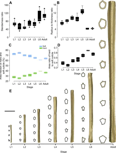
Fig. 3 represents the geometric parameters of the hindleg tibia at each postembryonic developmental stage. A gradual change occurred in the length and cross-sectional shape of the tibia during insect growth (Fig. 3E). The data obtained from µCT analysis (available on request) were used to calculate the slenderness ratio of the tibia, presented in Fig. 3A. Statistical analysis revealed no significant difference between slenderness ratios of the tibia at the first five nymphal stages (L1–L5) (N=67,P>0.05). We also found
no significant difference between the slenderness ratios at the last nymphal stage (L6) and the adult stage (N=26,P>0.05). However, there was a significant difference between the slenderness ratios of the tibiae in the first five nymphal stages (L1–L5) and the last two stages (L6 and adult) (H=58.23,N=93,P<0.05).
The radius to thickness (r/t) ratio of the tibia, shown in Fig. 3B, was found to increase from 6.84±0.67 at the first nymphal stage (L1) to 13.13±1.21 at the fifth nymphal stage (L5). From L5 to the sixth
E
B
Radius to thickness ratio
0
L1 L2 L3 L4 L5 L6 Adult L1 L2 L3 L4 L5 L6 Adult
Slenderness ratio
0 40 80 120
160
A
C
L1 L2 L3 L4 L5 L6 Adult
4 8 12 16
80
70
60
50
40
30
20
L1 L2 L3 L4 L5 L6 Adult
Area fraction of hard and
soft cuticle (%)
Stage
Soft
Hard
D
Stage
L1 L2 L3 L4 L5 L6 Adult
Area ratio of hard
to soft cuticle
0.3 0.4 0.5 0.6 0.7
[image:5.612.116.494.167.664.2]Stage
Fig. 3. Data from geometric analysis of the hindleg tibia at all postembryonic developmental stages.(A) Slenderness ratio of the tibia. Although the
slenderness ratio remains almost constant at the first five nymphal stages (L1–L5), a significant increase is observed from the fifth (L5) to the sixth (L6) nymphal and the adult stage. (B) Ratio of the radius to the thickness of the hindleg tibia at each developmental stage. (C) Area of the hard, sclerotized cuticle and soft, resilin-dominated cuticle as a fraction of the total cross-sectional area. (D) Area ratio of the hard to soft cuticle, which appears to follow the same trend as Young’s modulus. The borders of the boxes indicate the 25th and 75th percentiles, the line within them marks the median, and the whiskers (error bars) define the 10th and 90th percentiles. (E) Three-dimensionally (3D) reconstructed models of the hindleg tibia at all developmental stages. The cross-section of the tibia at several points is
shown beside each model. A gradual change can be seen in the length and cross-sectional shape of the tibia during growth. Scale bar: 1 mm.
Journal
of
Experimental
nymphal stage (L6), ther/tratio suddenly dropped to 4.32±0.37, but it remained almost constant from the sixth nymphal stage (L6) to the adult stage (N=16,P>0.05).
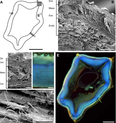
The results of CLSM revealed the presence of two distinguishable layers with different autofluorescence characteristics in the hindleg tibia cuticle (Fig. 4D,E). According to Michels and Gorb (2012), the blue autofluorescence of the inner layer indicates a high proportion of resilin, the pliant protein. The outer layer, in contrast, shows a strong green autofluorescence, which suggests that this layer mainly consists of sclerotized chitinous material. Fig. 3C shows the area of the hard sclerotized and soft resilin-dominated cuticle as a fraction of total area obtained from measurements on tibia cross-sections. Using these data, we calculated the area ratio of the hard to soft cuticle at all stages (Fig. 3D). Interestingly, the obtained data seem to follow the same trend as that observed for the effective Young’s
modulus. The area ratio of the hard to soft cuticle slightly decreased from 0.39±0.03 at the first nymphal stage (L1) to 0.34±0.02 at the second nymphal stage (L2). After this, the area ratio continuously increased, reaching a maximum value of 0.65±0.01 at the sixth nymphal stage (L6). The area ratio of the hard to soft cuticle at the adult stage (0.57±0.03) was slightly lower than that measured in L6. Fig. 4B,C,F shows representative SEM images of the hindleg tibia cuticle. The obtained images suggest that the tibia cuticle consists of up to five main layers: (i) epicuticle, (ii) outer exocuticle, (iii) mesocuticle, (iv) inner exocuticle and (v) endocuticle. The epicuticle is the outermost layer, which is usually very thin, with a thickness of up to 0.5 µm in adult individuals. It covers a very dense exocuticular layer with a lamellar organization. The thickness of this layer in adult specimens was 1.0–2.8 µm. Below the exocuticle, there is a less dense layer, which is probably the mesocuticle, with a
A
B
C
D
F
E
B
C
D
F
Exo
Exo
Endo Meso
Epi
Exo Exo
[image:6.612.111.504.238.654.2]Meso
Fig. 4. Microstructure and material composition of the hindleg tibia cuticle.(A) Scheme of a tibia cross-section (see E) showing the approximate position of
each given image (B,C,D,F). (B) Scanning electron microscopy (SEM) image showing the outer exocuticle (Exo), mesocuticle (Meso), inner exocuticle and endocuticle (Endo) in a cross-section of a specimen at the fifth nymphal stage (L5). (C) SEM image illustrating the lamellar microstructure of the outer and inner exocuticle and the unorganized mesocuticle in a cross-section of the tibia at the fifth nymphal stage (L5). The thin epicuticle layer covering the tibia is also visible in this image. (D) Confocal laser scanning microscopy (CLSM) image of a cross-section of an adult specimen showing the presence of a material gradient along the thickness of the tibia. (E) CLSM image of the whole cross-section of an adult specimen showing the hard sclerotized and the soft resilin-dominated cuticle in green and blue, respectively. (F) SEM image showing the perpendicularly arranged chitin fibres within the endocuticle of a specimen at the adult stage.
Scale bars: A, 100 µm; B, 10 µm; C, 2 µm; D, 10 µm; E, 100 µm; and F, 5 µm.
Journal
of
Experimental
thickness ranging from 2.0 to 3.6 µm in adult individuals. Because of its less-organized structure, it was not possible to unambiguously determine the arrangement of chitin microfibres in this layer. We found another exocuticle layer just beneath the mesocuticle. Although less dense than the outer exocuticle, the two seem to have a similar layered structure. The inner exocuticle reached a thickness of up to 2 µm in the adult stick insects. The innermost layer in the cuticle microstructure is a less-dense endocuticle, consisting of alternating thin helicoidal and thicker unidirectional layers. In some regions, the unidirectional layers within the endocuticle had the same orientation and, together with helicoidal layers, formed so-called locust-like layering (Neville and Luke, 1969) (Fig. 4B). In some other regions, however, they changed orientation by 90 deg, resulting in the formation of a pseudo-orthogonal layering (Neville and Luke, 1969) (Fig. 4F). The endocuticle, with a thickness of 8–16 µm in adult individuals, is the thickest layer in the tibia cuticle. Although we were able to confirm the presence of the aforementioned layers in tibiae at all postembryonic developmental stages, their thickness was found to be very variable. The thickness of each particular layer was also highly dependent on the measurement point on the tibia, even in a single individual.
The measured mechanical and geometric characteristics of the tibia during development of the stick insect C. morosus are summarized in Table 1.
DISCUSSION
A variety of biological factors may influence the mechanical behaviour of tibiae of the stick insectC. morosus. An example is the increase in the body size (Fig. S1B), which has an inevitable impact on the length of the tibia (Fig. 3E; Fig. S1C). The increase in body size further results in an increase in body mass (Fig. S1A) and, consequently, raises the force acting on each tibia. Although one may expect that the added body mass at each postembryonic developmental stage increases the mechanical stress in the tibiae, a simple estimation suggests that this is not the case (Fig. 2C). Indeed, we found no significant difference between the stresses applied to the tibiae by the weight of the insect at different developmental stages. This may be a major reason why the buckling resistance of the hindleg tibia also remains constant during insect growth (Fig. 2B).
A remarkable difference, however, was observed between the stress at buckling and the stress imposed on the hindleg tibia by the body weight (Fig. 2B,D). Comparison of the two stresses at the adult stage, for instance, suggests that the tibia possesses a large factor of safety of 127.2±23.2 in standing posture. Using kinematic data from the literature, we additionally determined the factor of safety of the tibia when subject to compressive stresses during daily activities. Data from ground reaction force measurements on freely walking adult stick insects showed that compressive forces in the insect hindlegs can reach a peak value of 9.6 mN (Dallmann et al., 2016). Assuming the entire force to be transmitted to the tibia, this results in a factor of safety of 13.7±2.2 for the tibia when walking. This is about twice the maximum factor of safety measured for the tibia of other arthropods, which ranged from 1.7 to 7 (Parle et al., 2016a). Therefore, this finding raises the question of why the tibia needs such a high load-carrying capacity.
A recent study reported frequent intentional drops or accidental falls of the wingless stick insect Extatosoma tiaratum, while climbing vegetation (Zeng et al., 2015). This behaviour, which may serve as a defence reaction, is also likely to be valid for the wingless speciesC. morosus. Immediately after falling from a height, the stick insects are able to reorientate and stabilize their body during
the plunge (Zeng et al., 2017), and land on their tarsi (M.S., T.H.B., S.N.G. and H.R., unpublished observation). However, landing on the tarsi may induce much higher stresses in the tibiae than those applied in normal standing posture (Heitler, 1977). Although further investigations are needed, this may explain the large factor of safety considered in the design of the hindleg tibia.
It is well known that the resistance of a structure to buckling is essentially influenced by its geometric characteristics, in particular slenderness, and the stiffness of the material of which the structure is made (Cedolin, 2010). The increase of the slenderness ratio, leading to a reduced buckling resistance, seems to be an inevitable part of insect growth. In the case of the stick insectC. morosus, this is reflected in the significant increase of the slenderness ratio of the tibia from the first five nymphal stages (L1–L5) to the last two stages (L6 and adult) (Fig. 3A). However, except for the slight decrease in the effective Young’s modulus, the development of the stick insect is accompanied by a significant increase in the stiffness of its tibia cuticle (Fig. 2D). The increase in the effective Young’s modulus appears to compensate for the negative effect of the increasing slenderness, enabling the tibia to retain its buckling resistance during insect growth (Fig. 2B).
The change of the effective Young’s modulus during insect postembryonic development was found to follow the same trend as that of the area proportion of the hard sclerotized to soft resilin-dominated cuticle (Figs 2D and 3D). Taking into account the predominant role of the sclerotized cuticle in stiffening insect cuticle (Hepburn, 1985; Rajabi et al., 2017b), the similar trend between these two parameters appears to be very reasonable. The slight decrease in the effective Young’s modulus from the sixth nymphal stage (L6) to the adult stage (Fig. 2D) can also be explained by the presence of a lower area proportion of the hard to soft cuticle at the adult compared with the L6 stage (Fig. 3C,D). Taking into account the remarkably longer life period ofC. morosusat the adult stage than at all nymphal stages together (Roth, 1917), it is reasonable to hypothesize that the deposition of the resilin-dominated endocuticle at the adult stage is also more prolonged.
We were able to distinguish two distinctly different force– displacement curves in the results from the uniaxial compression tests on tibia specimens. While the specimens at the first five nymphal stages (L1–L5) suddenly lost their load-carrying capacity after buckling, those at the last nymphal (L6) and adult stages showed a gradual reduction in their ability to support the external load (red and black dashed lines in Fig. 1A, respectively). This difference appears to be essentially the result of the occurrence of two different buckling modes in these two groups of specimens (i.e. local buckling and Euler buckling, respectively). This finding is also consistent with the observation of the considerably lowerr/t ratio of the tibiae at the sixth nymphal (L6) and adult stage when compared with that of the other living stages (L1–L5) (Fig. 3B). Although the higherr/tratio of the tibiae in the L1–L5 group causes them to become more resistant to Euler buckling, this subsequently makes them more prone to local buckling.
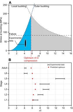
Here, we adapted the method suggested by Taylor and Dirks (2012) to find the optimum value of ther/tratio of the hingleg tibia at each postembryonic developmental stage. This optimum ratio should provide the highest load-carrying capacity for a given weight. As stated, the failure of the tibia under compression is essentially the result of either Euler or local buckling. Fig. 5A shows graphs of these two buckling modes (blue and grey dashed lines), plotted as a function of r/tfor adult stick insects. The buckling stress, given by the Euler curve, increases with the increase of ther/t ratio. In contrast, the stress at buckling predicted by the local
Journal
of
Experimental
buckling curve decreases as ther/tratio increases. The optimumr/t is reached where the two curves intersect. For the tibia of adult specimens, this optimum value occurs at a point where ther/tratio is equal to 5.72. As seen in Fig. 5A, the experimentally measured values of r/t ratio, 4–4.46 (black ellipse), are close to the theoretically predicted optimum value. Fig. 5B compares the experimentalr/tvalues with those calculated theoretically at each postembryonic developmental stage. A good agreement was found between the two sets of data at all stages, except at the fifth nymphal stage (L5). Our analysis, in general, suggests the presence of a biomechanical adaptation in the hindleg tibia towards an optimum r/t ratio to reduce the probability of failure by buckling. The optimization of ther/tratio has already been employed in the design
of steel circular hollow tubes used in wind turbine towers to control and improve their buckling behaviour (Guo et al., 2013).
As shown in Fig. 5B, the experimentalr/tratios of tibiae at the first five nymphal stages (L1–L5) are higher than the theoretically predicted optimum values. In contrast, ther/tratios obtained from measurements on the tibiae at the sixth nymphal (L6) and adult stages are lower than the optimum ones. This finding highlights the dependency of the failure mode on ther/tratio at each stage and further supports our observation that the buckling mode changes during insect growth.
In the CLSM images, we were able to observe the presence of a material gradient along the thickness of the tibia cuticle (Fig. 4D,E). In addition to toughening the whole structure of the tibia, the soft resilin-dominated core can improve its resistance to buckling (Karam and Gibson, 1995; Suresh, 2001). The hard sclerotized coverage, in contrast, enhances the stability of the tibia and protects it against mechanical damage (Rajabi et al., 2016). Interestingly, in many regions, it was possible to identify a clear border between the hard and soft cuticle layers (Fig. 4E). This observation confirms our previous finding that a realistic model of insect cuticle, in addition to consideration of a material gradient, should take the discontinuity at the interface of layers into account (Rajabi et al., 2017b). Such a material discontinuity in the insect cuticle may be a biomechanical strategy to avoid the propagation of an already existing crack from one layer to another.
Natural fibre-reinforced composites, such as the tibia cuticle, are well known for their light weight, high structural integrity, reasonable stiffness, strength and toughness (Mayer and Sarikaya, 2002; Dirks and Taylor, 2012; Keogh et al., 2015). However, the behaviour of such composites under loading is also influenced by their fibre orientation (Laranjeira et al., 2010; Martinie and Roussel, 2011). It has previously been reported that hollow cylinders with fibres oriented in the axial direction have an optimum bending strength (compared with cylinders with fibres at a particular angle). In contrast, hollow cylinders reinforced with fibres in the circumferential direction show optimum compression resistance (Li et al., 2006). In the tibia cuticle, we demonstrated the presence of both fibre orientations (Fig. 4F). During the insect lifetime, the tibia experiences a combination of bending and compression. Therefore, it is reasonable to expect that the cuticle in this body part is adapted to resist both loading conditions by aligning fibres in these two orientations.
Comparison of our results with those of previous measurements on tibiae of the adult stick insects loaded in bending (Parle et al., 2016b) indicates that the bending stiffness of the tibia cuticle is larger than its stiffness in compression (5.5±4.13 and 3.65± 0.79 GPa, respectively). The difference between the measured properties is even more striking when comparing the strength of the tibia in bending and compression: 116.0±66.1 and 37.4±15.9 MPa, respectively. This suggests that, although compression is a typical loading condition for the tibia, especially when an insect is standing upright, bending stresses in the leg during daily activities may exceed those caused by compression. Future studies are planned to assess macro- and micro-morphological differences between forelegs, midlegs and hindlegs of the stick insectC. morosusand to evaluate their potential influence on the buckling resistance of each leg under combined bending and compression.
Conclusions
In this study, we tracked changes in the material, structural and geometric properties of the hindleg tibia of the stick insect C. morosusat all postembryonic developmental stages. We aimed to
0 50 100 150 200 250
0 2 4 6 8 10 12 14 16
Stress at buckling (MPa)
r/t
Optimum r/t in compression
0 2 4 6 8 10 12 14 16
L1 L6
L5 Adult
L4
L3
L2
r/t
Stage
A
B
Predicted optimum Euler buckling
Experimental data
Experimental data Failure
[image:8.612.50.300.56.438.2]Local buckling
Fig. 5. Theoretical prediction of the optimal radius to thickness (r/t) ratio
of the hindleg tibia when loaded in compression.Comparisons are shown
between the theoretical and experimental data at all postembryonic developmental stages. (A) Stress at buckling versusr/tratio for the tibia at the adult stage. The load-carrying capacity of the tibia predicted by Euler buckling (blue dashed line) and local buckling (grey dashed line) is plotted as a function ofr/tratio. The optimum value ofr/toccurs at the point where the two curves intersect (red dashed line). Assuming a yield strength of about 85 MPa (Parle et al., 2015), the horizontal black dashed line gives the stress at which the tibia cuticle may fail as a result of plasticity. The experimental data obtained from measurements on tibiae of adult stick insects fall within the black ellipse. (B) Comparison of the theoretically predicted optimal (red) and experimentally measured (black)r/tvalues at each developmental stage. Except at the fifth nymphal stage (L5), the experimentalr/tvalues are close to the optimum value.
Journal
of
Experimental
understand how the observed changes can influence the load-carrying capacity of the tibia under compression. Based on the obtained data, the following conclusions can be drawn. (i) Although the geometric characteristics of the hindleg tibia change remarkably during growth, the tibia is able to maintain its resistance to buckling. This is the result of a compromise between the increasing slenderness of the tibia and its increasing stiffness. (ii) The failure of the tibia under compression mainly occurs in two different buckling modes: local buckling, in the first five nymphal stages, and Euler buckling, in the sixth nymphal and the adult stage. (iii) The occurrence of either of these buckling modes at each developmental stage is likely to be influenced by the ratio of radius to thickness (r/t) of the tibia at that stage. This ratio at most developmental stages is close to an optimum value, representing a good capability to resist both Euler and local buckling. (iv) The presence of a soft, resilin-dominated endocuticle with fibres orientated in both longitudinal and circumferential directions is likely to be a biomechanical adaptation that provides the tibia with an enhanced buckling resistance.
Acknowledgements
The authors would like to thank Dr Clemens Schaber (Kiel University) and Dr Alexander Kovalev (Kiel University) for their technical help. We thank Dr Jan Michels (Kiel University) for his helpful comments on the CLSM images. We acknowledge the helpful discussions with Prof. David Taylor (Trinity College Dublin), Halvor T. Tramsen (Kiel University), Mohsen Jafarpoor (Guilan University), Prof. Tobias Seidl (Westphalian University of Applied Sciences) and Ahmad R. Mojdehi (Virginia Polytechnic Institute & State University). Thanks also to Prof. Volker Dürr (Bielefeld University) and Marek Noheijl (Řečany nad Labem, Czech Republic) for providing specimens for this study.
Competing interests
The authors declare no competing or financial interests.
Author contributions
Conceptualization: S.N.G., H.R.; Methodology: T.H.B., S.N.G., H.R.; Software: M.S.; Validation: M.S., T.H.B.; Formal analysis: M.S.; Investigation: M.S.; Resources: T.H.B., S.N.G.; Data curation: M.S., T.H.B., H.R.; Writing - original draft: M.S., H.R.; Writing - review & editing: T.H.B., S.N.G., H.R.; Visualization: M.S., T.H.B., H.R.; Supervision: T.H.B., S.N.G., H.R.; Project administration: S.N.G., H.R.; Funding acquisition: S.N.G., H.R.
Funding
This study was financially supported by Federal State Funding at Kiel University (Christian-Albrechts-Universität zu Kiel) to H.R.
Data availability
3D reconstructed models of the hindleg are available on request from the corresponding author.
Supplementary information
Supplementary information available online at
http://jeb.biologists.org/lookup/doi/10.1242/jeb.173047.supplemental
References
Beer, F. P., Johnston, R., Dewolf, J. and Mazurek, D.(2006).Mechanics of
Materials. New York: McGraw-Hill.
Budynas, R. G. and Nisbett, J. K. (2015).Shigley’s Mechanical Engineering
Design, 10th edn. New York: McGraw-Hill.
Büscher, T. H. and Gorb, S. N.(2017). Subdivision of the neotropical Prisopodinae Brunner von Wattenwyl, 1893 based on features of tarsal attachment pads (Insecta, Phasmatodea).ZooKeys645, 1-11.
Cedolin, L.(2010).Stability of Structures: Elastic, Inelastic, Fracture and Damage
Theories. Singapore: World Scientific.
Clausen, T.(1851). Über die Form architektonischer Säulen.Bulletin de la Classe
physico-mathématique de l’Académie impériale des sciences de
Saint-Pétersbourg9, 368-379.
Dallmann, C. J., Dürr, V. and Schmitz, J.(2016). Joint torques in a freely walking insect reveal distinct functions of leg joints in propulsion and posture control.
Proc. R. Soc. B283, 20151708.
Dean, J.(1991). Effect of load on leg movement and step coordination of the stick insectCarausius morosus.J. Exp. Biol.159, 449-471.
Dirks, J.-H. and Taylor, D.(2012). Fracture toughness of locust cuticle.J. Exp. Biol.
215, 1502-1508.
Doube, M., Kłosowski, M. M., Arganda-Carreras, I., Cordelières, F. P., Dougherty, R. P., Jackson, J. S., Schmid, B., Hutchinson, J. R. and Shefelbine, S. J.(2010). BoneJ: free and extensible bone image analysis in ImageJ.Bone47, 1076-1079.
Galambos, T. V.(ed.) (1998).Guide to Stability Design Criteria for Metal Structures. New York: John Wiley & Sons.
Graham, D.(1972). A behavioural analysis of the temporal organisation of walking movements in the 1st instar and adult stick insect (Carausius morosus).J. Comp.
Physiol. A Neuroethol. Sens. Neural Behav. Physiol.81, 23-52.
Grote, K.-H. and Feldhusen, J.(2011).Dubbel: Taschenbuch für den technischen
Maschinenbau. Heidelberg: Springer.
Guo, L., Yang, S. and Jiao, H.(2013). Behavior of thin-walled circular hollow section tubes subjected to bending.Thin-Walled Structures73, 281-289.
Harries, K. A., Bumstead, J., Richard, M. and Trujillo, D.(2017). Geometric and material effects on bamboo buckling behaviour.Proc. Inst. Civil Eng. Structures
Buildings170, 236-249.
Heitler, W. J. (1977). The locust jump: III. Structural specializations of the metathoracic tibiae.J. Exp. Biol.67, 29-36.
Hepburn, H. R.(1985). The integument. InFundamentals of Insect Physiology(ed. M. S. Blum), pp. 139-183. Hoboken, NJ: Wiley.
Kappel, R., Mattheck, C., Bethge, K. and Tesari, I. (2004). Bamboo as a composite structure and its mechanical failure behaviour.WIT Trans. Ecol.
Environ.73, 285-293.
Karam, G. N. and Gibson, L. J.(1994). Biomimicking of animal quills and plant stems: natural cylindrical shells with foam cores.Mater. Sci. Eng. C2, 113-132.
Karam, G. N. and Gibson, L. J.(1995). Elastic buckling of cylindrical shells with elastic cores - II. Experiments.Int. J. Solids Struct.32, 1285-1306.
Keller, J. B.(1960). The shape of the strongest column.Arch. Ration. Mech. Anal.5, 275-285.
Keogh, L., O’Hanlon, P., O’Reilly, P. and Taylor, D.(2015). Fatigue in bamboo.
Int. J. Fatigue75, 51-56.
Laranjeira, F., Aguado, A. and Molins, C.(2010). Predicting the pullout response of inclined straight steel fibers.Mater. Structures43, 875-895.
Levy, M. and Salvadori, M.(1992).Why Buildings Fall Down - How Structures Fail. New York: W.W. Norton & Company.
Li, G., Maricherla, D., Singh, K., Pang, S.-S. and John, M.(2006). Effect of fiber orientation on the structural behavior of FRP wrapped concrete cylinders.
Compos. Struct.74, 475-483.
Martinie, L. and Roussel, N.(2011). Simple tools for fiber orientation prediction in industrial practice.Cement Concrete Res.41, 993-1000.
Mayer, G. and Sarikaya, M.(2002). Rigid biological composite materials: structural examples for biomimetic design.Exp. Mech.42, 395-403.
Michels, J. and Gorb, S. N.(2012). Detailed three-dimensional visualization of resilin in the exoskeleton of arthropods using confocal laser scanning microscopy.
J. Microsc.245, 1-16.
Nayak, B. K. and Hazra, A.(2011). How to choose the right statistical test?Indian
J. Ophthalmol.,59, 85.
Neville, A. C. and Luke, B. M.(1969). A two-system model for chitin-protein complexes in insect cuticles.Tissue Cell1, 689-707.
Parle, E. and Taylor, D.(2017). The effect of aging on the mechanical behaviour of cuticle in the locustSchistocerca gregaria. J. Mech. Behav. Biomed. Mater.68, 247-251.
Parle, E., Herbaj, S., Sheils, F., Larmon, H. and Taylor, D.(2015). Buckling failures in insect exoskeletons.Bioinspir. Biomim.11, 016003.
Parle, E., Larmon, H. and Taylor, D. (2016a). Biomechanical factors in the adaptations of insect tibia cuticle.PloS One11, e0159262.
Pfeiffer, F., Weidemann, H.-J. and Danowski, P.(1991). Dynamics of the walking stick insect.IEEE Control Systems11, 9-13.
Rajabi, H., Ghoroubi, N., Darvizeh, A., Dirks, J.-H., Appel, E. and Gorb, S. N.
(2015). A comparative study of the effects of vein-joints on the mechanical behaviour of insect wings: I. Single joints.Bioinspir. Biomim.10, 056003.
Rajabi, H., Shafiei, A., Darvizeh, A., Dirks, J.-H., Appel, E. and Gorb, S. N.
(2016). Effect of microstructure on the mechanical and damping behaviour of dragonfly wing veins.J. R. Soc. Open Sci.3, 160006.
Rajabi, H., Ghoroubi, N., Stamm, K., Appel, E. and Gorb, S. N.(2017a). Dragonfly wing nodus: a one-way hinge contributing to the asymmetric wing deformation.
Acta Biomaterialia60, 330-338.
Rajabi, H., Jafarpour, M., Darvizeh, A., Dirks, J. H. and Gorb, S. N.(2017b). Stiffness distribution in insect cuticle: a continuous or a discontinuous profile?
J. R. Soc. Interface,14, 20170310.
Rajabi, H., Stamm, K., Appel, E. and Gorb, S. N.(2018). Micro-morphological adaptations of the wing nodus to flight behaviour in four dragonfly species from the family Libellulidae (Odonata: Anisoptera).Arthropod Struct. Dev.
Rees, D. W. A.(1997).Basic Solid Mechanics. London: Macmillan.
Roth, H. L.(1917). Observations on the growth and habits of the stick insect.
Trans. R. Entomol. Soc. London64, 345-386.
Journal
of
Experimental
Sakes, A., Dodou, D. and Breedveld, P.(2016). Buckling prevention strategies in nature as inspiration for improving percutaneous instruments: a review.Bioinspir.
Biomim.11, 021001.
Schütz, C. and Dürr, V.(2011). Active tactile exploration for adaptive locomotion in the stick insect.J. R. Soc. Proc. B366, 2996-3005.
Suresh, S.(2001). Graded materials for resistance to contact deformation and damage.Science292, 2447-2451.
Taylor, D. and Dirks, J.-H. (2012). Shape optimization in exoskeletons and endoskeletons: a biomechanics analysis.J. R. Soc. Interface9, 3480-3489.
Tichy, H.(1979). Hygro-and thermoreceptive triad in antennal sensillum of the stick insect,Carausius morosus.J. Comp. Physiol. A Neuroethol. Sens. Neural. Behav
Physiol.132, 149-152.
Timoshenko, S. P.(1953).History of Strength of Materials: with a Brief Account of
the History of Theory of Elasticity and Theory of Structures. New York:
McGraw-Hill.
Timoshenko, S. and Gere, J. M.(1961).Theory of Elastic Stability, 2nd edn. New York: McGraw-Hill.
Vilhelmsen, L., Isidoro, N., Romani, R., Basibuyuk, H. H. and Quicke, D. L. J.
(2001). Host location and oviposition in a basal group of parasitic wasps: the subgenual organ, ovipositor apparatus and associated structures in the Orussidae (Hymenoptera, Insecta).Zoomorphology121, 63-84.
Vincent, J. F. V. and Owers, P.(1986). Mechanical design of hedgehog spines and porcupine quills.J. Zool.210, 55-75.
Zeng, Y., Lin, Y., Abundo, A. and Dudley, R.(2015). Visual ecology of directed aerial descent in first-instar nymphs of the stick insectExtatosoma tiaratum.
J. Exp. Biol.218, 2305-2314.
Zeng, Y., Lam, K., Chen, Y., Gong, M., Xu, Z. and Dudley, R. (2017). Biomechanics of aerial righting in wingless larval stick insects.Interface Focus,
7, 20160075.
Journal
of
Experimental