Original Article
SASH1 inhibits proliferation and invasion of thyroid
cancer cells through PI3K/Akt signaling pathway
Dawei Sun1*, Rui Zhou2*, Huamin Liu1, Wenhai Sun1, Anbing Dong1, Hongmei Zhang1
1Department of Thyroid Surgery, The Affiliated Hospital of Qingdao University, Qingdao 266003, China; 2Medical College of Qingdao University, Qingdao 266021, China. *Equal contributors.
Received August 17, 2015; Accepted September 23, 2015; Epub October 1, 2015; Published October 15, 2015
Abstract: The SASH1 (SAM- and SH3-domain containing 1) gene, a member of the SLY-family of signal adapter proteins, has an important regulatory role in tumorigenesis, but its implication in thyroid carcinoma has not been yet investigated. In this study, we investigated the role of SASH1 in proliferation and invasion of thyroid cancer cells and the underlying mechanism. Our results demonstrated that SASH1 is down-regulated in thyroid cancer cells. Overexpression of SASH1 inhibits thyroid cancer cell proliferation, migration and invasion with decreased epithelial-mesenchymal transition (EMT). Mechanistically, overexpression of SASH1 inhibits thyroid cancer cell proliferation and invasion through down-regulation of PI3K and Akt phosphorylation. Taken together, the present study showed that the loss or inhibition of SASH1 expression may play an important role in thyroid cancer development, invasion, and metastasis and that SASH1 may be a potential therapeutic target for the treatment of thyroid cancer.
Keywords: SASH1, thyroid cancer, proliferation, invasion
Introduction
Thyroid cancer is the most common malignant tumor in endocrine system, and its incidence has been steadily increasing in the world [1, 2]. The major type of thyroid cancer is papillary thy-roid carcinoma (PTC). In most cases, patients with thyroid cancer have a good prognosis. However, patients with local invasion outside the thyroid capsule and distant metastases fre-quently poor clinical outcome [3, 4]. Thus, a good understanding of the molecular mecha-nisms underlying thyroid cancer invasion and metastasis is still imperative.
The SASH1 (SAM- and SH3-domain containing 1) gene, a member of the SLY (SH3 domain-containing expressed in lymphocytes) family of signal adapter proteins, is a recently discov-ered tumor suppressor [5]. SASH1 is composed of one Src homology 3 (SH3) domain, which is necessary for protein-protein interaction and mediates the formation of signaling complexes [6]. Among these protein-protein interaction domains, the SAM domain can exhibit more complex functions. The SAM domain could mediate protein-protein interactions through homologous or heterogonous oligomerization with the SAM domains of other proteins, and it
could regulate transcription by mediating the binding of the Smaug protein and mRNA [7]. The reduction or absence SASH1 is closely related to tumor growth, invasion, metastasis, and poor prognosis [8-10]. Meng et al. showed that the expression of SASH1 was down-regu-lated in osteosarcoma tissues compared to normal bone tissue, and overexpression of SASH1 significantly reduced osteosarcoma cell viability, proliferation, and invasive ability [11]. Chen et al. reported that SASH1 inhibited A549 human lung cancer cell growth and proliferation as well as promote cellular apoptosis [12].Most recently, one study reported that SASH1 mRNA expression was significantly decreased in pri-mary thyroid cancers [5]. However, the role of SASH1 in thyroid cancer remains unclear. In this study, we show that SASH1 is lowly expressed in thyroid cancer cell lines. SASH1 inhibits thyroid cancer cell proliferation, migra-tion and invasion.
Materials and methods
Cell culture
SASH1 inhibits thyroid cancer cell proliferation and invasion
Culture Collection (ATCC, USA). All cells were cultured in Dulbecco’s modified Eagle’s medi-um (DMEM) (Gibco Laboratories, Grand Island, NY, USA) supplemented with 10% fetal bovine serum (FBS), 100 U/ml penicillin, and 100 mg/ ml streptomycin (Gibco Laboratories, Grand Island, NY, USA) at 37°C in humidified air with 5% CO2.
Quantitative real-time polymerase chain reac-tion (qRT-PCR) analysis
Total RNA was isolated from thyroid cancer cells using the RNA plus kit (Invitrogen, Carlsbad, CA, USA). One microgram of total RNA was reverse-transcribed to complementa-ry DNA (cDNA) using the Transcriptor First Strand cDNA Synthesis Kit (Invitrogen, Carlsbad, CA, USA). The levels of gene mRNA transcripts were analyzed by using the specific primers and SYBR Green I reagent and the RT-PCR kit, according to the manufacturer’s instructions, on Bio-Rad iQ5 Quantitative PCR System (Takara Bio Inc., Otsu, Japan). The spe-cific primers for SASH1 were sense, 5’-TCCCGTCACAGGAAGAAACG-3’, and antisen- se, 5’-GATACCCATCACGTCGGTCC-3’; and for β-actin were sense, 5’-AAATCGTGCGTGACATCA- AAGA-3’ and antisense, 5’-GGCCATCTCCTGCTC- GAA-3’. The PCR procedure was as follows: 94°C for 4 min; 94°C for 20 s, 55°C for 30 s, and 72°C for 20 s; 2 s for plate reading for 35 cycles; and melting curve from 65 to 95°C. β-actin was used as a control for normalizing gene expression. Experiments were performed independently at least three times. The data obtained were calculated by 2-ΔΔCt [13]. All experiments were repeated at least three times.
Western blot
Total protein extracts were lysed in lysis buffer (20 mM HEPES [pH 7.6], 350 mM NaCl, 20% glycerol, 0.5 mM EDTA, 0.1 mM EGTA, 1% NP-40, 50 mM NaF, 0.1 mM DTT, 0.1 mM PMSF and a protease inhibitor cocktail). The protein concentration was determined using a Bradford protein assay (Takara Biotechnology, Dalian, China). Equal amounts of protein were electro-phoresed on SDS-PAGE and blotted onto PVDF membranes (Millipore Corp, Billerica, MA, USA). After the membranes were blocked by a 5% skim milk solution, the membranes were incu-bated overnight at 4°C with various primary
antibodies (rabbit polyclonal SASH1, E-cadherin, N-cadherin, Vimentin, phospho-PI3K, PI3K, phospho-Akt, Akt or GAPDH from Santa Cruz Biotechnology, Santa Cruz, CA, USA) at 4°C overnight. The membranes were washed and incubated at room temperature for 1 h with the secondary antibodies, which were conjugated with horseradish peroxidase (HRP). The immu-noreactive protein bands were visualized by ECL kit (Pierce, Rockford, USA).
Construction of the pcDNA3.1-SASH1 vector and cell transfection
The full-length SASH1 open reading frame was amplified from human thyroid cancer cells by RT-PCR, and cloned into the pcDNA3.1 expres-sion vector to construct the pcDNA3.1-SASH1 recombinant expression vector. FTC133 cells were transfected with pcDNA3.1-SASH1 or pcDNA3.1 (empty vector) using Lipofectamine 2000 (Invitrogen, Carlsbad, CA), according to the manufacturer’s instructions. After 48 h of transfection, the transfectants were selected in a medium containing 0.5 mg/ml of G418 for 2 to 3 weeks to generate the stable pools. Cell proliferation assay
FTC133 cells transfected with pcDNA3.1-SASH1 or empty vector were seeded into 96-well plates (5×10³ cells/well) and incubated for 24 h, 48 h and 72 h, respectively. Then, cells were stained with an equal volume (100 μl) of fresh medium containing 0.456 mg/ml 3-[4,5-dimethylthiazol-2-yl]-2,5-diphenyl tetra-zolium bromide (MTT) and incubated for 4 h at 37°C in the dark, followed by removal of the cul-ture medium and addition of 100 ml of dimethyl sulfoxide (Sigma). Cell growth was determined by the colorimetric comparison of optical den-sity (OD) values from a microplate reader at an absorption wavelength of 570 nm.
Determination of cell cycle by flow cytometry
cells and incubated for 30 min at 37°C in the dark. The cell cycle was detected by flow cytom-etry (Invitrogen, Carlsbad, CA, USA). The experi-ment was repeated three times.
Cell migration and invasion assays
The invasive and migration behaviors of indi-cated cells were analyzed by Transwell cham-ber (Corning Costar Corp., Cambridge, MA, USA) assay with or without coated Matrigel (BD Biosciences, Bedford, MA, USA). FTC133 trans-fected with overexpession-SASH1 or vector (5×10⁴ cells/ml) suspended in RPMI medium were added to the upper chamber. The lower chamber of the Transwell was filled with 500 μl DMEM containing 10% FBS as a chemoattrac-tant. After 24 h incubation, cells on the surface of upper chamber were removed by scraping with a cotton swab. The invaded/migrated cells on the lower surface of the filter were washed, fixed, stained with Giemsa, and counted under a microscope. Experiments were repeated at least three times.
Statistical analysis
Data are expressed as mean ± standard devia-tion (SD) and analyzed by one-way analysis of variance, Student’s t test or ANOVA. The
differ-ence between means was considered signifi-cant when the P value was <0.05.
Results
SASH1 expression in thyroid cancer cell lines
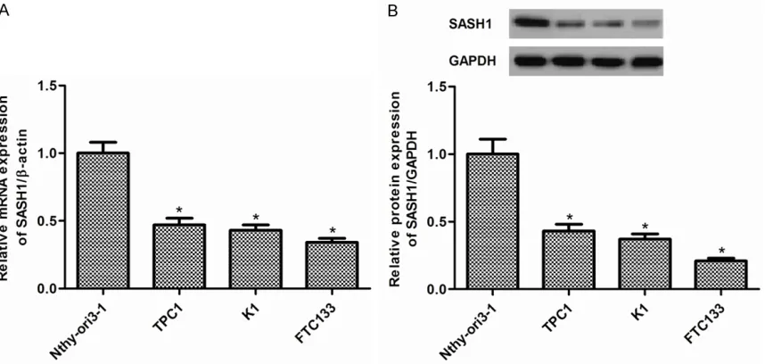
We firstly examined levels of SASH1 in TPC1, K1, and FTC133 thyroid cancer cell lines and Nthy-ori3-1 normal thyroid cell line. As shown in Figure 1A, the expression of SASH1 mRNA was significantly decreased in thyroid cancer cells compared with normal thyroid cells. In line with the results of qRT-PCR, Western blot analysis demonstrated that the expression of SASH1 protein was also obviously reduced in thyroid cancer cells (Figure 1B). FTC133 cells were selected because of expressing low level of SASH1.
Effect of SASH1 on thyroid cancer cell growth
[image:3.612.94.523.72.276.2]To examine the role of SASH1 in thyroid cancer cell growth, FTC133 cells were transduced with pcDNA3.1-SASH1. FTC133 cell line stably transfected with pcDNA3.1-SASH1 has a sig-nificant increase in SASH1 expression com-pared with the vector control (Figure 2A, 2B). Moreover, we evaluated the cell growth by MTT assay. We observed that overexpression of
Figure 1. SASH1 expression in thyroid cancer cell lines. A. The expression levels of SASH1 mRNA were significantly
decreased in TPC1, K1, and FTC133 thyroid cancer cell lines compared with that in Nthy-ori3-1 normal thyroid cell
SASH1 inhibits thyroid cancer cell proliferation and invasion
SASH1 resulted in a dramatic decrease in growth of the tumors cell lines (Figure 2C). Effect of SASH1 on thyroid cancer cell cycle
Then, we examined the effect of SASH1 on thy-roid cancer cell cycle using flow cytometry. As shown in Figure 3, flow cytometry assay indi-cated that overexpression of SASH1 increased the percentage of cells in the G1-G0 phase and decreased the percentage of S-phase in FTC133 cells. There was no significant differ-ence between the blank control group and the empty vector control group.
Effect of SASH1 on thyroid cancer cell migra-tion and invasion
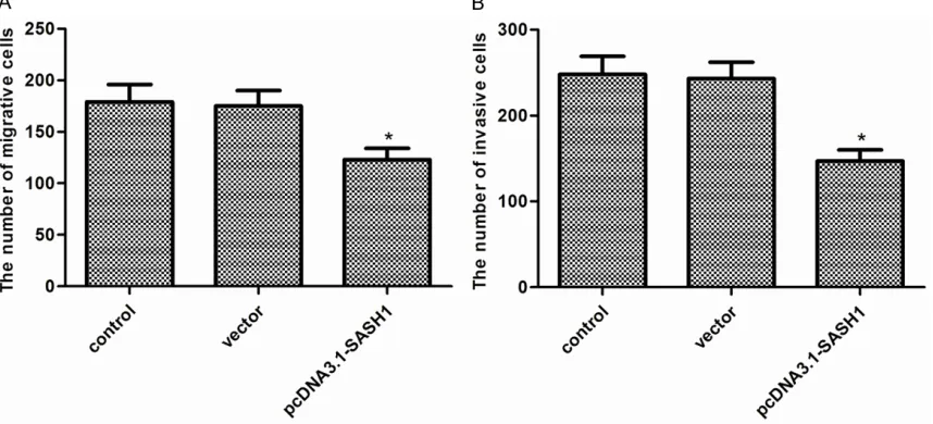
To test the effects of SASH1 on thyroid cancer cell migration and invasion, transwell assays were used. As shown in Figure 4A,
overexpres-Figure 2.Effect of SASH1 on thyroid cancer cell growth. A. Overexpression of SA1SH1 mRNA in FTC133 cells stably transfected with pcDNA3.1-SASH1. B. Overexpression of SA1SH1 protein in FTC133 cells stably transfected with pcDNA3.1-SASH1. C. MTT assay of cell growth in FTC133 cells. The re-sults are expressed as mean ± SD and n=3 per group. *P<0.05 compared with the con-trol and vector groups.
[image:4.612.88.526.66.644.2]cell migration. Transwell invasion assay showed that overexpression of SASH1 in FTC133 cells suppressed the number of invaded cells (Figure 4B).
Effect of SASH1 on EMT of thyroid cancer cells
[image:5.612.94.523.76.271.2]It is well known that epithelial-mesenchymal transition (EMT) plays a critical role in cancer cell migration and invasion [14]. As shown in Figure 5, overexpression of SASH1 increased expression of the epithelial marker E-cadherin
and decreased that of N-cadherin and Vimentin, two mesenchymal markers in FTC133 cells. SASH1 inhibits thyroid cancer cell prolifera-tion, migration and EMT through suppressing PI3K/Akt signaling pathway
PI3K/Akt signaling pathway plays an important role in the development of tumor [15]. Therefore, we investigated the effect of SASH1 on the expression of certain molecules involved in the PI3K/Akt signaling pathway. As shown in Figure
[image:5.612.91.523.334.511.2]Figure 4. Effect of SASH1 on thyroid cancer cell migration and invasion. A. Cellmigration of FTC133cells with SASH1 overexpression.B. Cellinvasion of FTC133cells with SASH1 overexpression. The results are expressed as mean ± SD and n=3 per group. *P<0.05 compared with the control and vector groups.
Figure 5.Effect of SASH1 on EMT of thyroid cancer cells. A. The levels of E-cadherin, N-cadherin and Vimentin were detected in vector, pcDNA3.1-SASH1-transfected FTC133 cells by western blot analysis. B. Quantification of E-cadherin, N-cadherin and Vimentin. The results are expressed as mean ± SD and n=3 per group. *P<0.05 compared
SASH1 inhibits thyroid cancer cell proliferation and invasion
6, overexpression of SASH1 obviously decreased levels of PI3K and Akt phosphoryla-tion in FTC133 cells.
Discussion
SASH1 can inhibit proliferation and migration/ invasion of several cancer cells. However, the role of SASH1 in thyroid cancer is unknown. In this study, we found that SASH1 is down-regu-lated in thyroid cancer cells. Overexpression of SASH1 inhibits thyroid cancer cell proliferation, migration and invasion with decreased EMT. Mechanistically, overexpression of SASH1 inhibits thyroid cancer cell proliferation and migration through down-regulation of PI3K and Akt phosphorylation.
Several studies have demonstrated that down-regulated expression of SASH1 was involved in tumorigenesis. Study by Zeller et al. showed that decreased or absent SASH1 mRNA expres-sion in six breast cancer cell lines [5]. It also has been observed that SASH1 expression in colon cancers is also significantly decreased [8]. In line with these results, in this study, we found that SASH1 is down-regulated in thyroid
cancer cells, suggesting that SASH1 functioned as a tumor suppressor in thyroid cancer cells. Previous studies have demonstrated that SASH1 could inhibit cell proliferation [11, 12, 16]. In line with these results, in this study, we found that overexpression of SASH1 inhibits thyroid cancer cell proliferation. In addition, overexpression of SASH1 induced G1-S-phase arrest. These results suggest that overexpres-sion of SASH1 inhibits thyroid cancer cell prolif-eration via inducing G1-S-phase arrest in thy-roid cancer cells.
Previous studies have reported that SASH1 downregulation is closely related to tumor inva-sion, metastasis, and poor prognosis, indicat-ing that SASH1 may also play an important role in these processes. In current study, we strong-ly demonstrated that overexpression of SASH1 significantly inhibited thyroid cancer cell migra-tion and invasion.
[image:6.612.91.519.70.376.2]Epithelial-mesenchymal transition (EMT) has been found to be closely related with carcino-ma progression, and acts as a carcino-major driver of tumor invasion [17]. Reduction or a loss of
Figure 6. SASH1 inhibits thyroid can-cer cell proliferation, migration and EMT through suppressing PI3K/Akt signaling pathway. (A) The levels of phosphorylated PI3K, total PI3K, phosphorylated Akt, total Akt, were detected in vector, pcDNA3.1-SASH1-transfected FTC133 cells by western blot analysis. Quantification of (B)
p-PI3K/PI3K and (C) p-Akt/Akt. The re-sults are expressed as mean ± SD and n=3 per group. *P<0.05 compared
E-cadherin expression has a crucial role in tumor progression to invasive cancer and is one of the well-established hallmarks of EMT [17]. In this study, we found that overexpression of SASH1 increased expression of the epithelial marker E-cadherin and decreased that of N-cadherin and Vimentin, two mesenchymal markers in FTC133 cells. These results suggest that we assumed that SASH1 may promote cancer migration and invasion by reducing EMT in thyroid cancer cells.
PI3K/Akt signaling has been shown to play a central role in cancer cell proliferation, migra-tion and invasion [15, 18-20]. PI3K is activated by oncogenes, and activated PI3K promoted cancer cell growth and survival [21]. Akt (pro-tein kinase B) is a central signaling molecule in the PI3K pathway that is frequently activated in human cancer [22]. Activated Akt could stimu-late the phosphorylation and impact various downstream targets, including GSK-3β, BAD, IKK, p27, MDM2, and the FOXO family of tran-scription factors [23]. Pharmacological and molecular inhibition of PI3K and Akt isoforms has been shown to reduce proliferation and invasion in a variety of human thyroid cancer cell lines in vitro, and PI3K and more specific Akt inhibitors reduce thyroid cancer cell cycle progression at G2/M phase transition and induce apoptosis [24, 25]. In the current study, we observed that overexpression of SASH1 obviously decreased levels of PI3K and Akt phosphorylation in thyroid cancer cells. These results suggest that SASH1 inhibits thyroid can-cer cell proliferation, migration and EMT through suppressing PI3K/Akt signaling path- way.
In conclusion, the present study showed that the loss or inhibition of SASH1 expression may play an important role in thyroid cancer devel-opment, invasion, and metastasis and that SASH1 may be a potential therapeutic target for the treatment of thyroid cancer.
Acknowledgements
This research was funded by the Medical and Health Technology Development program of Shandong Province (grant number: 2014WS- 0457).
Disclosure of conflict of interest
None.
Address correspondence to: Wenhai Sun and Huamin Liu, Department of Thyroid Surgery, The
Affiliated Hospital of Qingdao University, Qingdao
266003, China. Tel: +86-532-82911847; Fax:
+86-532-82911847; E-mail: wenhai_sun@sina.com (WHS); liu_huaminhm@sina.com (HML)
References
[1] Xing M, Haugen BR and Schlumberger M. Progress in molecular-based management of differentiated thyroid cancer. Lancet 2013; 381: 1058-1069.
[2] Davies L and Welch HG. Increasing incidence of thyroid cancer in the United States, 1973-2002. JAMA 2006; 295: 2164-2167.
[3] Vasko VV and Saji M. Molecular mechanisms involved in differentiated thyroid cancer inva-sion and metastasis. Curr Opin Oncol 2007; 19: 11-17.
[4] Yang Q, Ji M, Guan H, Shi B and Hou P. Shikonin inhibits thyroid cancer cell growth and inva-siveness through targeting major signaling pathways. J Clin Endocrinol Metab 2013; 98: E1909-E1917.
[5] Zeller C, Hinzmann B, Seitz S, Prokoph H, Burkhard-Goettges E, Fischer J, Jandrig B, Schwarz LE, Rosenthal A and Scherneck S. SASH1: a candidate tumor suppressor gene on chromosome 6q24. 3 is downregulated in breast cancer. Oncogene 2003; 22: 2972-2983.
[6] Buday L. Membrane-targeting of signalling molecules by SH2/SH3 domain-containing adaptor proteins. Biochim Biophys Acta 1999; 1422: 187-204.
[7] Aviv T, Lin Z, Lau S, Rendl LM, Sicheri F and Smibert CA. The RNA-binding SAM domain of
Smaug defines a new family of post-transcrip -tional regulators. Nat Struct Mol Biol 2003; 10: 614-621.
[8] Rimkus C, Martini M, Friederichs J, Rosenberg R, Doll D, Siewert J, Holzmann B and Janssen
K. Prognostic significance of downregulated
expression of the candidate tumour suppres-sor gene SASH1 in colon cancer. Brit J Cancer 2006; 95: 1419-1423.
[9] Lin S, Zhang J, Xu J, Wang H, Sang Q, Xing Q and He L. Effects of SASH1 on melanoma cell proliferation and apoptosis in vitro. Mol Med Rep 2012; 6: 1243-1248.
[10] Yang L, Liu M, Gu Z, Chen J, Yan Y and Li J. Overexpression of SASH1 related to the de-creased invasion ability of human glioma U251 cells. Tumor Biol 2012; 33: 2255-2263. [11] Meng Q, Zheng M, Liu H, Song C, Zhang W, Yan
SASH1 inhibits thyroid cancer cell proliferation and invasion
[12] Chen EG, Chen Y, Dong LL and Zhang JS. Effects of SASH1 on lung cancer cell prolifera-tion, apoptosis, and invasion in vitro. Tumor Biol 2012; 33: 1393-1401.
[13] Livak KJ and Schmittgen TD. Analysis of rela-tive gene expression data using real-time quantitative PCR and the 2-ΔΔCT method.
Methods 2001; 25: 402-408.
[14] Huber MA, Kraut N and Beug H. Molecular re-quirements for epithelial-mesenchymal transi-tion during tumor progression. Curr Opin Cell Biol 2005; 17: 548-558.
[15] Vivanco I and Sawyers CL. The phosphati-dylinositol 3-kinase-AKT pathway in human cancer. Nat Rev Cancer 2002; 2: 489-501. [16] Martini M, Gnann A, Scheikl D, Holzmann B
and Janssen KP. The candidate tumor sup-pressor SASH1 interacts with the actin cyto-skeleton and stimulates cell-matrix adhesion. Int J Biochem Cell B 2011; 43: 1630-1640. [17] Thiery JP, Acloque H, Huang RY and Nieto MA.
Epithelial-mesenchymal transitions in develop-ment and disease. Cell 2009; 139: 871-890. [18] Shukla S, MacLennan GT, Hartman DJ, Fu P,
Resnick MI and Gupta S. Activation of PI3K-Akt signaling pathway promotes prostate cancer cell invasion. Int J Cancer 2007; 121: 1424-1432.
[19] Brader S and Eccles SA. Phosphoinositide 3-ki-nase signalling pathways in tumor progression, invasion and angiogenesis. Tumori 2004; 90: 2-8.
[20] Dillon R, White D and Muller W. The phosphati-dyl inositol 3-kinase signaling network: impli-cations for human breast cancer. Oncogene 2007; 26: 1338-1345.
[21] Wong KK, Engelman JA and Cantley LC. Targeting the PI3K signaling pathway in can-cer. Curr Opin Genet Dev 2010; 20: 87-90. [22] Shinohara M, Chung YJ, Saji M and Ringel MD.
AKT in thyroid tumorigenesis and progression. Endocrinology 2007; 148: 942-947.
[23] Kazi AA, Molitoris KH and Koos RD. Estrogen rapidly activates the PI3K/AKT pathway and hypoxia-inducible factor 1 and induces vascu-lar endothelial growth factor A expression in luminal epithelial cells of the rat uterus. Biol Reprod 2009; 81: 378-387.
[24] Mandal M, Kim S, Younes M, Jasser S, El-Naggar A, Mills G and Myers J. The Akt inhibitor KP372-1 suppresses Akt activity and cell pro-liferation and induces apoptosis in thyroid can-cer cells. Brit J Cancan-cer 2005; 92: 1899-1905. [25] Braga-Basaria M, Hardy E, Gottfried R, Burman