Original Article
A novel long non-coding RNA FOXCUT and mRNA FOXC1
pair promote progression and predict poor prognosis in
esophageal squamous cell carcinoma
Fei Pan1*, Jie Yao2*, Yang Chen1*, Changxi Zhou3, Peiliang Geng1, Hui Mao1, Xiangqun Fang3
1Key Laboratory of Oncology, Cancer Center, Division of Internal Medicine, Chinese PLA General Hospital &
Chinese PLA Medical School, Beijing, China; 2Department of Oncology, The 161th Hospital of PLA, Wuhan, China; 3Nanlou Department of Respiratory Disease, Chinese PLA General Hospital, Beijing, China. *Equal contributors.
Received April 3, 2014; Accepted May 25, 2014; Epub May 15, 2014; Published June 1, 2014
Abstract: Accumulating evidences demonstrated that many long non-coding RNAs (lncRNAs) can cooperate with the adjacent coding genes, forming into “lncRNA-mRNA gene pairs” in multiple biological cellular processes. Here, we showed that a novel long non-coding RNA FOXCUT (FOXC1 promoter upstream transcript) and its neighboring gene FOXC1 played a similar important role in the oncogenesis and progression of esophageal squamous cell carcinoma (ESCC). In this study, the expression of FOXCUT/FOXC1 was measured in 82 ESCC tissues and adjacent noncan-cerous tissues by real-time quantitative PCR (qPCR). The prognostic significance of the lncRNA-mRNA gene pair was evaluated using Kaplan-Meier survival analysis and log-rank test. Cell biological experiments were performed in ESCC cell lines to explore their functions in tumor progression. Notably elevated FOXCUT and FOXC1 expres-sion levels were observed in cancerous tissues compared to adjacent noncancerous tissues (86.6% and 84.1%, respectively; P < 0.01), showing strong correlations with poor differentiation, advanced lymph node classification and metastasis (P < 0.05). Moreover, patients with upregulated FOXCUT or FOXC1 experienced a significantly worse prognosis than those with downregulated FOXCUT or FOXC1 (P < 0.001 and P = 0.014, respectively). In addition, the expression of FOXCUT was positively correlated with expression of FOXC1 in ESCC specimens. And the expression of FOXC1 was also decreased as the FOXCUT expression was silenced by siRNA. Assays in vitro demonstrated that knockdown of either FOXCUT or FOXC1 remarkably inhibited cell proliferation, colony formation, migration, invasion in ESCC cells. In conclusion, FOXCUT may be functionally involved in the tumor progression and survival of ESCC pa-tients, at least in part, by modulating FOXC1. FOXCUT and FOXC1 may function as a lncRNA-mRNA gene pair, which may represent a potential prognostic biomarker and therapeutic target for ESCC patients.
Keywords: ESCC, lncRNA, FOXC1, FOXCUT, progression, prognosis
Introduction
Esophageal cancer (EC) is one of the most agg- ressive malignant tumors, ranking eighth by global morbidity and sixth by global mortality rate among all types of cancers [1, 2]. His- tologically, the two main types of esophageal cancer, esophageal squamous cell carcinoma (ESCC) and esophageal adenocarcinoma (EAC), exhibit different etiologic and pathologic char-acteristics. In China, ESCC is the predominant subtype and contributes to nearly 90% of all ECs [3, 4]. Despite recent considerable advanc-es in diagnosis and treatment, the overall prog-nosis for ESCC remains still poor, with 5-year survival rate of 5-45% [5-8]. The treatment
fail-ure can be attributed to the extensive local invasion and regional lymph node metastasis [9]. Therefore, to identify more accurate bio-markers for early diagnosis, therapeutic strate-gies and prognosis of ESCC is urgently needed. Recently, accumulating evidences demonstrat-ed that long non-coding RNAs (lncRNAs), the largest transcript class in human genome, may play an important role in the tumorigenesis and
tumor progression [10-13]. LncRNAs are
molecular mechanisms in gene networks [16, 17]. Studies showed that a large number of lncRNAs can functionally contact with their adjacent mRNAs developing into a new form of “lncRNA-mRNA pairs” in the regulatory net-works. This new model indicated that transcrip-tion of lncRNAs may often be co-regulated with the adjacent protein-coding gene [18, 19]. Here, with bioinformatics analysis, we found a novel lncRNA TCONS_00011636 (http://gen- ome.ucsc.edu/), which is situated at chromo-some 6p25 and transcribed from the upstream side of FOXC1 promoter. So, we denominated it as “FOXC1 promoter upstream transcript, FOXCUT”. And the lncRNA-mRNA pair (FOXCUT and FOXC1) may be a new functional form. FOXC1, a member of the Forkhead Box, is fea-tured as a conserved 110 amino-acid DNA-binding domain. FOXC1 proteins are key regula-tors of diverse biological processes including the development of many organ systems [20], embryogenesis, tumorigenesis, and tumor pro-gression [21-23]. Recent studies have demon-strated that FOXC1 was overexpressed in many kinds of cancer tissues and the high expression level of FOXC1 had a strong association with the poor prognosis in patients of multiple malig-nant cancers, such as gastric cancer, breast cancer, hepatocellular carcinoma, non-small cell lung cancer, pancreatic ductal adenocarci-noma [22, 24-27]. However, it hasn’t been reported in ESCC yet. Considering the extensive clinical value of FOXC1, we speculated FOXCUT, the other part of lncRNA-mRNA pair, may be functionally involved in the tumor progression and survival of ESCC patients with FOXC1 together.
In the present study, we reported the expres-sion patterns of FOXC1 and FOXCUT in ESCC tissues and adjacent non-cancerous tissues and analyzed the correlation between FOXCUT/ FOXC1 and clinicopathological characteristics
for the first time. Then, we explored their func -tional role in ESCC cells. In all, this study was to offer a new functional lncRNA-mRNA pair in ESCC and to evaluate the lncRNA-mRNA pair as a new biomarker that predicts poor prognosis in ESCC patients.
Materials and methods
Patient samples
A total of 82 fresh ESCC tissue samples and paired adjacent noncancerous tissue samples
(> 1.5 cm away from cancer) were collected from patients who underwent surgery at Chi- nese PLA General Hospital (Beijing, China) between 2007 and 2012. The ESCC diagnosis
was histopathologically confirmed. None of the
patients received preoperative therapy such as radiotherapy or chemotherapy before surgical resection. All tissue specimens were immedi-ately frozen and stored in liquid nitrogen after surgery until the extraction of total RNA. The data from all subjects were obtained from med-ical records, pathology reports and personal interviews. The acquired clinical information for all of the samples is summarized in Table 1. Follow-up periods ranged from 1 month to 72 months, and the result of patients who were lost to follow-up or died from other etiology instead of ESCC were regarded as censored data. The research was approved by the ethical committee of PLA General Hospital. Written informed consent was signed by all partici- pants.
Cell line and cell culture
Human ESCC cell lines (KYSE30, KYSE70, KYSE140, KYSE150, and KYSE180) were cul-tured at 37°C with 5% CO2 in RPMI 1640 medi-um (Gibco, USA) supplemented with 10% fetal bovine serum, 100 units/ml penicillin and 100
μg/ml streptomycin (Hyclone, Logan, UT). One
normal esophageal cell line Het-1A was cul-tured in LHC-9 medium supplemented with 2% fetal bovine serum (Hyclone, Logan, UT).
RNA extraction and real-time quantitative PCR
Total RNA was extracted from ESCC cancerous, matched adjacent noncancerous specimens and ESCC cells using the Trizol Total RNA Reagent (Invitrogen, Carlsbad CA). The concen-tration and A260/280 ratio were measured by NanoVue Plus (GE Healthcare, USA). The quality assessment of RNA was evaluated in the 28S and 18 S bands using agarose gel electropho-resis. 1 μg RNA was reversely transcribed into cDNAs using the PrimeScript® RT reagent Kit (TAKARA, Dalian, China), according to the man-ufacturer’s protocol. Real-time quantitative PCR (qPCR) was performed using the SYBR® Premix Ex TaqTM (Takara, Dalian, China), in an Applied Biosystems 7500 Fluorescent Quantitative PCR System (Applied Biosystems, Foster City, CA). The reaction mixtures were incubated at 95°C for 30 s, followed by 40
amplification cycles of 95°C for 5 s and 60°C
lied to quantify relative expression of mRNA and lncRNA. Results were normalized to the expression of house-keeping gene GAPDH. The primers used in this study were as follows:
FOXC1 (Forward) 5′- GGCGAGCAGAGCTACTACC -3′, (Reverse) 5′- TGCGAGTACACGCTCATGG -3′; FOXCUT (Forward) 5′- GTCGCACCGATGACTAACG -3′, (Reverse) 5′- GCCCTGAAAGCCGAACTG -3′.
Transfection of siRNA
The siRNA sequences were designed by us and synthesised by GenePharma (Shanghai, China), including one negative control siRNA (NC siRNA) sequence, two FOXCUT siRNA sequences and two FOXC1 sequence. The sequences were as follows: si-FOXC1-1 sense strand 5’rCrCrArG- rArUrArArCrArCrGrUrArArGrUrUrUrCrUrUrCTT, antisense strand
[image:3.612.92.522.97.463.2]5’rArArGrArArGrArArArCrUrU-rArCrGrUrGrUrUrArUrCrUrGrGrArG; si-FOXC1-2 sense strand 5’rCrGrUrUrArArArUrUrGrCrCrUr-GrArArArCrUrUrUrArAAT, antisense strand 5’rAr- UrUrUrArArArGrUrUrUrCrArGrGrCrArArUrU- rUrArArCrGrUrC. si-FOXCUT-1 sense strand 5’rG- rArArUrGrGrArGrArArCrUrArArGrArCrArArUrUrA-rUCT, antisense strand 5’rArGrArUrArArUrUrGrU- rCrUrUrArGrUrUrCrUrCrCrArUrUrCrGrG; si-FOX-CUT-2 sense strand 5’rCrArGrCrCrUrCrCrCrUrCr- CrUrGrUrGrUrGrUrGrCrArGAG, antisense strand 5’rCrUrCrUrGrCrArCrArCrArCrArGrGrArGrGr-GrArGrGrCrUrGrCrA. Transfection of siRNA was conducted by X-tremeGENE transfection reag- ent (Roche) according to the manufacturer’s instructions. After transfection, total cells were collected for RNA isolation, cell proliferation assay, colony formation assay and scratch wound healing assay.
Table 1. Correlations of FOXCUT and FOXC1 expression with clinicopathological characteristics in ESCC patients
Characteristics FOXCUT expression P value FOXC1 expression P value
Low expression High expression Low expression High expression
Age (mean = 59.48) 0.022* 0.246
< 60 25 19 23 21
≥ 60 12 26 15 23
Gender 0.864 0.158
Male 24 30 22 32
Female 13 15 16 12
Tumor location 0.164 0.23
Upper 0 2 0 2
Middle 10 18 11 17
Lower 27 25 27 25
Differentiation 0.001* 0.001*
Well 2 23 3 22
Moderate 19 19 21 17
Poor 16 3 14 5
T classification 0.259 0.839
T1-2 27 26 25 28
T3-4 11 18 13 16
N classification 0.007* 0.045*
N0 12 4 11 5
N1-2 25 41 27 39
Metastasis 0.001* 0.001*
M0 31 15 30 16
M1 6 30 8 28
Clinical Stage 0.120 0.376
I-II 22 19 21 20
III-IV 15 26 17 24
Cell proliferation assay
After 24 hours (h) of transfection, cell prolifera-tion was measured by MTS assay (Promega) fol-lowing the manufacturer’s protocol. KYSE30 (1,000 cells per well) were seeded in 96-well plates. The cells were incubated for 0, 1, 2, 3 or
4 days, respectively. And 20 μl of the MTS
reagent was added to each well containing 100
μl culture medium. The plate was incubated for
2 h at 37°C in a humid, 5% CO2 atmosphere. The absorbance values of each well were detected with a universal microplate reader at the wavelength of 490 nm.
Colony formation assay
After 24 h of transfection, the cells (KYSE30) were reseeded into 6-well plates at 1000 cells
per well. The culture medium was replaced every 5 days. Cells were stopped after 10 days’ incubation at 37°C, and were washed twice
with PBS, fixed and stained with 0.5% crystal
violet. Colonies were counted by under an opti-cal microscope.
Scratch wound healing assay
KYSE30 were seeded on plastic 6-well plates.
When cell confluence reached approximately
90% at 24 h of transfection, wounds were made
[image:4.612.89.524.74.407.2]in confluent cells using a 10 μl pipette tip. Wound healing was observed at 0 h and 48 h respectively under optical microscope. Dupl- icate wells for each condition were examined, and each experiment was repeated in tripli- cate.
Migration and invasion assay
The cell migration assay was carried out by us- ing Transwell® Permeable Supports with 8 mm pores in 24-well tissue culture plates (Corning, USA). A cell invasion assay was performed
using modified BD BioCoat™ Matrigel™
Inva-sion Chamber with 8 mm pores in 24-well tis-sue culture plates (BD, USA). 1 x 10⁵ cells in 200 μl serum-free RPMI 1640 medium were added to the upper chambers of the inserts of a 24-well culture plate. In contrast, culture medium containing 20% fetal bovine serum in the lower chamber served as the chemoattrac-tant. The cells that had migrated through the
filter to the lower sides of the chambers were
stained with crystal violet, air-dried, photogr- aphed and counted.
Statistical analysis
All statistical analyses were performed by using SPSS version 18.0 (SPSS, Chicago, IL). Diff-
erences between groups were analyzed using Student’s t test, one-way ANOVA, chi-square test. Correlation between gene expressions was analyzed by using Pearson’s correlation
coefficient. Oveall survival probability was cal -culated by the Kaplan-Meier methods and was evaluated by log-rank test. For all statistical analyses, P < 0.05 was considered statistically
significant.
Results
FOXC1 and FOXCUT were co-overexpressed in ESCC tissue specimens and ESCC cell lines
The FOXC1 mRNA and FOXCUT lncRNA expres-sion levels were detected in a total of 82 paired ESCC cancerous and adjacent noncancerous tissues from ESCC patients by qPCR. Using GAPDH as the normalization control, 69 of the 82 ESCC patients (84.1%, P < 0.01) exhibited remarkably higher expression of FOXC1 mRNA in cancerous tissues than in noncancerous
[image:5.612.93.524.72.377.2]sues (Figure 1A) and 71 of the 82 ESCC patients (86.6%, P < 0.01) showed significantly
higher expression FOXCUT lncRNA in cancerous tissues compared to the levels in noncancer-ous tissues (Figure 1B). In particular, the rela-tive expression of FOXC1 was posirela-tively corre-lated with that of FOXCUT in ESCC tissue specimens (R2 = 0.7305, P < 0.0001, Figure
1C). Then, the expression of FOXC1 and FOXCUT were assessed in ESCC cell lines, including KYSE30, KYSE70, KYSE140, KYSE150, and KYSE180 and in the normal esophageal cell line Het-1A. The expression of FOXC1 and FOXCUT were remarkably higher in these ESCC
cell lines than Het-1A. Of the five ESCC cell
lines, KYSE30 cell lines expressed the highest levels of FOXC1 and FOXCUT (P < 0.05, Figure 1D).
FOXC1 and FOXCUT were correlated respec-tively with clinicopathological characteristics in ESCC
According to the mean value of relative FOXC1 and FOXCUT expression (1.438 and 1.488, respectively) in tumor tissues, the 82 ESCC patients were divided into two groups including the high expression of FOXC1 (n = 44)/FOXCUT (n = 45) and the low expression of FOXC1 (n = 38)/FOXCUT (n = 37). We then evaluated the correlation of FOXC1 and FOXCUT expression levels with clinicopathological characteristics in ESCC patients (Table 1). FOXC1 upregulation was correlated with poor differentiation (P = 0.001, Table 1), advanced lympth node classifi -cation (P = 0.045, Table 1) and metastasis (P = 0.001, Table 1), however, statistical analyses showed no correlation of FOXC1 with age, gen-der, tumor location, tumor size, and clinical stage. Similarly, high expression of FOXCUT was correlated with age (P = 0.022), poor differen-tiation (P = 0.001), advanced lymph node
clas-sification (P = 0.007) and metastasis (P = 0.001) and has no association with gender,
tumor location, tumor classification, and clini -cal stage. Furthermore, we discovered that FOXC1 and FOXCUT expression levels were remarkably higher in metastatic ESCC tumor tissues (n = 36) than in non-metastatic ESCC tumor tissues (n = 46) (P < 0.01, Figure 2A,
2B). And FOXC1 and FOXCUT expression levels
were significantly elevated in poorly differenti -ated tumor tissues (P < 0.01, Figure 2C, 2D). Combined with all these above results, it sh- owed that both of the elevated expression
lev-els of FOXC1 and FOXCUT were related to the progression of ESCC respectively.
Upregulation of FOXC1 and FOXCUT were cor-related with poor prognosis in ESCC patients
Kaplan-Meier survival analysis and log-rank tests were conducted to further evaluate the relationship between FOXC1/FOXCUT and prog-nosis of ESCC patients. From the Kaplan-Meier survival curve, we found that the median sur-vival time of patients with high and low expres-sion levels of FOXC1 were 20 months and 32
months, respectively. The five-year survival rate
of high expression group (15.2%) was remark-ably lower than that of low expression group (33.3%). The patients with upregulation of
FOXC1 (n = 44) had significantly shorter surviv -al time than those with downregulation of FOXC1 (P = 0.014, Figure 3A). Similarly, the median survival time of patients with high and low expression levels of FOXCUT were 20
months and 48 months, respectively. The
five-year survival rate of high expression group
(11.10%) was significantly lower than that of
low expression group (39.0%). The patients with upregulation of FOXCUT (n = 45) had remarkably shorter survival time than those with downregulation of FOXCUT (P < 0.001,
Figure 3B). These findings supported that
upregulation of FOXC1 and FOXCUT were corre-lated with poor prognosis in ESCC patients.
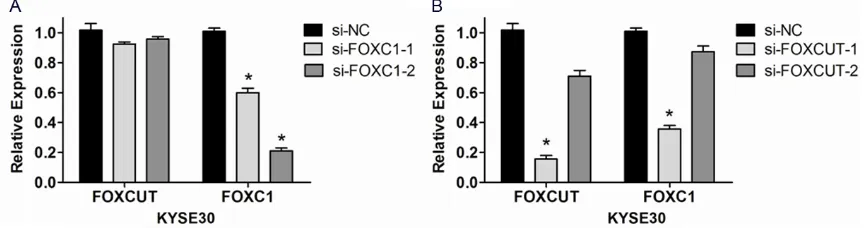
FOXC1 expression in KYSE30 cells was sup-pressed by FOXC1 siRNA and FOXCUT siRNA
In ESCC cell line KYSE30, RNAi technique was executed to further demonstrate the correla-tion between the expression of lncRNA-FOXCUT and mRNA FOXC1. The results proved that the FOXC1 expression level was apparently kn- ocked down by two kinds of FOXC1 siRNAs (P < 0.05, Figure 4A). And si-FOXC1-2 played more
significant effect than si-FOXC1-1 (Figure 4A). Moreover, the FOXC1 expression level was also down-regulated in both FOXCUT siRNAs com-pared with the control siRNA (Figure 4B). Particularly, as the lncRNA-FOXCUT expression was knocked down up to 86% by si-FOXCUT-1, the FOXC1 mRNA expression was co-suppr- essed by 64% (Figure 4B). Whereas, the FOXCUT expression levels did not decrease together with the FOXC1 downregulation caused by FOXC1 siRNAs (Figure 4A). Consider-
-sion of mRNA FOXC1 might be regulated by lncRNA FOXCUT.
Knockdown of FOXC1 inhibited cell prolifera-tion, migraprolifera-tion, invasion abilities in KYSE30
Cell proliferation, migration, invasion abilities were important aspects of cancer progression. To clarify whether FOXC1 has a functional role in facilitating ESCC cell progression, a series of cell function experiments were conducted in KYSE30 after siRNA transfection. MTS assay and colony formation assay showed that sup-pression of FOXC1 notably repressed the cell proliferation of KYSE30 in contrast with the negative control (Figure 5A) and the number of cell colonies in the knockdown of FOXC1 groups
was also significantly reduced compared with
the NC siRNA group (Figure 5B). And si-FOXC1-2
played more significant value than si-FOXC1-1
in KYSE30 which was in accordance with down-regulation of FOXC1 by the two FOXC1 siRNAs (Figures 4A, 5A and 5B). Moreover, both scratch wound healing assay and migration assay proved that knockdown of FOXC1 inhibit-ed cell migration by 52% and 54% respectively (Figure 5C, 5D). In addition, the matrigel inva-sion assay similarly indicated the silence of FOXC1 in KYSE30 cells declined cell invasion in the Matrigel substrate by 56% (Figure 5E). These data demonstrated that FOXC1 promot-ed cell proliferation, enhancpromot-ed cell migration, invasion abilities in KYSE30.
Knockdown of FOXCUT inhibited cell prolif-eration, migration and invasion abilities in KYSE30
[image:7.612.90.524.75.229.2]To further testify the function of FOXCUT on the growth characteristics of the ESCC cell lines,
Figure 3. Kaplan-Meier overall survival curve for ESCC patients (n = 82) with different FOXC1 and FOXCUT expres-sion levels. A: Difference in overall survival for ESCC patients with high expresexpres-sion and low expresexpres-sion of FOXC1 was analyzed by log-rank test (P = 0.014). Patients with high expression of FOXC1 had a significantly worse prognosis than those with low expression of FOXC1. B: Difference in overall survival for ESCC patients with high expression and low expression of FOXCUT was analyzed by log-rank test (P < 0.001). Patients with high expression of FOXCUT had a remarkably worse prognosis than those with low expression of FOXCUT.
[image:7.612.92.525.324.438.2]the same series of cell function experiments were performed in KYSE30 after siRNA trans-fection. The results showed that cell
prolifera-tion ability of KYSE30 was also efficiently sup -pressed by si-FOXCUT (Figure 6A) and the nu- mber of cell colonies was apparently decreased through knockdown of FOXCUT compared with the NC siRNA (Figure 6B). Similarly, si-FOXCUT-1 played more vital part than si-FOXCUT-2 in KYSE30 corresponding to downregulation of FOXCUT by the two FOXCUT siRNAs (Figures 4B,
6A and 6B). Furthermore, silence of FOXCUT inhibited cell migration by 44% and 48% respectively (Figure 6C, 6D) by scratch wound healing assay and migration assay. Besides, the matrigel invasion assay similarly proved the downregulation of FOXCUT in KYSE30 cells
retarded cell invasion significantly in the Matr-
igel substrate by 43% (Figure 6E). Taking these remarkable results into account, FOXCUT may promote cell proliferation, enhance cell migra-tion, invasion abilities in KYSE30.
Discussion
In the present study, we first identified a new
lncRNA-mRNA pair, lncRNAFOXCUT and its adjacent mRNA FOXC1, as a new form of can-cer-related gene compound correlated clinically with aggressive biological behaviors and poor survival in ESCC.
[image:8.612.93.522.69.425.2]ESCC is a kind of tumor involved in complex dynamic biological processes and it initiates from multiple steps of genetic and epigenetic alterations [28]. Previous studies about ESCC
related genes mainly focused on protein-coding genes. Recently, advances in high-throughput sequencing has helped revealing great func-tions of lncRNAs in cancers [18]. Numerous new lncRNA molecules have been proved to
play significant roles in the tumorigenesis, pro -gression of various malignant tumors [29-31].
In ESCC, several lncRNAs have been identified
as new biomarkers and therapeutic targets such as PlncRNA-1, HOTAIR [32, 33]. However, the expression and functional roles of most lncRNAs are still unknown in ESCC.
Emerging evidences have demonstrated that FOXC1 was overexpressed in a wide variety of malignant cancers and it can promote tumori-genesis, and tumor progression [21-23]. In our
current study, we found a new lncRNA FOXCUT transcribed from the upstream side of FOXC1 promoter through bioinformatics analysis. FOXCUT, an adjacent lncRNA of FOXC1, belongs to promoter upstream transcripts (PROMPTs) [34, 35]. The PROMPTs are often associated with the adjacent protein coding transcripts in their functions [19]. So, we speculated that lncRNA FOXCUT and mRNA FOXC1 may consti-tute into a new functional lncRNA-mRNA gene pair.
In our study, it is the first time to show that the
expression of FOXCUT and FOXC1
(lncRNA-mRNA gene pair) were both identified to be
upregulated in 82 ESCC tissues compared with adjacent noncancerous tissues. Linear
[image:9.612.92.522.70.427.2]sion analysis revealed that FOXCUT and FOXC1 had a positive correlation with each other in ESCC. Furthermore, the expression of FOXC1 was also decreased as the FOXCUT expression was downregulated by siRNA. Whereas, the
silence of FOXC1 did not influence the expres -sion of FOXCUT. It indicates that lncRNA FOXCUT may regulate the expression of FOXC1 by some
specific mechanisms, which needs further
researches to be elucidated completely.
Additionally, our study demonstrated that FOXC1 and FOXCUT expression levels were
sig-nificantly elevated in tumor samples from
patients with the presence of metastasis and poor differentiation. Upregulation of FOXC1 and FOXCUT were both correlated with aggressive clinicopathological characteristics, such as poor differentiation, advanced lymph node
classification, metastasis. And, patients with
high expression of FOXCUT or FOXC1 had a
sig-nificantly poor prognosis. These data suggests
that high expression of FOXCUT or FOXC1 might play an important role in the tumorigenesis, development, progression of ESCC, and both of them may serve as prognostic biomarkers for ESCC patients.
To further explore the functional role of this new lncRNA-mRNA gene pair in ESCC cells, we designed siRNAs to knock down the expression of FOXC1 or FOXCUT. Functional assays in vitro
demonstrated that knockdown of FOXCUT or FOXC1 remarkably impeded cell proliferation, migration and invasion abilities in ESCC cells which were consistent with the reported func-tions of FOXC1 in other cancers [22]. The results indicate that this new lncRNA-mRNA gene pair is an important compound function-ing in the ESCC cell aggressive biological behaviours, similar to the well-know lncRNA, HOTAIR in ESCC [6, 32].
In conclusion, FOXCUT and FOXC1, the new lncRNA-mRNA gene pair, are both novel upregu-lated functional molecules in ESCC. Upregula- tion of the lncRNA-mRNA pair may be a prog-nostic factor for ESCC patients, indicating short survival and high risk for metastasis and poor differentiation. FOXCUT may inhibit ESCC cell proliferation, migration and invasion abilities, partially by the regulation of FOXC1 expression. And both FOXCUT and FOXC1 may serve as potential diagnostic markers and therapeutic targets for future ESCC treatment.
Acknowledgements
This study was supported by the National Nat- ural Science Foundation of China (81301781). We would like to thank the Key Laboratory of Oncology, Cancer Center, Division of Internal Medicine, Chinese PLA General Hospital & Chinese PLA Medical School.
Disclosure of conflict of interest
None.
Address correspondence to: Dr. Xiangqun Fang, Nanlou Department of Respiratory Disease, Chinese PLA General Hospital, 28 Fuxing Road, Beijing 100853, China. E-mail: xiangqunf@163.com
References
[1] Enzinger PC and Mayer RJ. Esophageal cancer. N Engl J Med 2003; 349: 2241-2252. [2] Kano M, Seki N, Kikkawa N, Fujimura L,
Hoshi-no I, Akutsu Y, Chiyomaru T, EHoshi-nokida H, Nak-agawa M and Matsubara H. 145, miR-133a and miR-133b: Tumor-suppressive miRNAs target FSCN1 in esophageal squa-mous cell carcinoma. Int J Cancer 2010; 127: 2804-2814.
[3] Hiyama T, Yoshihara M, Tanaka S and Chaya-ma K. Genetic polymorphisms and esophageal cancer risk. Int J Cancer 2007; 121: 1643-1658.
[4] Li T, Lu ZM, Chen KN, Guo M, Xing HP, Mei Q, Yang HH, Lechner JF and Ke Y. Human papillo-mavirus type 16 is an important infectious fac-tor in the high incidence of esophageal cancer in Anyang area of China. Carcinogenesis 2001; 22: 929-934.
[5] Sato F, Shimada Y, Watanabe G, Uchida S, Makino T and Imamura M. Expression of vas-cular endothelial growth factor, matrix metal-loproteinase-9 and E-cadherin in the process of lymph node metastasis in oesophageal can-cer. Br J Cancer 1999; 80: 1366-1372. [6] Lv XB, Lian GY, Wang HR, Song E, Yao H and
Wang MH. Long noncoding RNA HOTAIR is a prognostic marker for esophageal squamous cell carcinoma progression and survival. PLoS One 2013; 8: e63516.
[7] Roder JD, Busch R, Stein HJ, Fink U and Siew-ert JR. Ratio of invaded to removed lymph nodes as a predictor of survival in squamous cell carcinoma of the oesophagus. Br J Surg 1994; 81: 410-413.
TNM staging in esophageal cancer: a patho-logical review of resected specimens. Ann Surg Oncol 2008; 15: 3447-3458.
[9] Wang LS, Chow KC, Chi KH, Liu CC, Li WY, Chiu JH and Huang MH. Prognosis of esophageal squamous cell carcinoma: analysis of clinico-pathological and biological factors. Am J Gas-troenterol 1999; 94: 1933-1940.
[10] Huarte M and Rinn JL. Large non-coding RNAs: missing links in cancer? Hum Mol Genet 2010; 19: R152-161.
[11] Prensner JR and Chinnaiyan AM. The emer-gence of lncRNAs in cancer biology. Cancer Discov 2011; 1: 391-407.
[12] Tsai MC, Spitale RC and Chang HY. Long inter-genic noncoding RNAs: new links in cancer progression. Cancer Res 2011; 71: 3-7. [13] Zhou Y, Zhang X and Klibanski A. MEG3
non-coding RNA: a tumor suppressor. J Mol Endo-crinol 2012; 48: R45-53.
[14] Qi P and Du X. The long non-coding RNAs, a new cancer diagnostic and therapeutic gold mine. Mod Pathol 2013; 26: 155-165.
[15] Ma L, Bajic VB and Zhang Z. On the classifica-tion of long non-coding RNAs. RNA Biol 2013; 10: 925-933.
[16] Guttman M, Amit I, Garber M, French C, Lin MF, Feldser D, Huarte M, Zuk O, Carey BW, Cas-sady JP, Cabili MN, Jaenisch R, Mikkelsen TS, Jacks T, Hacohen N, Bernstein BE, Kellis M, Regev A, Rinn JL and Lander ES. Chromatin signature reveals over a thousand highly con-served large non-coding RNAs in mammals. Nature 2009; 458: 223-227.
[17] Perez DS, Hoage TR, Pritchett JR, Ducharme-Smith AL, Halling ML, Ganapathiraju SC, Streng PS and Smith DI. Long, abundantly ex-pressed non-coding transcripts are altered in cancer. Hum Mol Genet 2008; 17: 642-655. [18] Cao W, Wu W, Shi F, Chen X, Wu L, Yang K, Tian
F, Zhu M, Chen G, Wang W, Biddle FG and Gu J. Integrated analysis of long noncoding RNA and coding RNA expression in esophageal squa-mous cell carcinoma. Int J Genomics 2013; 2013: 480534.
[19] Sigova AA, Mullen AC, Molinie B, Gupta S, Or-lando DA, Guenther MG, Almada AE, Lin C, Sharp PA, Giallourakis CC and Young RA. Diver-gent transcription of long noncoding RNA/ mRNA gene pairs in embryonic stem cells. Proc Natl Acad Sci U S A 2013; 110: 2876-2881.
[20] Hannenhalli S and Kaestner KH. The evolution of Fox genes and their role in development and disease. Nat Rev Genet 2009; 10: 233-240. [21] Myatt SS and Lam EW. The emerging roles of
forkhead box (Fox) proteins in cancer. Nat Rev Cancer 2007; 7: 847-859.
[22] Xia L, Huang W, Tian D, Zhu H, Qi X, Chen Z, Zhang Y, Hu H, Fan D, Nie Y and Wu K.
Overex-pression of forkhead box C1 promotes tumor metastasis and indicates poor prognosis in he-patocellular carcinoma. Hepatology 2013; 57: 610-624.
[23] Xu ZY, Ding SM, Zhou L, Xie HY, Chen KJ, Zhang W, Xing CY, Guo HJ and Zheng SS. FOXC1 con-tributes to microvascular invasion in primary hepatocellular carcinoma via regulating epi-thelial-mesenchymal transition. Int J Biol Sci 2012; 8: 1130-1141.
[24] Ray PS, Wang J, Qu Y, Sim MS, Shamonki J, Bagaria SP, Ye X, Liu B, Elashoff D, Hoon DS, Walter MA, Martens JW, Richardson AL, Giulia-no AE and Cui X. FOXC1 is a potential progGiulia-nos- prognos-tic biomarker with functional significance in basal-like breast cancer. Cancer Res 2010; 70: 3870-3876.
[25] Wang L, Gu F, Liu CY, Wang RJ, Li J and Xu JY. High level of FOXC1 expression is associated with poor prognosis in pancreatic ductal ade-nocarcinoma. Tumour Biol 2013; 34: 853-858. [26] Wei LX, Zhou RS, Xu HF, Wang JY and Yuan MH.
High expression of FOXC1 is associated with poor clinical outcome in non-small cell lung cancer patients. Tumour Biol 2013; 34: 941-946.
[27] Xu Y, Shao QS, Yao HB, Jin Y, Ma YY and Jia LH. Overexpression of FOXC1 correlates with poor prognosis in gastric cancer patients. Histopa-thology 2014; 64: 963-70.
[28] Hao JJ, Gong T, Zhang Y, Shi ZZ, Xu X, Dong JT, Zhan QM, Fu SB and Wang MR. Characteriza-tion of gene rearrangements resulted from ge-nomic structural aberrations in human esoph-ageal squamous cell carcinoma KYSE150 cells. Gene 2013; 513: 196-201.
[29] Kim K, Jutooru I, Chadalapaka G, Johnson G, Frank J, Burghardt R, Kim S and Safe S. HO-TAIR is a negative prognostic factor and exhib-its pro-oncogenic activity in pancreatic cancer. Oncogene 2013; 32: 1616-1625.
[30] Nakagawa T, Endo H, Yokoyama M, Abe J, Ta-mai K, Tanaka N, Sato I, Takahashi S, Kondo T and Satoh K. Large noncoding RNA HOTAIR en-hances aggressive biological behavior and is associated with short disease-free survival in human non-small cell lung cancer. Biochem Biophys Res Commun 2013; 436: 319-324. [31] Spizzo R, Almeida MI, Colombatti A and Calin
GA. Long non-coding RNAs and cancer: a new frontier of translational research? Oncogene 2012; 31: 4577-4587.
[32] Li X, Wu Z, Mei Q, Guo M, Fu X and Han W. Long non-coding RNA HOTAIR, a driver of malignan-cy, predicts negative prognosis and exhibits oncogenic activity in oesophageal squamous cell carcinoma. Br J Cancer 2013; 109: 2266-2278.
Shi WH and Cao XF. Upregulation of the Long Non-coding RNA PlncRNA-1 Promotes Esopha-geal Squamous Carcinoma Cell Proliferation and Correlates with Advanced Clinical Stage. Dig Dis Sci 2014; 59: 591-597.
[34] Preker P, Almvig K, Christensen MS, Valen E, Mapendano CK, Sandelin A and Jensen TH. PROMoter uPstream Transcripts share charac-teristics with mRNAs and are produced up-stream of all three major types of mammalian promoters. Nucleic Acids Res 2011; 39: 7179-7193.