organic papers
o1164
Lokajet al. C11H10N2O doi:10.1107/S1600536807004746 Acta Cryst.(2007). E63, o1164–o1166
Acta Crystallographica Section E
Structure Reports
Online
ISSN 1600-5368
3-Acetyl-4-aminoquinoline
Jan Lokaj,aViktor Kettmann,b* Petra Cˇ ernuchova´,aViktor Milataaand Marek Fronca
aFaculty of Food and Chemical Technology, Slovak Technical University, Radlinskeho 9, SK-81237 Bratislava, Slovak Republic, and bFaculty of Pharmacy, Comenius University, Odbojarov 10, SK-83232 Bratislava, Slovak Republic
Correspondence e-mail: [email protected]
Key indicators
Single-crystal X-ray study
T= 296 K
Mean(C–C) = 0.004 A˚
Rfactor = 0.063
wRfactor = 0.210
Data-to-parameter ratio = 16.3
For details of how these key indicators were automatically derived from the article, see http://journals.iucr.org/e.
Received 13 October 2006 Accepted 29 January 2007
#2007 International Union of Crystallography All rights reserved
The title compound, C11H10N2O, a potential antitumour drug, crystallizes with two molecules in the asymmetric unit. The 4-amino N atom is strongly conjugated with the quinoline ring involving the 3-carbonyl group. As a result, the molecule is almost planar. The amino group is involved in both intra-molecular N—H O and intermolecular N—H N hydrogen bonds.
Comment
Quinoline derivatives are known to possess a variety of biological properties (Balasubramanian & Key, 1996); recently it has been reported that addition of a substituent containing a carbonyl function (acetyl, ester or amide group) to the 3-position of 4-aminoquinoline endows the molecule with cytotoxic activity (Repicky´et al., 2005). The cytotoxic activity of such molecules is related to intercalation of their planar chromophore between adjacent base pairs of DNA, whereas the effect of the substituents can be connected to an additional intercalation component or to interaction with minor groove functionalities of DNA. The former effect requires coplanarity and the latter non-coplanarity of the substituent with respect to the plane of the chromophore. Thus, we were interested in detailed knowledge of the molecular structure of the above derivatives. In this communication we report the crystal structure of the 3-acetyl-4-amino derivative, (I) (Fig. 1).
The structure determination reveals two molecules (Aand B) in the asymmetric unit. Their geometrical parameters are similar. Thus, only moleculeAis shown in Fig. 1. The analysis is focused on the planarity of the molecules; the acetyl group and the quinoline heterocycle form dihedral angles of 2.7 (4) and 8.7 (4) for molecules AandB, respectively. The
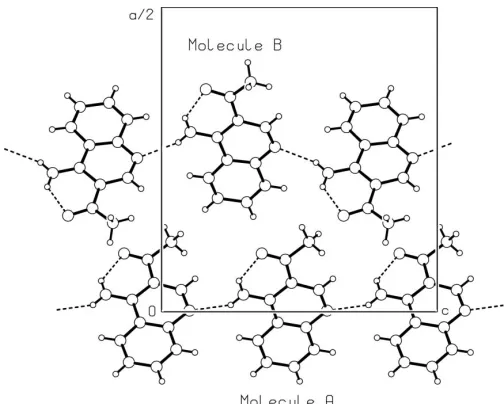
atom also contributes to the coplanarity of the acetyl group (Fig. 1 and Table 2). The other amino H atom (of both inde-pendent molecules) is involved in an intermolecular hydrogen bond with the quinoline N atom (Table 2), generating chains (Fig. 2) along thecaxis.
Experimental
The synthesis of the title compound, (I), has been described previously (Cˇ ernuchova´et al., 2005). In short, aniline (2 g, 21.6 mmol) and 2-ethoxymethylene-3-oxobutanenitrile (3 g, 21.6 mmol) were heated at 343 K for 2 min. The reaction mixture was cooled to room temperature, EtOH formed during the reaction was evaporated and the product recrystallized from toluene. Next, aluminium trichloride (2.1 g, 16.2 mmol) was added to 2-anilinomethylene-3-oxobutane-nitrile (1 g, 5.4 mmol) formed in the previous step and the mixture was stirred at 453 K for 1 h. The reaction mixture was poured on to ice, saturated with K2CO3 powder and extracted with
dichloro-methane (320 ml). After evaporation, the residue was purified by flash chromatography (hexane–AcOEt, 8:2) to afford the title compound (yield 45%; m.p. 426–4-27 K) as yellow crystals.
Crystal data
C11H10N2O
Mr= 186.21 Orthorhombic,Pccn a= 30.815 (6) A˚
b= 8.884 (2) A˚
c= 13.305 (3) A˚
V= 3642.4 (14) A˚3
Z= 16
Dx= 1.358 Mg m
3
MoKradiation
= 0.09 mm1
T= 296 (2) K Prism, yellow 0.300.200.15 mm
Data collection
Oxford Diffraction Gemini R CCD diffractometer
!and’scans
Absorption correction: none 10828 measured reflections
4160 independent reflections 1954 reflections withI> 2(I)
Rint= 0.041 max= 27.5
Refinement
Refinement onF2 R[F2> 2(F2)] = 0.063
wR(F2) = 0.210
S= 0.98 4160 reflections 255 parameters
H-atom parameters constrained
w= 1/[2(F
o2) + (0.1154P)2]
whereP= (Fo2+ 2Fc2)/3
(/)max= 0.003
max= 0.58 e A˚3
min=0.29 e A˚3
Table 1
Selected geometric parameters (A˚ ,).
C3A—C4A 1.432 (3) C3A—C11A 1.453 (3) C4A—N14A 1.323 (3) C11A—O12A 1.225 (3)
C3B—C4B 1.420 (4) C3B—C11B 1.471 (4) C4B—N14B 1.333 (3) C11B—O12B 1.233 (4)
C2A—C3A—C4A 117.0 (2) C2A—C3A—C11A 121.7 (2) C4A—C3A—C11A 121.3 (2) N14A—C4A—C3A 121.4 (2) N14A—C4A—C10A 120.6 (2) C3A—C4A—C10A 117.9 (2)
C2B—C3B—C4B 117.3 (2) C2B—C3B—C11B 122.0 (3) C4B—C3B—C11B 120.6 (3) N14B—C4B—C3B 121.6 (3) N14B—C4B—C10B 121.6 (3) C3B—C4B—C10B 116.8 (2)
C11A—C3A—C4A—N14A 1.5 (3) C4A—C3A—C11A—O12A 0.5 (3)
[image:2.610.59.273.73.209.2]C11B—C3B—C4B—N14B 1.0 (4) C4B—C3B—C11B—O12B 5.7 (4)
Table 2
Hydrogen-bond geometry (A˚ ,).
D—H A D—H H A D A D—H A
N14A—H14A O12A 0.86 1.99 2.639 (3) 132 N14A—H14B N1Ai
0.86 2.10 2.879 (3) 150 N14B—H14C O12B 0.86 1.96 2.615 (3) 132 N14B—H14D N1Bii
0.86 2.14 2.921 (3) 151
Symmetry codes: (i)x;yþ1 2;z
1 2; (ii)xþ
3 2;y;zþ
1 2.
H atoms were treated as riding, with C—H = 0.93 (aromatic CH) or 0.96 A˚ (CH3) and N—H = 0.86 A˚ (NH2);Uiso(H) values were set at
1.2 (1.5 for the methyl H atoms) times Ueq of the parent atom.
Because of the indication from the Hirshfeld test (Hirshfeld, 1976), 72 rigid-bond restraints on anisotropic displacement parameters for all bonds involving the non-H atoms were applied during the least-squares refinement.
Data collection: CrysAlis CCD (Oxford Diffraction, 2001); cell refinement: CrysAlis RED (Oxford Diffraction, 2003); data reduc-tion:CrysAlis RED; program(s) used to solve structure:SHELXS97 (Sheldrick, 1997); program(s) used to refine structure:SHELXL97
organic papers
Acta Cryst.(2007). E63, o1164–o1166 Lokajet al. C
11H10N2O
o1165
Figure 1
[image:2.610.44.296.261.463.2]The molecular structure of one of the two independent molecules of (I), showing the atom-labelling scheme for the non-H atoms. Displacement ellipsoids are drawn at the 35% probability level. The intramolecular hydrogen bond is shown as a dashed line.
Figure 2
[image:2.610.314.565.538.596.2](Sheldrick, 1997); molecular graphics:PLUTON(Spek, 2003); soft-ware used to prepare material for publication:SHELXL97.
This work was supported by the Grant Agency of the Slovak Republic, project Nos. 1/1167/04, 1/2448/05 and 1/3587/ 06, as well as by the Science and Technology Assistance Agency (grant No. APVT-20–00734).
References
Balasubramanian, M. & Key, J. G. (1996).Pyridines and their Benzoderivatives: Applications, In The Chemistry of Heterocyclic Compounds, Vol. 5, edited by A. R. Katritzky, C. W Rees & E. F. V. Scriven, pp. 245–300. Oxford: Pergamon–Elsevier.
Cˇ ernuchova´, P., Vo-Thanh, G., Milata, V., Loupy, A., Jantova´, S. & Theiszova´, M. (2005).Tetrahedron,61, 5379–5388.
Davies, J. E. & Bond, A. D. (2001).Acta Cryst.E57, o947–o949. Hirshfeld, F. L. (1976).Acta Cryst.A32, 239–244.
Oxford Diffraction (2001).CrysAlis CCD. Version 169.3. Oxford Diffraction Ltd, Abingdon, Oxfordshire, England.
Oxford Diffraction (2003).CrysAlis RED. Version 170.17. Oxford Diffraction Ltd, Abingdon, Oxfordshire, England.
Repicky´, A., Jantova´, S., Theiszova´, M. & Milata, V. (2005).Biomed. Pap. Med. Fac. Univ. Palacky Olomouc Czech. Rep.49, 345–347.
Sheldrick, G. M. (1997). SHELXS97 and SHELXL97. University of Go¨ttingen, Germany.
Shmueli, U., Shanan-Atidi, H., Horwitz, H. & Shvo, Y. (1973).J. Chem. Soc. Perkin Trans. 2, pp. 657–662.
Spek, A. L. (2003).J. Appl. Cryst.36, 7–13.
organic papers
o1166
Lokajet al. Csupporting information
sup-1 Acta Cryst. (2007). E63, o1164–o1166
supporting information
Acta Cryst. (2007). E63, o1164–o1166 [https://doi.org/10.1107/S1600536807004746]
3-Acetyl-4-aminoquinoline
Jan Lokaj, Viktor Kettmann, Petra
Č
ernuchov
á
, Viktor Milata and Marek Fronc
3-Acetyl-4-aminoquinoline
Crystal data
C11H10N2O
Mr = 186.21 Orthorhombic, Pccn
Hall symbol: -P 2ab 2ac
a = 30.815 (6) Å
b = 8.884 (2) Å
c = 13.305 (3) Å
V = 3642.4 (14) Å3
Z = 16
F(000) = 1568
Dx = 1.358 Mg m−3
Melting point: 426 K
Mo Kα radiation, λ = 0.71073 Å Cell parameters from 20 reflections
θ = 7–20°
µ = 0.09 mm−1
T = 296 K Prism, yellow
0.30 × 0.20 × 0.15 mm
Data collection
Oxford Difraction Gemini R CCD diffractometer
Radiation source: fine-focus sealed X-ray tube Graphite monochromator
ω and φ scans
10828 measured reflections 4160 independent reflections
1954 reflections with I > 2σ(I)
Rint = 0.041
θmax = 27.5°, θmin = 3.1°
h = 0→39
k = −11→11
l = −17→17
Refinement
Refinement on F2
Least-squares matrix: full
R[F2 > 2σ(F2)] = 0.063
wR(F2) = 0.210
S = 0.98 4160 reflections 255 parameters 72 restraints
Primary atom site location: structure-invariant direct methods
Secondary atom site location: difference Fourier map
Hydrogen site location: inferred from neighbouring sites
H-atom parameters constrained
w = 1/[σ2(F
o2) + (0.1154P)2]
where P = (Fo2 + 2Fc2)/3
(Δ/σ)max = 0.003
Δρmax = 0.58 e Å−3
supporting information
sup-2 Acta Cryst. (2007). E63, o1164–o1166
Special details
Geometry. All e.s.d.'s (except the e.s.d. in the dihedral angle between two l.s. planes) are estimated using the full covariance matrix. The cell e.s.d.'s are taken into account individually in the estimation of e.s.d.'s in distances, angles and torsion angles; correlations between e.s.d.'s in cell parameters are only used when they are defined by crystal symmetry. An approximate (isotropic) treatment of cell e.s.d.'s is used for estimating e.s.d.'s involving l.s. planes.
Least-squares planes (x,y,z in crystal coordinates) and deviations from them (* indicates atom used to define plane) 18.2732 (0.0119) x - 7.1393 (0.0029) y + 0.6729 (0.0090) z = 7.7599 (0.0086)
* 0.0042 (0.0017) N1A * 0.0026 (0.0018) C2A * 0.0011 (0.0018) C3A * -0.0006 (0.0017) C4A * -0.0032 (0.0018) C5A * 0.0091 (0.0019) C6A * 0.0020 (0.0019) C7A * -0.0083 (0.0019) C8A * -0.0009 (0.0020) C9A * -0.0061 (0.0019) C10A 0.0311 (0.0029) C11A -0.0040 (0.0028) N14A
Rms deviation of fitted atoms = 0.0048
17.5837 (0.0928) x - 7.2512 (0.0194) y + 1.2046 (0.0188) z = 7.7392 (0.0415) Angle to previous plane (with approximate e.s.d.) = 2.72 (0.35)
* 0.0000 (0.0000) C11A * 0.0000 (0.0000) O12A * 0.0000 (0.0000) C13A Rms deviation of fitted atoms = 0.0000
18.8594 (0.0128) x - 7.0254 (0.0031) y - 0.1183 (0.0100) z = 6.9644 (0.0129) Angle to previous plane (with approximate e.s.d.) = 6.34 (0.35)
* 0.0032 (0.0020) N1B * 0.0108 (0.0021) C2B * -0.0117 (0.0019) C3B * -0.0077 (0.0020) C4B * 0.0131 (0.0021) C5B * -0.0107 (0.0022) C6B * 0.0038 (0.0020) C7B * -0.0080 (0.0022) C8B * -0.0052 (0.0022) C9B * 0.0124 (0.0021) C10B -0.0461 (0.0035) C11B -0.0553 (0.0034) N14B
Rms deviation of fitted atoms = 0.0093
20.4498 (0.1097) x - 6.5536 (0.0265) y + 1.6519 (0.0275) z = 9.6706 (0.0792) Angle to previous plane (with approximate e.s.d.) = 8.73 (0.43)
* 0.0000 (0.0000) C11B * 0.0000 (0.0000) O12B * 0.0000 (0.0000) C13B Rms deviation of fitted atoms = 0.0000
Refinement. Refinement of F2 against ALL reflections. The weighted R-factor wR and goodness of fit S are based on F2,
conventional R-factors R are based on F, with F set to zero for negative F2. The threshold expression of F2 > σ(F2) is used
only for calculating R-factors(gt) etc. and is not relevant to the choice of reflections for refinement. R-factors based on F2
are statistically about twice as large as those based on F, and R- factors based on ALL data will be even larger.
Fractional atomic coordinates and isotropic or equivalent isotropic displacement parameters (Å2)
x y z Uiso*/Ueq
N1A 0.49820 (7) 0.2443 (2) 0.60074 (15) 0.0344 (5) C2A 0.46573 (9) 0.1584 (3) 0.57123 (19) 0.0336 (6)
H2A 0.4483 0.1186 0.6216 0.040*
C3A 0.45456 (8) 0.1203 (3) 0.47153 (19) 0.0301 (5) C4A 0.48109 (8) 0.1815 (3) 0.39309 (19) 0.0277 (5) C5A 0.54515 (8) 0.3417 (3) 0.3503 (2) 0.0316 (6)
H5A 0.5407 0.3247 0.2821 0.038*
C6A 0.57914 (9) 0.4298 (3) 0.3812 (2) 0.0363 (6)
H6A 0.5978 0.4711 0.3336 0.044*
C7A 0.58609 (9) 0.4581 (3) 0.4818 (2) 0.0379 (6)
H7A 0.6092 0.5188 0.5014 0.045*
C8A 0.55904 (9) 0.3971 (3) 0.5534 (2) 0.0369 (6)
H8A 0.5638 0.4174 0.6210 0.044*
supporting information
sup-3 Acta Cryst. (2007). E63, o1164–o1166
H13A 0.3673 −0.0946 0.5095 0.060*
H13B 0.3780 0.0528 0.5694 0.060*
H13C 0.4076 −0.0896 0.5813 0.060*
N14A 0.47301 (7) 0.1523 (2) 0.29715 (15) 0.0346 (6)
H14A 0.4515 0.0956 0.2810 0.041*
H14B 0.4893 0.1901 0.2511 0.041*
N1B 0.74114 (9) 0.9880 (3) 0.58896 (19) 0.0516 (6) C2B 0.71028 (10) 0.9036 (3) 0.6225 (2) 0.0472 (7)
H2B 0.6932 0.8557 0.5745 0.057*
C3B 0.69967 (9) 0.8764 (3) 0.7228 (2) 0.0427 (6) C4B 0.72594 (10) 0.9451 (3) 0.7975 (2) 0.0417 (6) C5B 0.78981 (10) 1.1131 (3) 0.8287 (2) 0.0480 (7)
H5B 0.7862 1.0995 0.8975 0.058*
C6B 0.82163 (11) 1.2024 (4) 0.7964 (3) 0.0584 (8)
H6B 0.8394 1.2529 0.8418 0.070*
C7B 0.82795 (10) 1.2192 (3) 0.6907 (3) 0.0495 (7)
H7B 0.8507 1.2784 0.6675 0.059*
C8B 0.80178 (11) 1.1518 (3) 0.6244 (3) 0.0535 (8)
H8B 0.8062 1.1662 0.5559 0.064*
C9B 0.76645 (9) 1.0559 (3) 0.6588 (2) 0.0414 (6) C10B 0.76163 (9) 1.0388 (3) 0.7622 (2) 0.0374 (6) C11B 0.66261 (11) 0.7815 (4) 0.7511 (3) 0.0568 (8) O12B 0.65085 (8) 0.7670 (3) 0.83945 (19) 0.0685 (7) C13B 0.63948 (11) 0.6905 (4) 0.6767 (3) 0.0684 (10)
H13D 0.6478 0.5869 0.6838 0.103*
H13E 0.6467 0.7248 0.6104 0.103*
H13F 0.6088 0.7002 0.6871 0.103*
N14B 0.71774 (9) 0.9281 (3) 0.89539 (19) 0.0596 (8)
H14C 0.6961 0.8743 0.9146 0.071*
H14D 0.7341 0.9708 0.9392 0.071*
Atomic displacement parameters (Å2)
U11 U22 U33 U12 U13 U23
supporting information
sup-4 Acta Cryst. (2007). E63, o1164–o1166
N1B 0.0609 (16) 0.0521 (16) 0.0419 (12) 0.0082 (11) −0.0021 (10) 0.0020 (12) C2B 0.0531 (17) 0.0424 (17) 0.0462 (12) 0.0095 (11) −0.0099 (12) −0.0104 (13) C3B 0.0365 (15) 0.0344 (15) 0.0571 (12) 0.0085 (10) 0.0003 (11) 0.0039 (13) C4B 0.0472 (16) 0.0384 (15) 0.0395 (11) 0.0077 (10) 0.0054 (11) 0.0040 (12) C5B 0.0470 (17) 0.0448 (17) 0.0520 (15) 0.0019 (12) −0.0064 (12) −0.0013 (14) C6B 0.0512 (19) 0.0538 (19) 0.0701 (15) −0.0038 (13) −0.0055 (16) 0.0069 (17) C7B 0.0385 (17) 0.0370 (16) 0.0730 (16) 0.0053 (12) 0.0059 (14) 0.0066 (15) C8B 0.059 (2) 0.0453 (18) 0.0565 (16) 0.0034 (12) 0.0081 (13) 0.0094 (14) C9B 0.0363 (15) 0.0437 (16) 0.0442 (12) 0.0101 (10) 0.0046 (11) 0.0060 (12) C10B 0.0363 (14) 0.0335 (14) 0.0425 (11) 0.0103 (9) −0.0048 (10) 0.0010 (12) C11B 0.0473 (18) 0.0466 (18) 0.0764 (18) 0.0022 (12) 0.0062 (14) −0.0071 (16) O12B 0.0649 (16) 0.0548 (14) 0.0859 (16) −0.0136 (12) 0.0252 (13) −0.0042 (13) C13B 0.055 (2) 0.0394 (17) 0.111 (2) 0.0077 (14) −0.017 (2) −0.0160 (18) N14B 0.070 (2) 0.0594 (17) 0.0490 (12) −0.0191 (15) 0.0031 (13) 0.0010 (13)
Geometric parameters (Å, º)
N1A—C2A 1.321 (3) N1B—C2B 1.291 (4)
N1A—C9A 1.396 (3) N1B—C9B 1.355 (4)
C2A—C3A 1.410 (3) C2B—C3B 1.396 (4)
C2A—H2A 0.9300 C2B—H2B 0.9300
C3A—C4A 1.432 (3) C3B—C4B 1.420 (4)
C3A—C11A 1.453 (3) C3B—C11B 1.471 (4)
C4A—N14A 1.323 (3) C4B—N14B 1.333 (3)
C4A—C10A 1.439 (3) C4B—C10B 1.457 (4)
C5A—C6A 1.369 (3) C5B—C6B 1.336 (4)
C5A—C10A 1.408 (3) C5B—C10B 1.403 (3)
C5A—H5A 0.9300 C5B—H5B 0.9300
C6A—C7A 1.375 (3) C6B—C7B 1.428 (6)
C6A—H6A 0.9300 C6B—H6B 0.9300
C7A—C8A 1.380 (3) C7B—C8B 1.338 (4)
C7A—H7A 0.9300 C7B—H7B 0.9300
C8A—C9A 1.408 (3) C8B—C9B 1.456 (4)
C8A—H8A 0.9300 C8B—H8B 0.9300
C9A—C10A 1.418 (3) C9B—C10B 1.392 (4)
C11A—O12A 1.225 (3) C11B—O12B 1.233 (4) C11A—C13A 1.511 (3) C11B—C13B 1.464 (4)
C13A—H13A 0.9600 C13B—H13D 0.9600
C13A—H13B 0.9600 C13B—H13E 0.9600
C13A—H13C 0.9600 C13B—H13F 0.9600
N14A—H14A 0.8600 N14B—H14C 0.8600
N14A—H14B 0.8600 N14B—H14D 0.8600
C2A—N1A—C9A 116.3 (2) C2B—N1B—C9B 116.3 (2) N1A—C2A—C3A 127.3 (2) N1B—C2B—C3B 127.1 (3)
N1A—C2A—H2A 116.3 N1B—C2B—H2B 116.5
C3A—C2A—H2A 116.3 C3B—C2B—H2B 116.5
supporting information
sup-5 Acta Cryst. (2007). E63, o1164–o1166
C2A—C3A—C11A 121.7 (2) C2B—C3B—C11B 122.0 (3) C4A—C3A—C11A 121.3 (2) C4B—C3B—C11B 120.6 (3) N14A—C4A—C3A 121.4 (2) N14B—C4B—C3B 121.6 (3) N14A—C4A—C10A 120.6 (2) N14B—C4B—C10B 121.6 (3) C3A—C4A—C10A 117.9 (2) C3B—C4B—C10B 116.8 (2) C6A—C5A—C10A 120.5 (2) C6B—C5B—C10B 121.8 (3)
C6A—C5A—H5A 119.8 C6B—C5B—H5B 119.1
C10A—C5A—H5A 119.8 C10B—C5B—H5B 119.1 C5A—C6A—C7A 121.2 (2) C5B—C6B—C7B 119.0 (3)
C5A—C6A—H6A 119.4 C5B—C6B—H6B 120.5
C7A—C6A—H6A 119.4 C7B—C6B—H6B 120.5
C6A—C7A—C8A 120.2 (2) C8B—C7B—C6B 121.2 (3)
C6A—C7A—H7A 119.9 C8B—C7B—H7B 119.4
C8A—C7A—H7A 119.9 C6B—C7B—H7B 119.4
C7A—C8A—C9A 120.5 (2) C7B—C8B—C9B 120.5 (3)
C7A—C8A—H8A 119.8 C7B—C8B—H8B 119.8
C9A—C8A—H8A 119.8 C9B—C8B—H8B 119.8
N1A—C9A—C8A 117.9 (2) N1B—C9B—C10B 124.6 (3) N1A—C9A—C10A 123.1 (2) N1B—C9B—C8B 118.2 (3) C8A—C9A—C10A 119.0 (2) C10B—C9B—C8B 116.7 (3) C5A—C10A—C9A 118.7 (2) C9B—C10B—C5B 120.7 (3) C5A—C10A—C4A 122.7 (2) C9B—C10B—C4B 117.4 (2) C9A—C10A—C4A 118.6 (2) C5B—C10B—C4B 122.2 (2) O12A—C11A—C3A 122.8 (2) O12B—C11B—C13B 116.3 (3) O12A—C11A—C13A 118.6 (2) O12B—C11B—C3B 122.2 (3) C3A—C11A—C13A 118.6 (2) C13B—C11B—C3B 121.4 (3) C11A—C13A—H13A 109.5 C11B—C13B—H13D 109.5 C11A—C13A—H13B 109.5 C11B—C13B—H13E 109.5 H13A—C13A—H13B 109.5 H13D—C13B—H13E 109.5 C11A—C13A—H13C 109.5 C11B—C13B—H13F 109.5 H13A—C13A—H13C 109.5 H13D—C13B—H13F 109.5 H13B—C13A—H13C 109.5 H13E—C13B—H13F 109.5 C4A—N14A—H14A 120.0 C4B—N14B—H14C 120.0 C4A—N14A—H14B 120.0 C4B—N14B—H14D 120.0 H14A—N14A—H14B 120.0 H14C—N14B—H14D 120.0
supporting information
sup-6 Acta Cryst. (2007). E63, o1164–o1166
C7A—C8A—C9A—N1A 179.2 (2) C7B—C8B—C9B—N1B −179.0 (2) C7A—C8A—C9A—C10A −1.1 (3) C7B—C8B—C9B—C10B −0.1 (4) C6A—C5A—C10A—C9A 0.2 (3) N1B—C9B—C10B—C5B 179.4 (2) C6A—C5A—C10A—C4A −179.1 (2) C8B—C9B—C10B—C5B 0.6 (4) N1A—C9A—C10A—C5A −179.6 (2) N1B—C9B—C10B—C4B −2.2 (4) C8A—C9A—C10A—C5A 0.7 (3) C8B—C9B—C10B—C4B 179.0 (2) N1A—C9A—C10A—C4A −0.3 (3) C6B—C5B—C10B—C9B 0.4 (4) C8A—C9A—C10A—C4A −179.9 (2) C6B—C5B—C10B—C4B −177.9 (2) N14A—C4A—C10A—C5A −0.6 (3) N14B—C4B—C10B—C9B −176.9 (2) C3A—C4A—C10A—C5A 179.8 (2) C3B—C4B—C10B—C9B 1.4 (3) N14A—C4A—C10A—C9A −179.9 (2) N14B—C4B—C10B—C5B 1.5 (4) C3A—C4A—C10A—C9A 0.4 (3) C3B—C4B—C10B—C5B 179.8 (2) C2A—C3A—C11A—O12A −178.0 (2) C2B—C3B—C11B—O12B −173.9 (3) C4A—C3A—C11A—O12A 0.5 (3) C4B—C3B—C11B—O12B 5.7 (4) C2A—C3A—C11A—C13A 4.5 (3) C2B—C3B—C11B—C13B 10.3 (4) C4A—C3A—C11A—C13A −176.9 (2) C4B—C3B—C11B—C13B −170.0 (2)
Hydrogen-bond geometry (Å, º)
D—H···A D—H H···A D···A D—H···A
N14A—H14A···O12A 0.86 1.99 2.639 (3) 132 N14A—H14B···N1Ai 0.86 2.10 2.879 (3) 150
N14B—H14C···O12B 0.86 1.96 2.615 (3) 132 N14B—H14D···N1Bii 0.86 2.14 2.921 (3) 151