7-Chloro-4-[(
E
)-
N
000-(4-fluorobenzylidene)-hydrazinyl]quinoline monohydrate
Marcus V. N. de Souza,aR. Alan Howie,b Edward R. T. Tiekink,c* James L. Wardelld‡ and Solange M. S. V. Wardelle
aInstituto de Tecnologia em Farmacos, Fundac¸a˜o Oswaldo Cruz (FIOCRUZ), FarManguinhos, Rua Sizenando Nabuco, 100, Manguinhos, 21041-250 Rio de Janeiro, RJ, Brazil,bDepartment of Chemistry, University of Aberdeen, Old Aberdeen AB15 5NY, Scotland,cDepartment of Chemistry, University of Malaya, 50603 Kuala Lumpur, Malaysia,dCentro de Desenvolvimento Tecnolo´gico em Sau´de (CDTS), Fundac¸a˜o Oswaldo Cruz (FIOCRUZ), Casa Amarela, Campus de Manguinhos, Av. Brasil 4365, 21040-900 Rio de Janeiro, RJ, Brazil, andeCHEMSOL, 1 Harcourt Road, Aberdeen AB15 5NY, Scotland
Correspondence e-mail: edward.tiekink@gmail.com
Received 9 December 2009; accepted 11 December 2009
Key indicators: single-crystal X-ray study;T= 120 K; mean(C–C) = 0.004 A˚; Rfactor = 0.059;wRfactor = 0.131; data-to-parameter ratio = 16.1.
The molecule of the title hydrate, C16H11ClFN3H2O, is slightly
twisted, as indicated by the dihedral angle of 9.55 (10) formed between the quinoline ring system and the benzene ring. The conformation about the C N double bond isE, and the amine-H atom is oriented towards the quinoline residue. In the crystal structure, the water molecule accepts an N— H O and makes two O—H Nquinoline hydrogen bonds,
generating a two-dimensional array in the abplane, which is further stabilized by C—H O interactions. The most significant contacts between layers are of the type C—H F.
Related literature
For background information on the pharmacological activity of quinoline derivatives, see: Elslageret al.(1969); Fontet al. (1997); Kaminsky & Meltzer (1968); Musiol et al. (2006); Nakamura et al. (1999); Palmeret al. (1993); Ridley (2002); Sloboda et al. (1991); Tanenbaum & Tuffanelli (1980); Warshakoon et al.(2006). For recent studies into quinoline-based anti-malarials, see: Andradeet al.(2007); Cunicoet al. (2006); da Silvaet al.(2003); de Souzaet al.(2005). For crys-tallographic studies on molecules related to the title compound, see: Kaiseret al.(2009); de Souzaet al.(2009); de Ferreira et al. (2009). For the synthesis, see: Pellerano et al. (1976).
Experimental
Crystal data
C16H11ClFN3H2O
Mr= 317.74 Monoclinic,P21=c
a= 3.7795 (2) A˚
b= 15.4188 (11) A˚
c= 24.8576 (16) A˚
= 90.286 (4)
V= 1448.57 (16) A˚3
Z= 4
MoKradiation
= 0.28 mm1
T= 120 K
0.900.040.04 mm
Data collection
Nonius KappaCCD area-detector diffractometer
Absorption correction: multi-scan (SADABS; Sheldrick, 2007)
Tmin= 0.614,Tmax= 0.746
19494 measured reflections 3291 independent reflections 2009 reflections withI> 2(I)
Rint= 0.098
Refinement
R[F2> 2(F2)] = 0.059
wR(F2) = 0.131
S= 1.04 3291 reflections 205 parameters 3 restraints
H atoms treated by a mixture of independent and constrained refinement
max= 0.33 e A˚
3
min=0.37 e A˚
[image:1.610.312.565.487.569.2]3
Table 1
Hydrogen-bond geometry (A˚ ,).
D—H A D—H H A D A D—H A
O1w—H1w N1i
0.84 (2) 2.28 (2) 2.999 (3) 144 (2) O1w—H2w N1ii
0.85 (2) 1.93 (2) 2.761 (3) 166 (3)
N2—H2n O1wiii 0.88 2.01 2.865 (3) 165
C5—H5 O1wiii
0.95 2.45 3.379 (3) 164
C10—H10 O1wiii
0.95 2.50 3.302 (3) 142
C1—H1 F1iv 0.95 2.56 3.399 (3) 147
C6—H6 F1v
0.95 2.56 3.477 (3) 161
Symmetry codes: (i)xþ1;yþ1 2;zþ
1
2; (ii)x;yþ 1 2;zþ
1
2; (iii)xþ1;y;z; (iv)
x;y;z; (v)xþ1;yþ1 2;zþ
1 2.
Data collection: COLLECT (Hooft, 1998); cell refinement:
DENZO(Otwinowski & Minor, 1997) andCOLLECT; data reduc-tion:DENZO andCOLLECT; program(s) used to solve structure:
SHELXS97(Sheldrick, 2008); program(s) used to refine structure:
SHELXL97 (Sheldrick, 2008); molecular graphics: DIAMOND
(Brandenburg, 2006); software used to prepare material for publi-cation:publCIF(Westrip, 2009).
The use of the EPSRC X-ray crystallographic service at the University of Southampton, England and the valuable assis-tance of the staff there is gratefully acknowledged. JLW acknowledges support from CAPES (Brazil).
organic compounds
o152
Souzaet al. doi:10.1107/S1600536809053367 Acta Cryst.(2010). E66, o152–o153 Acta Crystallographica Section EStructure Reports Online
ISSN 1600-5368
Supplementary data and figures for this paper are available from the IUCr electronic archives (Reference: LH2970).
References
Andrade, A. A., Varotti, F. D., de Freitas, I. Q., de Souza, M. V. N., Vasconcelos, T. R. A., Boechat, N. & Krettli, A. U. (2007).Eur.J.Pharm. 558, 194–198.
Brandenburg, K. (2006).DIAMOND. Crystal Impact GbR, Bonn, Germany. Cunico, W., Cechinel, C. A., Bonacorso, H. G., Martins, G. M. A. P., Zanetta, N., de Souza, M. V. N., Freitas, I. Q., Soares, R. P. P. & Krettli, A. U. (2006).
Bioorg.Med.Chem.Lett.16, 649–653.
Elslager, E. F., Tendick, F. H. & Werbel, L. M. (1969).J.Med.Chem.12, 600– 607.
Ferreira, M. L. de, de Souza, M. V. N., Howie, R. A., Tiekink, E. R. T., Wardell, J. L. & Wardell, S. M. S. V. (2009).Acta Cryst.E65, o3239–o3240. Font, M., Monge, A., Ruiz, I. & Heras, B. (1997).Drug Des. Disc.14, 259–272. Hooft, R. W. W. (1998).COLLECT. Nonius BV, Delft, The Netherlands. Kaiser, C. R., Pais, K. C., de Souza, M. V. N., Wardell, J. L., Wardell, S. M. S. V.
& Tiekink, E. R. T. (2009).CrystEngComm,11, 1133–1140. Kaminsky, D. & Meltzer, R. I. (1968).J.Med.Chem.11, 160–163.
Musiol, R., Jampilek, J., Buchta, V., Silva, L., Halina, H., Podeszwa, B., Palka, A., Majerz-Maniecka, K., Oleksyn, B. & Polanski, J. (2006).Bioorg.Med.
Chem.14, 3592–3598.
K., Mizuta, Y., Noguchi, Y., Kimura, Y., Tsuruo, T. & Kohno, S. (1999).
Biochem.Biophys.Res.Commun.255, 618–624.
Otwinowski, Z. & Minor, W. (1997). Methods in Enzymology, Vol. 276,
Macromolecular Crystallography, Part A, edited by C. W. Carter Jr & R. M. Sweet, pp. 307–326. New York: Academic Press.
Palmer, K. J., Holliday, S. M. & Brogden, R. N. (1993).Drugs,45, 430–475. Pellerano, C., Savini, L. & Fiorini, I. (1976).Atti Accad.Fisiocritic Siena,8, 43–
57.
Ridley, R. G. (2002).Nature (London),415, 686–693.
Sheldrick, G. M. (2007).SADABS. Bruker AXS Inc., Madison, Wisconsin, USA.
Sheldrick, G. M. (2008).Acta Cryst.A64, 112–122.
Silva, A. D. da, de Almeida, M. V., de Souza, M. V. N. & Couri, M. R. C. (2003).
Curr.Med.Chem.10, 21–39.
Sloboda, A. E., Powell, D., Poletto, J. F., Pickett, W. C., Gibbons, J. J., Bell, D. H., Oronsky, A. L. & Kerwar, S. S. (1991).J.Rheumatol.18, 855–860. Souza, M. V. N. de (2005). Mini-Rev.Med.Chem.5, 1009–1017.
Souza, M. V. N. de, Tiekink, E. R. T., Wardell, J. L. & Wardell, S. M. S. V. (2009).Acta Cryst.E65, o3120–o3121.
Tanenbaum, L. & Tuffanelli, D. L. (1980).Arch.Dermatol.116, 587–591. Warshakoon, N. C., Sheville, J., Bhatt, R. T., Ji, W., Mendez-Andino, J. L.,
supporting information
sup-1
Acta Cryst. (2010). E66, o152–o153supporting information
Acta Cryst. (2010). E66, o152–o153 [doi:10.1107/S1600536809053367]
7-Chloro-4-[(E)-N
′
-(4-fluorobenzylidene)hydrazinyl]quinoline monohydrate
Marcus V. N. de Souza, R. Alan Howie, Edward R. T. Tiekink, James L. Wardell and Solange M. S.
V. Wardell
S1. Comment
The title compound, crystallized as a hydrate, (I), was prepared as part of continuing studies designed to develop
antimalarial compounds based on the quinoline nucleus (Andrade et al., 2007; Cunico et al., 2006; da Silva et al., 2003;
de Souza et al., 2005). The systematic examination of quinoline derivatives comes about owing to the fact that the
majority of antimalarial agents, including chloroquine (Tanenbaum & Tuffanelli, 1980), mefloquine (Palmer et al., 1993),
primaquine (Elslager et al., 1969) and amodiaquine (Ridley, 2002), have a quinoline ring substructure, the mainstay of
malaria chemotherapy for much of the past 40 years (Font et al., 1997; Kaminsky & Meltzer, 1968; Musiol et al., 2006;
Nakamura et al., 1999; Sloboda et al., 1991; Warshakoon et al., 2006). Allied with these investigations are structural
studies aimed at elucidating systematic structural trends in these molecules (Kaiser et al. 2009; de Souza et al. 2009; de
Ferreira et al. 2009).
The molecule in (I), Fig. 1, features an effectively planar quinoline residue (maximum deviations of 0.018 (2) Å for
atom C4 and -0.025 (2) Å for atom C2) which forms a dihedral angle of 9.55 (10) ° with the C11–C16 benzene ring.
Twists in the molecule are evident about the N2–C3 and C10–C11 bonds as seen in the values of the N3–N2–C3–C2 and
N3–C10–C11—C12 torsion angles of 6.9 (4) and -6.6 (4) °, respectively. As observed in related systems, the amine-H is
orientated over the quinoline residue (Kaiser et al. 2009; de Souza et al. 2009; de Ferreira et al., 2009). The conformation
about the N3═C10 double bond is E. The molecule crystallizes as a hydrate and the latter species is pivotal in stabilizing
the crystal structure. Thus, the water-H atoms form donor O–H···N hydrogen bonds to quinoline-N atoms derived from
two molecules. At the same time, the water-O atom accepts a N–H···O hydrogen bond from the amine-N2 of another
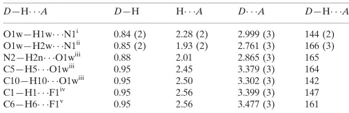
molecule. Thus, the water molecule provides links between three molecules, leading to the formation of a 2-D array, Fig.
2 and Table 1. The resultant layer in the ab plane is further stabilized by C–H···O interactions, Table 1, and weak π···π
contacts [ring centroid(N1,C1—C4,C9)···ring centroid(C4–C9)i = 3.7070 (14) Å, dihedral angle = 1.45 (11) ° for i: -1 + x,
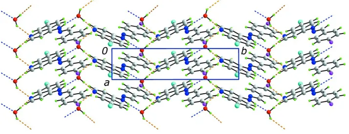
y, z]. Layers stack along the c direction with the most significant contacts between layers being of the type C–H···F
whereby the fluoride is bifurcated, Table 1 and Fig. 3.
S2. Experimental
A solution of 7-chloro-4-hydrazinoquinoline (0.20 g, 1.0 mmol) and 4-fluorobenzaldehyde (0.15 g, 1.2 mmol) in EtOH (5
ml) was maintained at room temperature overnight and rotary evaporated. The solid residue, was washed with cold Et2O
(3 x 10 ml) and recrystallized from EtOH m.pt. 518–519 K, lit. value 518 K (Pellerano et al., 1976), yield 74%. The
sample for the X-ray study was slowly grown from moist EtOH and was found to be the monohydrate. 1H NMR (400
MHz, DMSO-d6) δ: 7.28–7.32 (3H, m), 7.54 (1H, d, J = 8.4 Hz), 7.84–7.88 (3H, m), 8.34–8.40 (3H, m), 11.3 (1H, br.s,
NH). MS/ESI: [M+. - H]: 298. IR ν
The amine- and C-bound H atoms were geometrically placed (N–H = 0.88 Å and C–H = 0.95 Å) and refined as riding
with Uiso(H) = 1.2Ueq(C). The water-bound H atoms were located from a difference map and refined (O–H = 0.84 (1) Å)
[image:4.610.130.483.139.308.2]with Uiso(H) = 1.5Ueq(O).
Figure 1
The molecular structure of both components comprising the asymmetric unit of (I) showing the atom-labelling scheme
and displacement ellipsoids at the 50% probability level.
Figure 2
A view of the 2-D supramolecular array in (I) showing the O–H···N and N–H···O hydrogen bonds as orange and blue
[image:4.610.128.485.362.500.2]supporting information
[image:5.610.128.484.70.283.2]sup-3
Acta Cryst. (2010). E66, o152–o153Figure 3
A view in projection along the a axis of the unit-cell contents in (I) showing the stacking of layers along the c direction.
The O–H···N and N–H···O hydrogen bonds are shown as orange and blue dashed lines, respectively, and the C–H···F
contacts are represented by pink dashed lines. One of the 2-D arrays, as shown in Fig. 2, has been highlighted in
space-filling mode. Colour code: Cl, cyan; F, pink; O, red; N, blue; C, grey; and H, green.
7-Chloro-4-[(E)-N′-(4-fluorobenzylidene)hydrazinyl]quinoline monohydrate
Crystal data
C16H11ClFN3·H2O
Mr = 317.74
Monoclinic, P21/c
Hall symbol: -P 2ybc a = 3.7795 (2) Å b = 15.4188 (11) Å c = 24.8576 (16) Å β = 90.286 (4)° V = 1448.57 (16) Å3
Z = 4
F(000) = 656 Dx = 1.457 Mg m−3
Mo Kα radiation, λ = 0.71073 Å Cell parameters from 13530 reflections θ = 2.9–27.5°
µ = 0.28 mm−1
T = 120 K Needle, colourless 0.90 × 0.04 × 0.04 mm
Data collection
Enraf–Nonius KappaCCD area-detector diffractometer
Radiation source: Enraf Nonius FR591 rotating anode
10 cm confocal mirrors monochromator Detector resolution: 9.091 pixels mm-1
φ and ω scans
Absorption correction: multi-scan
(SADABS; Sheldrick, 2007)
Tmin = 0.614, Tmax = 0.746
19494 measured reflections 3291 independent reflections 2009 reflections with I > 2σ(I) Rint = 0.098
θmax = 27.5°, θmin = 3.1°
Refinement on F2
Least-squares matrix: full R[F2 > 2σ(F2)] = 0.059
wR(F2) = 0.131
S = 1.04 3291 reflections 205 parameters 3 restraints
Primary atom site location: structure-invariant direct methods
Secondary atom site location: difference Fourier map
Hydrogen site location: inferred from neighbouring sites
H atoms treated by a mixture of independent and constrained refinement
w = 1/[σ2(F
o2) + (0.0463P)2 + 0.5902P]
where P = (Fo2 + 2Fc2)/3
(Δ/σ)max < 0.001
Δρmax = 0.33 e Å−3
Δρmin = −0.37 e Å−3
Special details
Geometry. All s.u.'s (except the s.u. in the dihedral angle between two l.s. planes) are estimated using the full covariance matrix. The cell s.u.'s are taken into account individually in the estimation of s.u.'s in distances, angles and torsion angles; correlations between s.u.'s in cell parameters are only used when they are defined by crystal symmetry. An approximate (isotropic) treatment of cell s.u.'s is used for estimating s.u.'s involving l.s. planes.
Refinement. Refinement of F2 against ALL reflections. The weighted R-factor wR and goodness of fit S are based on F2,
conventional R-factors R are based on F, with F set to zero for negative F2. The threshold expression of F2 > 2σ(F2) is
used only for calculating R-factors(gt) etc. and is not relevant to the choice of reflections for refinement. R-factors based on F2 are statistically about twice as large as those based on F, and R- factors based on ALL data will be even larger.
Fractional atomic coordinates and isotropic or equivalent isotropic displacement parameters (Å2)
x y z Uiso*/Ueq
Cl1 0.85154 (18) −0.01273 (5) 0.43677 (3) 0.0319 (2) F1 0.1997 (4) 0.22958 (11) −0.11624 (6) 0.0367 (5) N1 0.3942 (5) −0.13713 (14) 0.26095 (9) 0.0214 (5) N2 0.6585 (5) 0.08516 (14) 0.17189 (8) 0.0221 (5)
H2N 0.7847 0.1268 0.1867 0.027*
N3 0.5476 (5) 0.09163 (15) 0.11914 (8) 0.0212 (5) C1 0.3270 (7) −0.12969 (18) 0.20855 (11) 0.0225 (6)
H1 0.2176 −0.1776 0.1911 0.027*
C2 0.4043 (6) −0.05737 (18) 0.17733 (10) 0.0210 (6)
H2 0.3436 −0.0566 0.1402 0.025*
C3 0.5701 (6) 0.01357 (17) 0.20052 (10) 0.0178 (6) C4 0.6457 (6) 0.01003 (16) 0.25750 (10) 0.0176 (6) C5 0.8024 (6) 0.07868 (18) 0.28696 (11) 0.0210 (6)
H5 0.8680 0.1303 0.2687 0.025*
C6 0.8613 (6) 0.07213 (18) 0.34118 (10) 0.0220 (6)
H6 0.9655 0.1189 0.3605 0.026*
C7 0.7663 (7) −0.00432 (18) 0.36790 (11) 0.0218 (6) C8 0.6151 (6) −0.07217 (18) 0.34132 (10) 0.0214 (6)
H8 0.5531 −0.1233 0.3604 0.026*
C9 0.5501 (6) −0.06654 (16) 0.28533 (10) 0.0179 (6) C10 0.6205 (7) 0.16293 (18) 0.09495 (11) 0.0219 (6)
H10 0.7507 0.2064 0.1136 0.026*
supporting information
sup-5
Acta Cryst. (2010). E66, o152–o153H12 0.3087 0.0586 0.0232 0.028*
C13 0.2471 (7) 0.13101 (18) −0.04411 (11) 0.0237 (6)
H13 0.1409 0.0871 −0.0656 0.028*
C14 0.3041 (7) 0.21294 (19) −0.06453 (11) 0.0256 (7) C15 0.4572 (7) 0.27824 (19) −0.03525 (11) 0.0269 (7)
H15 0.4911 0.3342 −0.0504 0.032*
C16 0.5614 (7) 0.26028 (18) 0.01715 (11) 0.0232 (6)
H16 0.6708 0.3044 0.0380 0.028*
O1W 0.0566 (5) 0.23633 (12) 0.20098 (8) 0.0287 (5) H1W 0.262 (3) 0.2503 (17) 0.2111 (12) 0.043* H2W −0.086 (5) 0.2775 (13) 0.2069 (12) 0.043*
Atomic displacement parameters (Å2)
U11 U22 U33 U12 U13 U23
Cl1 0.0381 (4) 0.0386 (5) 0.0191 (4) −0.0007 (3) −0.0065 (3) −0.0001 (3) F1 0.0475 (10) 0.0422 (11) 0.0205 (9) 0.0086 (8) −0.0082 (8) 0.0037 (8) N1 0.0218 (12) 0.0225 (13) 0.0200 (13) 0.0000 (10) −0.0006 (9) −0.0006 (10) N2 0.0269 (12) 0.0218 (13) 0.0177 (12) −0.0046 (10) −0.0046 (9) 0.0001 (10) N3 0.0219 (12) 0.0265 (14) 0.0153 (12) 0.0018 (10) −0.0022 (9) 0.0005 (10) C1 0.0185 (14) 0.0204 (16) 0.0286 (17) 0.0005 (11) −0.0024 (12) −0.0026 (13) C2 0.0204 (14) 0.0270 (16) 0.0156 (14) 0.0007 (12) −0.0052 (11) −0.0005 (12) C3 0.0150 (13) 0.0189 (15) 0.0195 (14) 0.0014 (11) −0.0008 (10) −0.0011 (12) C4 0.0151 (13) 0.0168 (14) 0.0209 (14) 0.0033 (11) −0.0002 (10) −0.0018 (12) C5 0.0204 (14) 0.0189 (15) 0.0237 (15) 0.0012 (11) 0.0001 (11) −0.0018 (12) C6 0.0242 (15) 0.0210 (16) 0.0208 (15) −0.0003 (12) −0.0035 (11) −0.0059 (12) C7 0.0204 (14) 0.0276 (17) 0.0175 (14) 0.0030 (12) −0.0016 (11) −0.0034 (12) C8 0.0198 (14) 0.0223 (16) 0.0220 (15) 0.0050 (12) −0.0007 (11) 0.0046 (12) C9 0.0154 (13) 0.0149 (14) 0.0233 (15) 0.0003 (11) −0.0026 (10) −0.0030 (12) C10 0.0222 (15) 0.0215 (16) 0.0221 (16) 0.0019 (12) 0.0000 (11) −0.0046 (13) C11 0.0185 (14) 0.0253 (16) 0.0209 (15) 0.0030 (12) 0.0004 (11) 0.0010 (12) C12 0.0261 (15) 0.0194 (15) 0.0238 (16) −0.0004 (12) 0.0010 (12) −0.0001 (12) C13 0.0240 (15) 0.0241 (16) 0.0229 (16) 0.0009 (12) −0.0018 (12) −0.0045 (13) C14 0.0273 (15) 0.0350 (18) 0.0145 (14) 0.0051 (13) −0.0025 (11) 0.0012 (13) C15 0.0265 (15) 0.0256 (17) 0.0287 (17) 0.0025 (13) −0.0004 (12) 0.0043 (13) C16 0.0244 (15) 0.0240 (16) 0.0213 (15) −0.0016 (12) −0.0012 (11) −0.0037 (13) O1W 0.0255 (11) 0.0227 (11) 0.0377 (13) −0.0003 (9) −0.0020 (9) −0.0045 (9)
Geometric parameters (Å, º)
Cl1—C7 1.745 (3) C6—H6 0.9500
F1—C14 1.367 (3) C7—C8 1.362 (4)
N1—C1 1.331 (3) C8—C9 1.414 (3)
N1—C9 1.377 (3) C8—H8 0.9500
N2—C3 1.356 (3) C10—C11 1.458 (4)
N2—N3 1.378 (3) C10—H10 0.9500
N2—H2N 0.8800 C11—C16 1.389 (4)
C1—H1 0.9500 C12—H12 0.9500
C2—C3 1.385 (4) C13—C14 1.379 (4)
C2—H2 0.9500 C13—H13 0.9500
C3—C4 1.445 (3) C14—C15 1.369 (4)
C4—C5 1.415 (4) C15—C16 1.387 (4)
C4—C9 1.416 (4) C15—H15 0.9500
C5—C6 1.369 (3) C16—H16 0.9500
C5—H5 0.9500 O1W—H1W 0.841 (10)
C6—C7 1.401 (4) O1W—H2W 0.845 (10)
C1—N1—C9 116.2 (2) C7—C8—H8 120.0
C3—N2—N3 118.9 (2) C9—C8—H8 120.0
C3—N2—H2N 120.5 N1—C9—C8 117.2 (2)
N3—N2—H2N 120.5 N1—C9—C4 123.6 (2)
C10—N3—N2 116.3 (2) C8—C9—C4 119.2 (2)
N1—C1—C2 125.2 (3) N3—C10—C11 121.6 (2)
N1—C1—H1 117.4 N3—C10—H10 119.2
C2—C1—H1 117.4 C11—C10—H10 119.2
C3—C2—C1 119.8 (2) C16—C11—C12 118.7 (2)
C3—C2—H2 120.1 C16—C11—C10 119.3 (2)
C1—C2—H2 120.1 C12—C11—C10 122.0 (3)
N2—C3—C2 122.4 (2) C13—C12—C11 120.8 (3)
N2—C3—C4 119.8 (2) C13—C12—H12 119.6
C2—C3—C4 117.7 (2) C11—C12—H12 119.6
C5—C4—C9 118.5 (2) C12—C13—C14 118.3 (3)
C5—C4—C3 124.0 (2) C12—C13—H13 120.9
C9—C4—C3 117.4 (2) C14—C13—H13 120.9
C6—C5—C4 121.3 (3) F1—C14—C15 118.8 (3)
C6—C5—H5 119.3 F1—C14—C13 118.3 (2)
C4—C5—H5 119.3 C15—C14—C13 123.0 (3)
C5—C6—C7 119.2 (2) C14—C15—C16 118.0 (3)
C5—C6—H6 120.4 C14—C15—H15 121.0
C7—C6—H6 120.4 C16—C15—H15 121.0
C8—C7—C6 121.6 (2) C15—C16—C11 121.2 (3)
C8—C7—Cl1 119.6 (2) C15—C16—H16 119.4
C6—C7—Cl1 118.8 (2) C11—C16—H16 119.4
C7—C8—C9 120.0 (2) H1W—O1W—H2W 110.0 (16)
C3—N2—N3—C10 176.0 (2) C7—C8—C9—N1 −179.5 (2)
C9—N1—C1—C2 0.5 (4) C7—C8—C9—C4 0.3 (4)
N1—C1—C2—C3 1.3 (4) C5—C4—C9—N1 179.6 (2)
N3—N2—C3—C2 6.9 (4) C3—C4—C9—N1 0.7 (4)
supporting information
sup-7
Acta Cryst. (2010). E66, o152–o153N2—C3—C4—C9 −179.5 (2) C16—C11—C12—C13 0.5 (4) C2—C3—C4—C9 1.1 (3) C10—C11—C12—C13 −179.5 (2) C9—C4—C5—C6 −0.1 (4) C11—C12—C13—C14 −0.8 (4) C3—C4—C5—C6 178.7 (2) C12—C13—C14—F1 −179.2 (2) C4—C5—C6—C7 0.4 (4) C12—C13—C14—C15 0.2 (4) C5—C6—C7—C8 −0.3 (4) F1—C14—C15—C16 180.0 (2) C5—C6—C7—Cl1 178.65 (19) C13—C14—C15—C16 0.5 (4) C6—C7—C8—C9 0.0 (4) C14—C15—C16—C11 −0.8 (4) Cl1—C7—C8—C9 −178.98 (18) C12—C11—C16—C15 0.2 (4) C1—N1—C9—C8 178.3 (2) C10—C11—C16—C15 −179.7 (2) C1—N1—C9—C4 −1.5 (4)
Hydrogen-bond geometry (Å, º)
D—H···A D—H H···A D···A D—H···A
O1w—H1w···N1i 0.84 (2) 2.28 (2) 2.999 (3) 144 (2)
O1w—H2w···N1ii 0.85 (2) 1.93 (2) 2.761 (3) 166 (3)
N2—H2n···O1wiii 0.88 2.01 2.865 (3) 165
C5—H5···O1wiii 0.95 2.45 3.379 (3) 164
C10—H10···O1wiii 0.95 2.50 3.302 (3) 142
C1—H1···F1iv 0.95 2.56 3.399 (3) 147
C6—H6···F1v 0.95 2.56 3.477 (3) 161