Mycobacteria in the environment of pig farms
in the Czech Republic between 2003 and 2007
K. Krizova, L. Matlova, A. Horvathova, M. Moravkova, V. Beran,
T. Boisselet, V. Babak, I. Slana, I. Pavlik
Veterinary Research Institute, Brno, Czech Republic
ABSTRACT: In the Czech Republic, most mycobacterial infections in pigs are caused by the Mycobacterium
avium complex (MAC) and potentially pathogenic mycobacteria (PPM) derived from the environment. This study was undertaken after the isolation of PPM from various components of the environment of pig herds between 1996 and 2002 (Matlova et al., Veterinarni Medicina, 48, 2003, 343–357). Between 2003 and 2007, a total of 1114 environmentally-derived samples from 24 farms were examined. After staining according to Ziehl-Neelsen, acid-fast rods were found in 42 (3.8%) samples by direct microscopy, and PPM were isolated from 223 (20.0%) samples by culture. PPM occurred primarily in soil from the paddocks (53.8%), peat (53.2%), bedding (28.4%) and biofilm from the pipeline (21.0%). From MAC,M. avium subsp. hominissuis (MAH) genotype IS901– and IS1245+ was most frequent; M. avium subsp. avium (MAA) genotype IS901+ and IS1245+ and M. intracellulare genotype IS901– and IS1245– were isolated from one (0.4%) and three (1.3%) samples, respectively. The remaining iso-lates were identified as 19 other mycobacterial species: M. gordonae (n = 8), M. triviale (n = 6), M. flavescens (n = 3), M. nonchromogenicum (n = 3), M. terrae (n = 3), M. xenopi (n = 3), M. fortuitum (n = 2), M. chelonae (n = 2), M. chitae (n = 2), M. abscessus (n = 1), M. gastri (n = 1), M. kumamotonense (n = 1), M. marinum (n = 1), M. parafortuitum (n = 1), M. peregrinum (n = 1), M. porcinum (n = 1), M. scrofulaceum (n = 1), M. smegmatis (n = 1) and M. simiae (n = 1). The remaining 41 isolates of unidentified mycobacterial species did not contain the sequences IS901 and/or IS1245, specific for medically important members of MAC (MAA and MAH); a further 44 isolates were not tested due to their contamination or loss of ability to grow in vitro. A farm where MAH was often detected in the lymph nodes of pigs and in the environment between 1996 and 2002 (Period I), was selected for further investigation between 2003 and 2007 (Period II). A comparison of the findings of mycobacteria on the investigated farm in Period I and in the following Period II showed a significant increase (P < 0.01) in the occur-rence of mycobacteria other than MAH, especially in peat samples.
Keywords: mycobacteria other than tuberculosis (MOTT); ecology; avian tuberculosis; avian mycobacteriosis; environmental saprophytic mycobacteria; cultivation
Supported by the Ministry of Agriculture of the Czech Republic (Grants No. MZE 0002716202, No. QH91240, and No. QH71054) and the Ministry of Education, Youth and Sports of the Czech Republic (AdmireVet; Grant No. CZ 1.05/2.1.00/01.0006).
The mycobacterial infection of pigs has resulted in severe financial losses to farmers in the Czech Republic. Between 1990 and 1999, the losses in-creased by 22 to 24% of slaughtered pig prices (Pavlik et al., 2003). Analysis of the occurrence of tuberculous lesions in pigs, performed in the period from 2000 to 2004, revealed that these were still being found in the tissues of slaughtered pigs. The
most important causative agents of these
tubercu-lous lesions were Mycobacterium avium complex
(MAC) organisms (Pavlik et al., 2005). Between 1990
and 1992, the causative agent of avian tuberculosis
agent of avian mycobacteriosis M. avium subsp.
hominissuis (MAH), which was only isolated from 18.3%, 9.7% and 30.3% of cases, during the respec-tive years. However in the following years, 1993 and
1995 to 1999, the isolation rates of MAH tended to
increase (Pavlik et al., 2003). These results were also confirmed by the study of all isolated members of
MAC between 1996 and 2004 (Shitaye et al.,2006),
by serotyping and IS901 PCR (Pavlik et al., 2000;
Bartos et al., 2006).
Molecular studies using a standardized IS901
RFLP method (Dvorska et al., 2003) identified MAA
isolates with similar RFLP types from pigs and con-taminated peat (Matlova et al., 2005), domestic and wild birds (Dvorska et al., 2007; Moravkova et al., 2007; Shitaye et al., 2008a,b) and in one case, a horse (Pavlik et al., 2008).
Studies performed in the Czech Republic showed that bedding materials, various feed supplements and drinking water for animals, etc., were the
sources of MAH rather than the infected animals
themselves. In the first half of the 1990’s, the source
of MAH was deep bedding, usually composed of
sawdust or other wood by-products, fermented by, e.g., ENVISTIM (Pavlik et al., 2003; Matlova et al., 2003a, 2004a). Results from the late 1990’s, showed that increased tuberculous lesion formation in pigs
was primarily elicited by MAH present in various
feed supplements, of which the most important was peat (Matlova et al., 2003a, 2005; Pavlik et al., 2003) and kaolin (Matlova et al., 2003a, 2004b).
In the following period (2000 to 2004), MAH was
increasingly isolated from the examined pigs rather
than MAA (Pavlik et al., 2005). Analysis of
sam-ples from the environment of pig herds between 1996 and 2002 determined that not only were the above-mentioned materials (above all sawdust used as bedding, peat and kaolin used as feed
supple-ments) sources of MAH, but also drinking water,
soil, invertebrate animals and dust, etc. (Matlova et al., 2003a).
With regard to the fact that the farm environment
is the primary source of MAH, attention was also
paid to the occurrence of PPM in the environment of pig herds in the Czech Republic. The objectives of this study were established to analyse the occur-rence of mycobacteria in 24 pig herds in the Czech Republic between 2003 and 2007. Beside that, the results for one of the examined pig herds previ-ously investigated by Matlova et al. (2003a), were compared with earlier data to assess implications for the risk of infection.
MATERIAL AND METHODS
Examined biological material
A total of 1114 samples from the environment (Table 1) of 24 pig farms in the Czech Republic were examined between 2003 and 2007. The selec-tion of the farms was done on the previous finding of tuberculosis lesions and positive reactions to avian tuberculin (data not shown). The samples were stored at +4°C for up to two days until they were analysed in the laboratory.
Microscopic examination
Before culture, samples were stained by the Ziehl-Neelsen (Z-N) technique and examined under an Olympus microscope at 1000× magnification for the presence of acid-fast rods (AFR). At least 100 fields of view were examined for each sample (Kubin et al., 1986).
Culture examination
Approximately 1 g of sample was homogenized and decontaminated by a previously described method (Fischer et al., 2000; Matlova et al., 2003a). A total volume of 100 µl of a decontaminated sample suspension was inoculated with sterile disposable tips and dispensed onto eight slopes of media: two tubes of egg-based media according to the method of Stonebrink (Stonebrink, 1978) four slopes of Herrold Egg Yolk Media (HEYM; two with, and two without Mycobactin J) and onto two tubes of liquid media according to Sula (Merkal et al., 1964; Kubin et al., 1986). Incubations were performed simultaneously at two temperatures: first set of the media at 25°C, and the second set at 37°C (i.e., every sample was cul-tured on four different media at each temperature). The cultures were checked for growth four times. The first reading was taken up to seven days for de-tection of fast-growing mycobacterial species; the subsequent readings were performed after 14 days, one, and finally, two months.
Identification of mycobacterial isolates by PCR
Cousins (1992) and modified by Moravkova et al. (2008). According to the above methods, isolates
were characterised as M. sp., M. avium sp. or
mem-bers of the MAC (Moravkova et al., 2008).
Mycobacteria, other than those identified as
M. avium species were identified by a biochemi-cal method according to Wayne and Kubica (1986)
or identification was based on 16S rRNA gene
se-quencing using the broad-range primers 16S-27f (5’-AGA GTT TGA TCM TGG CTC AG-3’) and 16S-907r (5’-CCG TCA ATT CMT TTR AGT TT-3’) according to Harmsen et al. (2003). Sequencing was performed at the MWG Biotech Company (Germany). Resulting sequences were analysed by Staden Package software (http://staden.source-forge.net/) and compared with the sequences in two databases, GenBank using the BLAST util-ity (http://www.ncbi.nlm.nih.gov/BLAST/) and RIDOM (http://rdna2.ridom.de/).
Statistical analysis
Statistical analysis was applied to the positive results obtained from one farm between 2003 and 2007 (Period II). These positive samples from Period II were statistically compared to the posi-tive samples examined previously by Matlova et al. (2003a) between 1996 and 2002 (Period I). Fisher’s exact test, which is a part of the GraphPad Prism v5.02 programme (GraphPad Software, Inc., USA) was used for the statistical evaluation.
RESULTS
From 1114 samples (collected from 24 farms), mi-croscopic examination revealed AFR by Z-N staining in 42 (3.8%) samples in nine of the 17 sample groups. By culture, mycobacteria were detected in 223 (20.0%) samples from 14 of the 17 sample groups (Table 1).
Microscopic detection of acid-fast rods in samples
AFR were most frequently detected in samples of biofilm from drinkers (10 samples; 12.3%) and in faeces (five samples; 8.6%), whereas the detec-tion rates in samples of feed, bedding, water from drinkers, dust and spider webs was lower (1.3 to 3.8%; Table 1).
Culture detection of mycobacteria in samples
Mycobacteria were most frequently detected by culture in soil from the paddocks (53.8%), peat (53.2%) and bedding (28.4%; Table 1).
Identification of mycobacterial isolates
From a total of 223 positive isolates, it was pos-sible to identify 138 strains classified as 22 species
of mycobacteria (including MAA). From the MAC,
92 (41.3%) isolates were identified as MAH, which
is a total of 8.3% of MAH from the 1114 samples
examined in this study. In contrast to this, only one
(0.4%) of the examined MAC isolates was identified
as MAA. Among the remaining PPM (223 isolates
in total), M. gordonae was found most frequently
(eight isolates, 3.6%), followed by M. triviale (six
isolates, 2.7%) and M. nonchromogenicum (three
isolates, 1.3%; Table 2).
Mycobacterial contamination of various samples from 24 farms
Water from the pipeline. Mycobacteria were detected in 35 (15.4%) samples (Table 1) with a wide species range; out of 22 detected mycobacte-rial species, nine where found to be contaminants of water. The most frequently found species were
MAH (seven isolates, 20.0%) and M. gordonae (four
isolates, 11.4%; Table 2).
Biofilm from the pipeline. Mycobacteria were detected in 17 (21.0%) samples (Table 1).
Feed. Six species of PPM were detected in 25
(13.6%) samples (Table 1). MAH was detected most
frequently (10 positive samples, 40.0%), followed by M. flavescens and M. triviale, each found in two (8.0%) samples (Table 2). By culture, feeding concentrates most frequently tested positive for PPM (22 positive samples, 17.9%), in comparison with feed supplements (Bentonit Zeo, Fibre cell and Sarbovet) and yeasts. In the feeding concen-trates, five mycobacterial species were found, of
which MAH was most frequent (eight isolates,
36.4%; Table 3). In other feed supplements, MAH
Feed leftovers. Mycobacteria were detected in
14.3% of samples of feed leftovers (Table 1). M.
fla-vescens, M. nonchromogenicum,M. gordonae and
M. kumamotonense were identified (Table 2). Peat and kaolin. The second highest mycobac-terial contamination was found in peat (Table 1). Out of 83 mycobacterial isolates, the following
members of the MAC were most often detected:
MAH (55 isolates), MAA (one isolate) and M.
intra-cellulare that was not further typed (two isolates; Table 2). No mycobacteria were detected in samples
of kaolin feed (Table 2). 5.9% of milk and whey samples were mycobacteria-positive (Table 1).
Bedding. The third highest mycobacterial con-tamination was found in bedding (28.4% positive samples; Table 1). Of seven mycobacterial species
identified, MAH was the most frequent, being
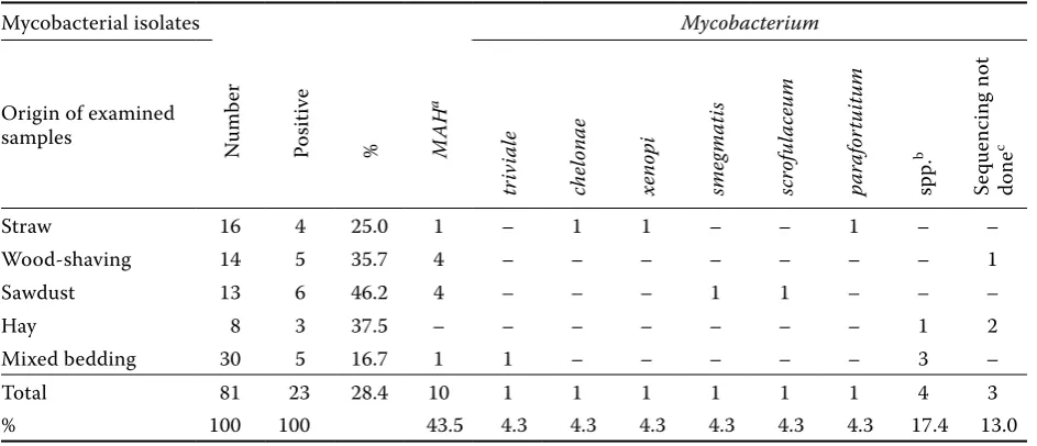
[image:4.595.62.532.102.369.2]de-tected in 10 samples (43.5%; Table 2). Of all types of bedding, sawdust was the most often contami-nated with mycobacteria (46.2% positive samples) followed by shavings (35.7% positive samples; Table 4).
Table 1. Environmental samples from pig farms collected between the years 2003 and 2007
Samples examineda Number Z-N stainingb Culture
positive % positive %
Water from pipeline 227 8 3.5 35 15.4
Feedc 184 7 3.8 25 13.6
Peat 156 4 2.6 83 53.2
Dust and spider websd 88 3 3.4 9 10.2
Biofilm from pipelinee 81 10 12.3 17 21.0
Beddingf 81 3 3.7 23 28.4
Scrapings from stablesg 79 1 1.3 8 10.1
Pig faecesh 58 5 8.6 6 10.3
Other samplesi 43 0 0 1 2.3
Feed leftoversj 42 1 2.4 6 14.3
Whey and milkk 17 0 0 1 5.9
Free living birdsl 17 0 0 0 0
Soil from paddocks 13 0 0 7 53.8
Kaolinm 9 0 0 0 0
Other feeding supplementsn 9 0 0 1 11.1
Small terrestrial mammalso 5 0 0 1 20.0
Invertebratesp 5 0 0 0 0
Total 1114 42 3.8 223 20.0
agroups of samples designated according to the previously published results (Matlova et al., 2003a) during the years 1996 to 2002
bdetection of acid-fast rods (AFR) after Ziehl-Neelsen (Z-N) staining
cfeed samples collected from the troughs in the stables and from the storage tanks (for more details see Table 3) dsamples collected from the stables and feed stores
ebiofilm samples collected from the drinking places and water-expanse reservoirs fsamples collected from the stables (for more details see in Table 4)
gscrapings collected in the stables: i.e. old pig faeces, feedstuff and dust from floors, wall, pen barriers and concrete pad-docks and pen barriers
hcollected from the floor in the boxes in the stables and paddocks
iincluded other samples, for example: waste water, samples from sewer, water filter, ventilation filter, skimming from the basin, rubbish mixture etc.
jsamples of leftovers of feed concentrates were collected from the troughs ksamples originating from the storage tanks
lparenchymatous organs were examined (for more details see Table 5) mkaolin was used as a feeding supplement for piglets under two months of age
nfeeding supplements other than peat and kaolin were examined (Mistral, Univit CT-80, Hemasoft, Stalosanf, Staldrom and Heamstal)
Ta
ble 2. M
yc ob ac ter ial sp ec ie s i sol at ed f rom en vir onmen tal sam ple s M yc ob ac ter ial i sol at es M yco bacter iu m Or ig
in of i
sol at es a Numb er MAH triv iale fortu itum chelonae gordonae flave scens xenopi sme gmati s simi ae scroful aceum absce ssus por cinum nonchromo-genicu m intr acellu lare c mar inum par afortu itum terrae kuma moto- nense chita e per egr inum gastr i spp. d Sequenc ing not e done
W at er f rom pip eline 35 7 1 1 1 4 – 1 – – – – – 1 – 1 – – – – 1 – 15 2 Feed 25 10 2 1 – – 2 – – – – 1 – – 1 – – – – – – – 2 6 Pe at 83 57 b – – – – – 1 – – – – – – 2 – – – – – – – 6 17 D
ust and spider we
bs 9 4 1 – – – – – – – – – – 1 – – – 1 – – – – – 2 Biofilm f rom pip eline 17 – – – – 3 – – – – – – – – – – – – – – – – 9 5 Be dding 23 10 1 – 1 – – 1 1 – 1 – – – – – 1 – – – – – 4 3 Sc ra ping s f rom st able s 8 – 1 – – – – – – – – – 1 – – – – 1 – – – 1 2 2 Pig f ae ce s 6 1 – – – – – – – – – – – – – – – – – 2 – – 2 1 O ther sam ple s 1 – – – – – – – – 1 – – – – – – – – – – – – – – Fe ed lef tovers 6 – – – – 1 1 – – – – – – 1 – – – – 1 – – – – 2 Whe
y and milk
1 – – – – – – – – – – – – – – – – – – – – – – 1 Soil f rom p addo ck s 7 3 – – – – – – – – – – – – – – – 1 – – – – 1 2 O ther f ee ding supplemen ts 1 1 – – – – – – – – – – – – – – – – – – – – – – Small t er re str ial mammal s 1 – – – – – – – – – – – – – – – – – – – – – – 1 Tot al 223 93 6 2 2 8 3 3 1 1 1 1 1 3 3 1 1 3 1 2 1 1 41 44 % 41.7 2.7 0.9 0.9 3.6 1.3 1.3 0.4 0.4 0.4 0.4 0.4 1.3 1.3 0.4 0.4 1.3 0.4 0.9 0.4 0.4 18.4 19.7 agr oup
s of sam
ple s de sig na te d ac cor ding t o pr ev iou sly publi she d r esult s (Ma tlov a e
t al., 2003a) dur
ing t
he ye
ars 1996 t
o 2002; f
or mor
e de
tail
s s
ee T
able 1 le
gend
bisol
at
es c
onsi
st
ed of one
M AA is ol at e ( genoty pe I S 901
+ and I
S
1245
+) and one i
sol
at
e whic
h w
as not suc
ce ssf ully ty pe d f ur ther cM. intr acel lu la re (genoty pe I S 901– and I S 1245– ) dis ol at
es not b
elong ing t o M AC af ter P C R e xamina tion (M ora vk ov a e
t al., 2008) and by s
equenc
ing not suc
ce ssf ully ty pe d t o t he sp ec ie s
eisol
at es not be long ing to M AC af ter PC R examina tion (M ora vk ov a et al., 2008); se quenc ing w as not done due to lac k of gr ow th in the sub cult ur es or due to the con tamina tion of t he i sol at e w ith anot her b ac ter
ial or mould sp
ec
ie
Scrapings from the stable and pig faeces. Mycobacteria contaminated 10.1% of samples of scrapings from floors in animal housing (Table 1),
among which four species were identified (M.
trivi-ale, M. porcinum, M. terrae and M. gastri; Table 2). Mycobacteria were found in 10.3% of pig faecal
samples (Table 1), where MAH and M. chitae were
identified (Table 2).
Dust and spider webs. Mycobacteria were iso-lated from nine (10.2%) samples of dust and spider webs (Table 1) of which four isolates were identified
as MAH (Table 2).
Soil from the paddocks. Mycobacterial contami-nations were confirmed in 53.8% of samples of soil
from the paddocks (Table 1). M. terrae and MAH
were identified (Table 2).
Other samples. Mycobacteria were detected in 2.3% of the other samples from pig herds, including swabs from basins, water filters, ventilation filters, cleaning devices, sewage water and samples from
sewers (Table 1). The isolate identified as M. simiae
originated from a sample of the rubbish mixture from the floor (Table 2).
No mycobacteria were found in free living birds and invertebrates (Tables 1 and 5). Regarding the
group of small terrestrial mammals, only one sam-ple was found to be positive. However, its exact identification was not possible (Table 2).
Statistical analysis of results obtained on one farm in Periods I and II
Analysis of data from Periods I and II showed a significantly decreased frequency of occurrence of
all positive isolates (P < 0.05) in comparison with
all tested samples (Table 6). The same tendency was
observed in MAH occurrence (P < 0.01; Table 7).
Mycobacterial contamination of different samples on the farm
Water from the pipeline. In this type of samples, a non-significant decrease in occurrence of
posi-tive samples was observed (Table 6). MAH was not
detected in any isolate, which is a statistically non-significant difference in comparison with Period I (Table 7). In Period II, the highest percentage of
[image:6.595.64.534.447.688.2]M. gordonae was found (Table 8).
Table 3. Mycobacterial species isolated from feeding concentrates and feed supplements
Mycobacterial isolates
N
umb
er
Po
sitive %
M
AH
a
Mycobacterium
Origin of examined samples
flave
scen
s
tr
iv
ia
le
ab
sce
ss
us
intr
acel
lu
la
re
b
for
tu
itu
m
spp.
c
Se
quenc
ing not
done
d
Feed
Feeding concentrates (finishing pigs) 123 22 17.9 8 2 2 1 – 1 2 6
Feeding concentrates COS 1 (piglets) 18 1 5.6 – – – – 1 – – –
Feed supplement (Bentonit Zeo) 15 2 13.3 2 – – – – – – –
Feed supplement (Fibre cell M1) 3 0 0 – – – – – – – –
Feed supplement (Sarbovet) 20 0 0 – – – – – – – –
Yeast 5 0 0 – – – – – – – –
Total 184 25 13.6 10 2 2 1 1 1 2 6
% 100 100 40.0 8.0 8.0 4.0 4.0 4.0 8.0 24.0
aMAH (genotype IS901– and IS1245+)
bM. intracellulare (genotype IS901– and IS1245–)
cisolates not belonging to the MAC after PCR examination (Moravkova et al., 2008) and by sequencing not successfully typed to the species level
Biofilm from the pipeline. In Period II, the oc-currence of mycobacteria non-significantly de-creased in comparison with Period I (Table 6). Among the isolates classified to the species level,
M. flavescens and M. terrae predominated. MAH
were not detected in the second period (Table 8). Feed. The occurrence of mycobacteria increased non-significantly (Table 6). The situation was
compa-rable for MAH, which was detected only in one case
per investigation period (Table 7). In the second period,
M. flavescens was identified in the majority of cases,
whereas in the first period, it was MAH (Table 8).
Peat. In peat samples, the occurrence of
my-cobacteria other than MAH increased
signifi-cantly (P < 0.01; Table 6). These were primarily
Mycobacterium spp., which could not be identi-fied. On the other hand, a significant decrease
(P < 0.01) in the occurrence of MAH in peat was
noted (Table 7). Species other than MAH were not
[image:7.595.64.536.101.304.2]detected in Period I. In Period II, M. xenopi was
Table 4. Mycobacterial species isolated from the different components of bedding
Mycobacterial isolates Mycobacterium
Origin of examined
samples Numb
er
Po
sitive
% MAH
a
tr
iv
ia
le
chelonae xenopi sme
gmati
s
scrof
ul
aceu
m
pa
rafor
tu
itu
m
spp.
b
Se
quenc
ing not
done
c
Straw 16 4 25.0 1 – 1 1 – – 1 – –
Wood-shaving 14 5 35.7 4 – – – – – – – 1
Sawdust 13 6 46.2 4 – – – 1 1 – – –
Hay 8 3 37.5 – – – – – – – 1 2
Mixed bedding 30 5 16.7 1 1 – – – – – 3 –
Total 81 23 28.4 10 1 1 1 1 1 1 4 3
% 100 100 43.5 4.3 4.3 4.3 4.3 4.3 4.3 17.4 13.0
aMAH (genotype IS901– and IS1245+)
bisolates not belonging to the MAC after PCR examination (Moravkova et al., 2008) and by sequencing not successfully typed to the species level
cisolates not belonging to the M. avium complex after PCR examination (Moravkova et al., 2008); sequencing not done due to a lack of growth in the subcultures or due to the contamination of isolate with another bacterial or mould species
Table 5. Mycobacterial species isolated from samples of birds, small terrestrial mammals and invertebrates
Group of samples Source of samples Number of examined
animals samples positive
Free living birds
sparrow (Passer domesticus) 5 10 0
swallow (Hirundo rustica) 1 2 0
black redstart (Phoenicurus ochruros) 1 0
great tit (Parus major) 1 2 0
faeces from birds (especially sparrows) nk 1 0 Small terrestrial
mammals
mouse (Mus musculus) 1 1 1a
faeces from mice nk 4 0
Invertebrates darkling beetle (Tenebrio sp.) 2 2 0
flies in the whey (Musca domestica) 3 3 0
nk = not known (mixture samples)
[image:7.595.72.533.542.730.2]identified and M. intracellulare was detected in two cases (Table 8).
Dust and spider webs. No positive samples were found in dust and spider webs in Period II. This was a non-significant decrease in comparison with Period I when one isolate was detected (Table 6). This isolate could not be identified at the species level (Table 8).
Scrapings from the stables and pig faeces. A non-significant increase in the occurrence of posi-tive samples of scrapings was noted in Period II (Table 6). Positive isolates were only found in Period II and only one of them was identified at the species
level, namely M. triviale (Table 8). In the samples of
pig faeces, MAH was not detected in Period II. On
the other hand, the occurrence of other mycobacte-rial species increased non-significantly (Table 6). These were not further identified (Table 8).
DISCUSSION
The environment is the main source of condition-ally pathogenic mycobacteria (i.e., primarily serious pathogens of animals with the following most
im-portant two MAC members: MAH, M.
intracellu-Table 6. Comparison of mycobacteria detection between Period I (1996–2002) and Period II (2003–2007) on one farm
Samples examined Period I
a Period II Comparison Period I vs. II
number positive % number positive % trend significanceb
Water from pipeline 60 20 33.3 68 13 19.1 decrease ns
Feed 49 1 2.0 78 8 10.3 increase ns
Peat 268 136 50.7 33 29 87.9 increase ++
Dust and spider webs 30 1 3.3 13 0 0 decrease ns
Biofilm from pipeline 229 87 38.0 38 12 31.6 decrease ns
Scraping from stables 3 0 0 8 5 62.5 increase ns
Pig faeces 55 5 9.1 12 3 25.0 increase ns
Total 694 250 36.0 250 70 28.0 decrease +
apublished previously (Matlova et al., 2003a)
bns = non-significant (P > 0.05), + = P < 0.05, ++ = P < 0.01 (P is two-sided P-value for Fisher’s exact test)
Table 7. Comparison of Mycobacterium avium subsp. hominissuis detection between Period I (1996–2002) and Period II (2003–2007) on one farm
Samples examined Period I
a Period II Comparison Period I vs. II
number positive % number positive % trend significanceb
Water from pipeline 20 2 10.0 13 0 0 decrease ns
Feed 1 1 100 8 1 12.5 decrease ns
Peat 136 94 69.1 29 11 37.9 decrease ++
Dust and spider webs 1 1 100 0 0 0 nt nt
Biofilm from pipeline 87 2 2.3 12 0 0 decrease ns
Scrapings from stables 0 0 0 5 0 0 nt nt
Pig faeces 5 1 20.0 3 0 0 decrease ns
Total 250 101 40.4 70 12 17.1 decrease ++
apublished previously (Matlova et al., 2003a)
[image:8.595.69.534.116.287.2] [image:8.595.67.533.557.728.2]lare, and also other species: M. xenopi, M. chelonae
etc.; Kazda, 2000; Kazda et al., 2009). Of the MAC
members, MAH predominated in our study, and was
identified in a total of 93 (40.8%) isolates (Table 2).
Also in other countries, MAH has been determined
as the most frequent causative agent of tuberculous lesions in pigs (Dalchow and Nassal, 1979; Windsor et al., 1984; Dalchow, 1988; Alfredsen and Skjerve, 1993; Leinemann et al., 1993; Morita et al., 1994; Nishimori et al., 1995; Balian et al., 1997; Ritacco et al., 1998; Komijn et al., 1999; Offermann et al., 1999; Ramasoota et al., 2001; Mijs et al., 2002).
[image:9.595.62.538.115.371.2]Between 1996 and 2002, the strong effect of a contaminated environment on the occurrence of tuberculin reactions to avian tuberculin and occur-rence of tuberculous lesions in the lymph nodes of pigs at slaughter was investigated (Matlova et al., 2003b,c). One of the herds under investigation in the above-mentioned study was also included in
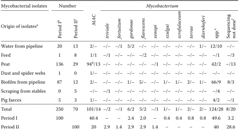
Table 8. Mycobacterial species isolated from environmental samples from one farm in Period I (1996–2002) and Period II (2003–2007)
Mycobacterial isolates Number Mycobacterium
Origin of isolatesa
Per
io
d I
b
Per
io
d I
I
c
M
AC
tr
iv
ia
le
for
tu
itu
m
gordonae flave
scen
s
xenopi szulg
ai
scrof
ul
aceu
m
ter
rae
dier
nhofer
i
spp.
e
Se
quenc
ing
not done
f
Water from pipeline 20 13 2/– –/– –/1 5/2 –/– –/– –/– –/– –/– 1/– 12/10 –/–
Feed 1 8 1/1 –/1 –/– –/– –/2 –/– –/– –/– –/– –/– –/1 –/3
Peat 136 29 94d/13 –/– –/– –/– –/– –/1 –/– –/– –/– –/– 42/2 –/13 Dust and spider webs 1 0 1/– –/– –/– –/– –/– –/– –/– –/– –/– –/– –/– –/– Biofilm from pipeline 87 12 2/– –/– –/– 1/– 5/– –/– 1/– 1/– 2/– 1/– 66/9 8/3 Scraping from stables 0 5 –/– –/1 –/– –/– –/– –/– –/– –/– –/– –/– –/4 –/–
Pig faeces 5 3 1/– –/– –/– –/– –/– –/– –/– –/– –/– –/– 4/2 –/1
Total 250 70 101/14 –/2 –/1 6/2 5/2 –/1 1/– 1/– 2/– 2/– 124/28 8/20
Period I 100 40.4 – – 2.4 2.0 – 0.4 0.4 0.8 0.8 49.6 3.2
Period II 100 20 2.9 1.4 2.9 2.9 1.4 – – – – 40 28.6
agroups of samples designated according to previously published results (Matlova et al., 2003a); for more details see Table 1 legend
b, ccomparison of all Mycobacterium detection in Period I (1996–2002) published previously (Matlova et al., 2003a) and Period II (2003–2007)
dMycobacterium avium complex isolates consisted of one M. a. avium isolate (genotype IS901+ and IS1245+) from peat,
92 isolates of M. a. hominissuis (genotype IS901– and IS1245+) and one isolate was not successfully typed further eisolates not belonging to the M. avium complex after PCR examination (Moravkova et al., 2008) and by sequencing not successfully typed to the species level
fisolates not belonging to the M.avium complex after PCR examination (Moravkova et al., 2008), sequencing not done due to lack of growth in the subcultures or due to contamination of the isolate with another bacterial or mould species
the current study. Special attention in this part of our study was paid to collection of samples, which were identified as positive in the previous study
(Matlova et al., 2003a). Emphasis was put on MAH
-infected samples in the previous study (Matlova
et al., 2003a) because MAH is an important swine
pathogen. Accordingly, the purpose of the present study was to monitor the occurrence of mycobac-teria in the environment of pig herds in the period between 2003 and 2007 and to compare the
occur-rence of MAH and other mycobacteria in Period I
(1996–2002) described by Matlova et al. (2003a) with their occurrence in Period II (2003–2007).
Statistical analysis of a one farm analysed between Periods I and II
In the pig herd investigated in the present study between 1998 and 2001, considerable impact of contaminated environment on the occurrence of tuberculin reactions to avian tuberculin and oc-currence of tuberculin lesions in the lymph nodes of slaughtered pigs was observed (Matlova et al., 2003b,c). Therefore, this herd was selected for a lon-gitudinal study. The results of monitoring the occur-rence of mycobacteria in this pig farm environment between 1996 and 2002 (Period I) were part of the study of Matlova et al. (2003a). Results obtained on this farm were also presented in the current study, because the most contaminated samples found in
the previous period (especially with MAH frequently
found in pigs) were selected for subsequent inves-tigation. The purpose of the current study was to monitor the occurrence of mycobacteria in the en-vironment of the pig farm in the following years, be-tween 2003 and 2007, and to evaluate the occurrence
of MAH and mycobacteria on the farm in Periods I
(1996–2002) and II (2003–2007).
Mycobacterial contamination of different samples
Water from the pipeline and biofilm from the pipeline. Water plays a significant role as a ve-hicle for the transmission of mycobacteria, and is regarded as a common source and milieu for multiplication under favourable conditions. In
this study, the finding of MAH was most frequent
(20%), followed by M. gordonae (11.4%; Table 2).
Expansion vessels are at risk of the accumulation and subsequent multiplication of PPM, especially in the summer months (Kazda, 2000; Hilborn et al., 2006; Kazda et al., 2009). Furthermore, the highest risk for pigs is the uptake of surface water because it can be contaminated from different sources. Mycobacteria were isolated from replicate samples of, e.g., sediments of sewage water where
concen-trations reached up to 10 × 105 CFU/ml (Brooks et
al., 1984). With regard to biofilm from the pipeline, as the detection rate of AFR by microscopy after Z-N staining is rather low (Margolis et al., 1994), this method was used here as an auxiliary tool for obtaining approximate results (Table 1). In many
cases, mycobacteria cannot be isolated in vitro
from Z-N positive samples (Matlova et al., 2003a),
increasing the high risk of mycobacterial infection for pigs via water contaminated (Tables 1 and 2). Between 2000 and 2001, the occurrence of myco-bacteria in the water main on one farm was investi-gated. Matlova et al. (2003b) revealed that the pH of the water decreased to 4.0 owing to disinfection and consequently, biofilm-containing mycobacteria was released into the water main. Consequently, this led to the formation of tuberculous lesions in up to 90% of slaughtered pigs from the above mentioned environment. Water and biofilm samples examined in the current study originated from the same water main, but were collected after this event. In both types of samples, the occurrence of mycobacteria (Table 6) non-significantly decreased from Period I (1996–2002) to Period II (2003–2007). This could be explained by regular mechanical cleaning of the drinkers, and steaming of the whole water main with hot water (70 to 80°C) for at least 20 min ac-cording to the recommendations (unpublished data).
Feed. Beerwerth and Schurmann (1969) isolated mycobacteria from 3.6% of 111 samples of unripe corn, as well as from 3.3% of 400 samples of feed-ing concentrates. They explained the relatively low detection rate by the distance of corn ears from the soil (up to 1 m) and a minimum risk of contamina-tion of the ears with mycobacteria present in the soil. Even though mycobacterial contamination of feeds was not very high in the current study (13.6%), 40% of pathogens were identified as the clinically
relevant MAH (Tables 1 and 2), followed by M.
fla-vescens and M. triviale (Table 3). The occurrence of these mycobacteria can also be explained by their transmission from the environment. Accordingly, we can suppose that inadequate handling and stor-age of feeding concentrates can contribute to my-cobacterial contamination of feeds. The present study documented that contamination of feed mixtures was non-significantly higher in Period II in comparison with Period I (Table 6). Therefore, we can assume that inappropriate handling prac-tices or poor storage conditions could contribute to the contamination of these feed mixtures with mycobacteria.
Accordingly, if stored properly, kaolin can be a safe material used to prevent diarrhoea in weaned pig-lets (Trckova et al., 2009), in contrast to other feed supplements among which 11.1% of samples exam-ined tested positive for mycobacteria (Tables 1, 2 and 3). However, farmers should be aware of the risks associated with the application of feed sup-plements.
Peat. In the mid-1990s, the feeding of piglets with peat as a supplement was introduced into some farms in the Czech Republic. Due to the fact that the occurrence of tuberculous lesions in pigs increased, farmers ceased feeding peat as a supplement in the late 1990s (Pavlik et al., 2003). An absolute ban on the use of antibiotic growth promoters came into force in 2006 (Trckova et al., 2009), and thus since that time, alternative sup-plements such as peat and kaolin have been used for the prevention of enteric diseases (Trckova et al., 2005; 2006a,b; 2009). In our study, peat was the second most contaminated material, with a
pre-dominatingoccurrence of MAH (Tables 1 and 2).
Its application as a feed supplement often caused formation of tuberculoid lesions in the lymph nodes of pigs (Matlova et al., 2003b, 2005; Pavlik et al., 2007). However, natural peat constitutes an im-portant source of atypical mycobacteria (Kazda, 2000; Kazda et al., 2009). Additionally, even though sterile upon extraction from the underground, it can be easily contaminated with mycobacteria af-terwards. Accordingly, it should be stressed that this feed supplement is highly risky. In Period I,
69.1% of samples were contaminated with MAH.
Between 1998 and 1999, MAH contaminated the
peat on this farm, with significant implications for the formation of tuberculous lesions in the lymph nodes of pigs (Matlova et al., 2003b, 2005).
In Period II, MAH occurrence declined by 31.2%,
and conversely, the proportion of other mycobac-terial species increased (Table 7). Hence, we can assume that tuberculous lesions were not formed
in pigs in Period II owing to the decline in MAH
occurrence in peat.
Bedding. Wood products have been considered as the most risky bedding material (Kazda et al., 2009) and were found to be highly contaminated with my-cobacteria when used as bedding, sawdust and wood shavings (Pavlas and Patlokova, 1985; Falkinham, 1989; Pavlas et al., 1991; Hanzlikova and Vilimek, 1992). In the present study, the detection rate of mycobacteria in bedding (above all in sawdust and shavings) was high (Tables 1, 2 and 3). In this group of samples,
MAC members were most common, with a
prepon-derant occurrence of MAH (Table 4). Mycobacteria
can propagate in wood materials if they are stored under poor conditions in a moist environment, pri-marily in summer (Zorawski et al., 1983; Pavlas et al., 1991; Kazda, 2000). Studies strongly indicate that pig herds kept on deep litter bedding consisting of wood products are at a high risk.
Scrapings from the stables and pig faeces. Faeces and environment contaminated with fae-ces provide conditions suitable for the survival and further spread of conditionally pathogenic
myco-bacteria in pig herds, e.g., M. triviale, M.
porci-num, M. terrae and M. gastri detected in our study (Table 2). Accordingly, the application of basic principles of animal hygiene is necessary. A non-significant increase in the frequency of
occur-rence of mycobacteria (Table 6) other than MAH
occurred in both groups of these samples (Table 8). The scrapings comprised above all faeces, feeds, peat and other materials. Due to the fact that myco-bacterial contamination was significantly increased in all of these types of samples (Table 6), it was also apparent here.
Dust and spider webs. The occurrence of myco-bacteria in dust and spider webs is less common. Dust present in the environment of animal hous-ing is closely associated with beddhous-ing and various activities on pig farms in general. Dust particles are often in motion and, as a vehicle for the trans-mission of mycobacteria, can easily contaminate water and feeds and also penetrate the respira-tory tract of animals and humans (Kazda et al., 2009). Therefore, general preventive measures to minimise the formation and spread of dust which can be contaminated with mycobacteria should be adopted (Tables 1 and 2), as dust contaminated with mycobacteria can cause infection under cer-tain conditions. In Period II, no dust and spider web samples were positive in the present study, which presents a non-significant decline in com-parison with Period I (Table 6). Dust and spider webs are less important sources of infection for pigs. However, the concentration of mycobacteria in the environment is very important. Accordingly, it is necessary to keep swine in clean conditions.
Rhodococcus equi that causes the formation of tu-berculous lesions in pigs (Dvorska et al., 1999). This causative agent considerably complicates the veterinary meat inspection in the slaughterhouses because it causes the formation of tuberculoid le-sions, especially in the head lymph nodes. These cannot often be discriminated by gross examination from tuberculous lesions caused by mycobacteria (Dvorska et al., 1999; Shitaye et al., 2006). All these results indicate that the external environment, es-pecially soil, is a source of causative agents of tu-berculous lesions in pigs.
Other samples. M. simiae was isolated from this group of samples (Tables 1 and 2), likewise from the faeces and parenchymatous organs of one wild boar (Machackova et al., 2003). Due to the fact that the boar was found in water (Kazda et al., 2009), this detection in organs and faeces is not surprising.
Free living birds, invertebrates and small ter-restrial mammals. Even though wild birds consti-tute a large reservoir and vector of the causative
agent of avian tuberculosis, MAA, their contact
with pigs has been prevented in the majority of pig
facilities. Thus, the risk of MAA spread to pig herds
by birds is low (Tables 1, 2 and 5). Invertebrate animals can also become a source of mycobacte-rial infection for pigs (Fischer et al., 2001, 2003a,b, 2004a,b, 2006). Thus, regular implementation of di-sinsection procedures in the summer months is rec-ommended. However, no positive isolate was found in this study. Although small terrestrial mammals constitute a low risk of mycobacteria spread into pig herds (Tables 1, 2 and 5), it is necessary to main-tain rodent control in herds. As documented in a previous study performed in the Czech Republic (Fischer et al., 2000), these small free living animals, potentially contaminated with mycobacteria, can penetrate pig housing when there is a depletion of their natural food source.
It follows from our study that the environment poses the highest risk to pigs. The most important components in this regard are soil contaminated with mycobacteria, feed supplements such as peat,
and bedding, especially wood materials. MAH,
which poses a risk of mycobacterial infection to pigs, predominated in all kinds of these samples. However, we confirmed that the occurrence of this
MAC subspecies on the investigated farm, where
such mycobacterial infections were previously detected (Matlova et al., 2003b,c, 2005; Pavlik et al., 2007) decreased from Period I (1996–2002) to Period II (2003–2007). Accordingly, we can
as-sume that this tendency will be maintained due to a general awareness of this problem among breeders and the relevant authorities. This pre-diction is supported by statistical data of the State Veterinary Administration of the Czech Republic, showing that the occurrence of tuberculous lesions in slaughtered pigs decreased during the period 2005–2008.
REFERENCES
Alfredsen S, Skjerve E (1993): An abattoir based case control study of risk factors for mycobacteriosis in Norwegian swine. Preventive Veterinary Medicine, 15, 253–259.
Balian SC, Ribeiro P, Vasconcellos SA, Pinheiro SR, Neto JSF, Guerra JL, Xavier JG, Morais ZM, Telles MAS (1997): Tuberculosis lymphadenitis in slaughtered swine from Sao Pau State (Brazil): gross lesions, his-topathology and demonstration of mycobacteria. Re-vista de Saude Publica, 31, 391–397.
Bartos M, Hlozek P, Svastova P, Dvorska L, Bull T, Mat-lova L, Parmova I, Kuhn I, Stubbs J, Moravkova M, Kintr J, Beran V, Melicharek I, Ocepek M, Pavlik I (2006): Identification of members of Mycobacterium avium species by Accu-Probes, serotyping, and single IS900, IS901, IS1245 and IS901–flanking region PCR with internal standards. Journal of Microbiological Methods, 64, 333–345.
Brooks RW, Parker BC, Gruft H, Falkinham JO (1984): Epidemiology of infection by nontuberculous myco-bacteria. 5. Numbers in Eastern-United-States soils and correlation with soil characteristics. American Review of Respiratory Disease, 130, 630–633. Beerwerth W, Schurmann J (1969): Contribution to
ecol-ogy of mycobacteria (in German). Zentralblatt fur Bakteriologie, Parasitenkunde, Infektionskranheiten und Hygiene, Abteilung 1-originale medizinisch hy-giensche Bakteriologie Virusforschung und Parasi-tologie, 211, 58–69.
Dalchow W (1988): Mycobacteriosis in pigs fed cereal waters. Tierarztliche Rundschau, 43, 62–74.
Dalchow W, Nassal J (1979): Mycobacterial disease in swine caused by use of sawdust for litter. Tierarztliche Umschau, 34, 253–261.
Dvorska L, Bull TJ, Bartos M, Matlova L, Svastova P, We-ston RT, Kintr J, Parmova I, van Soolingen D, Pavlik I (2003): A standardised restriction fragment length polymorphism (RFLP) method for typing Mycobacte-rium avium isolates links IS901 with virulence for birds. Journal of Microbiological Methods, 55, 11– 27.
Dvorska L, Matlova L, Ayele WY, Fischer OA, Amemori T, Weston RT, Alvarez J, Beran V, Moravkova M, Pav-lik I (2007): Avian tuberculosis in naturally infected captive water birds of the Ardeideae and Threskior-nithidae families studied by serotyping, IS901 RFLP typing, and virulence for poultry. Veterinary Micro-biology, 119, 366–374.
Falkinham JO (1989): Epidemiology of infection by non-tuberculous mycobacteria. VII. Absence of mycobac-teria in chicken litter. The American Review of Respiratory Disease, 139, 1347–1350.
Fischer O, Matlova L, Bartl J, Dvorska L, Melicharek I, Pavlik I (2000): Findings of mycobacteria in insecti-vores and small rodents. Folia Microbiologica, 45, 147–152.
Fischer O, Matlova L, Dvorska L, Svastova P, Bartl J, Melicharek I, Weston RT, Pavlik I (2001): Diptera as vectors of mycobacterial infections in cattle and pigs. Medical and Veterinary Entomology, 15, 208–211. Fischer OA, Matlova L, Bartl J, Dvorska L, Svastova P,
du Maine R, Melicharek I, Bartos M, Pavlik I (2003a): Earthworms (Oligochaeta, Lumbricidae) and myco-bacteria. Veterinary Microbiology, 91, 325–338. Fischer OA, Matlova L, Dvorska L, Svastova P, Pavlik I
(2003b): Nymphs of the Oriental cockroach (Blatta orientalis) as passive vectors of causal agents of avian tuberculosis and paratuberculosis. Medical and Vet-erinary Entomology, 17, 145–150.
Fischer OA, Matlova L, Dvorska L, Svastova P, Bartl J, Weston RT, Pavlik I (2004a): Blowflies Calliphora vicina and Lucilia sericata as passive vectors of My-cobacterium avium subsp. avium, M. a. paratubercu-losis and M. a. hominissuis. Medical and Veterinary Entomology, 18, 116–122.
Fischer OA, Matlova L, Dvorska L, Svastova P, Peral DL, Weston RT, Bartos M, Pavlik I (2004b): Beetles as pos-sible vectors of infections caused by Mycobacterium avium species. Veterinary Microbiology, 102, 247– 255.
Fischer OA, Matlova L, Dvorska L, Svastova P., Bartos M., Weston R.T., Pavlik I. (2006): Various stages in the life cycle of syrphid flies (Eristalis tenax; Diptera: Syr-phidae) as potential mechanical vectors of pathogens causing mycobacterial infections in pig herds. Folia Microbiologica, 51, 147–153.
Hanzlikova M, Vilimek L (1992): Occurrence of myco-bacteria in samples from Slovakia (in Slovak). Veteri-narstvi, 42, 339–342.
Harmsen D, Dostal S, Roth A, Niemann S, Rothganger J, Sammeth M, Albert J, Frosch M, Richter E (2003): RIDOM: Comprehensive and public sequence data-base for identification of Mycobacterium species. Bmc Infectious Diseases, 3, 26.
Hilborn ED, Covert TC, Yakrus MA, Harris SI, Donnelly SF, Rice EW, Toney S, Bailey SA, Stelma GN (2006): Persistence of nontuberculous mycobacteria in a drinking water system after addition of filtration treat-ment. Applied and Environmental Microbiology, 72, 5864–5869.
Kazda J (2000): The Ecology of the Mycobacteria. 1st ed. Kluwer Academic Publishers, Dordrecht/Boston/Lon-don. 72 pp.
Kazda J, Pavlik I, Falkinham III JO, Hruska K (eds.) (2009): The Ecology of Mycobacteria: Impact on Animal’s and Human’s Health. 1st ed. Springer. XVIII, 522 pp. Komijn RE, de Haas PEW, Schneider MME, Eger T,
Nieu-wenhuijs JHM, van den Hoek RJ, Bakker D, Erveld FGV, van Soolingen D (1999): Prevalence of Mycobac-terium avium in slaughter pigs in the Netherlands and comparison of IS1245 restriction fragment length polymorphism patterns of porcine and human isolates. Journal of Clinical Microbiology, 37, 1254–1259. Kubin M, Burianova B, Mezensky L, Slosarek M, Turzova
M (1986): Diagnosis of mycobacterial infections. In: Schindler J, Tichacek B, Potuznik V (eds.): Microbio-logical Examination Methods (in Czech). Vol. 3. 1st ed. Avicenum, Prague. 122 pp.
Leinemann H, Berner H, Gerlach H, Kalau H, Kosters J (1993): Mycobacteriosis on pigs due to Mycobacterium avium intracellulare (in German). Tierarztliche Rund-schau, 48, 713–718.
Machackova M, Matlova L, Lamka J, Smolik J, Meli-charek I, Hanzlikova M, Docekal J, Cvetnic Z, Nagy G, Lipiec M, Ocepek M, Pavlik I (2003): Wild boar (Sus scrofa) as a possible vector of mycobacterial infections: review of literature and critical analysis of data from Central Europe between 1983 to 2001. Veterinarni Medicina, 48, 51–65. http://old.vri.cz/docs/vetmed/48-3-51.pdf
Margolis MJ, Hutchinson LJ, Kephart KB, Hattel AL, Whitlock RH, Payeur JB (1994): Results staining to con-firm a diagnosis of swine mycobacteriosis made on the basis of gross examination. Journal of the American Veterinary Medical Association, 204, 1571–1572. Matlova L, Dvorska L, Bartl J, Bartos M, Ayele WY, Alexa
the years 1996 to 2002. Veterinarni Medicina, 48, 343– 357. http://old.vri.cz/docs/vetmed/48-12-343.pdf Matlova L, Alexa M, Pavlik I (2003b): Tuberculosis –
current threat for pig breeds in the Czech Republic (in Czech). Nas Chov, 63 16–21.
Matlova L, Dvorska L, Bartos M, Pavlik I (2003c): Tu-berculosis of pigs – the most frequent sources of infec-tion (in Czech). Slechtitel (Genoservis, a.s. Olomouc), 6, 50–52.
Matlova L, Dvorska L, Bartos M, Docekal J, Trckova M, Pavlik I (2004a): Tuberculous lesions in pig lymph nodes caused by kaolin fed as a supplement. Veteri-narni Medicina, 49, 379–388. http://old.vri.cz/docs/ vetmed/49-10-379.pdf
Matlova L, Dvorska L, Palecek K, Maurenc L, Bartos M, Pavlik I (2004b): Impact of sawdust and wood shavings in bedding on pig tuberculosis lesions in lymph nodes, and IS1245 RFLP analysis of Mycobacterium avium subsp.hominissuis of serotypes 6 and 8 isolated from pigs and environment. Veterinary Microbiology, 102, 227–236.
Matlova L, Dvorska L, Ayele WY, Bartos M, Amemori T, Pavlik I (2005): Distribution of Mycobacterium avium complex isolates in tissue samples of pigs fed peat naturally contaminated with mycobacteria as a supplement. Journal of Clinical Microbiology, 43, 1261–1268.
Merkal RS, Kopecky KE, Thurston JR, Larsen AB (1964): Improvements in techniques for primary cultivation of Mycobacterium paratuberculosis. American Journal of Veterinary Research, 25, 1290–1294.
Mijs W, de Haas P, Rossau R, Van der Laan T, Rigouts L, Portaels F, van Soolingen D (2002): Molecular evidence to support a proposal to reserve the designation Myco-bacterium avium subsp. avium for bird-type isolates and ‘M. avium subsp. hominissuis’ for the human/por-cine type of M. avium. International Journal of System-atic and Evolutionary Microbiology, 52, 1505–1518. Moravkova M, Bartos M, Dvorska-Bartosova L, Beran
V, Parmova I, Ocepek M, Pate M, Pavlik I (2007): Ge-netic variability of Mycobacterium avium subsp. avium of pig isolates. Veterinarni Medicina, 52, 430–436. http://old.vri.cz/docs/vetmed/52-10-430.pdf
Moravkova M, Hlozek P, Beran V, Pavlik I, Preziuso S, Cuteri V, Bartos M (2008): Strategy for the detection and differentiation of Mycobacterium avium species in isolates and heavily infected tissues. Research in Veterinary Science, 85, 257–264.
Morita Y, Maruyama S, Katsube Y (1994): Pathogenicity of Mycobacterium avium serovar-1 isolated from swine in Japan for the 1st time. Journal of Veterinary Medical Science, 56, 77–81.
Nishimori K, Eguchi M, Nakaoka Y, Onodera Y, Ito T, Tanaka K (1995): Distribution of IS901 in strains of Mycobacterium avium complex from swine by using IS901-detecting primers that discriminate between Mycobacterium avium and Mycobacterium intracellu-lare. Journal of Clinical Microbiology, 33, 2102–2106. Offermann U, Bodmer T, Audige L, Jemmi T (1999):
Prevalence of Salmonella, Yersinia and mycobacteria in Swiss slaughter pigs. Schweizer Archiv fur Tierheilkunde, 141, 509–515.
Pavlas M, Patlokova V (1985): Occurrence of mycobac-teria in sawdust, straw, hay and their epizootiological significance. Acta Veterinaria Brno, 54, 85–90. Pavlas M, Pavlik I, Kocianova I (1991): Occurrence of
atypical mycobacteria in environment. Final news, Veterinary Research Institute, Brno. 49 pp.
Pavlik I, Ayele WY, Dvorska L, Matlova L, Oktabcova L, Palece K, Svec V, Konopa M, Docekal J, Vraj L (2000): Mycobacterial infections of swine in the Czech Re-public in 1989–1999 (in Czech). Veterinarstvi, 50, 194–198.
Pavlik I, Matlova L, Dvorska L, Bartl J, Oktabcova L, Docekal J, Parmova I (2003): Tuberculous lesions in pigs in the Czech Republic during 1990–1999: occurrence, causal factors and economic losses. Veterinarni Medicina, 48, 113–125. http://old.vri.cz/docs/vetmed/48-5-113. pdf
Pavlik I, Matlova L, Dvorska L, Shitaye JE, Parmova I (2005): Mycobacterial infections in cattle and pigs caused by Mycobacterium avium complex members and atypical mycobacteria in the Czech Republic dur-ing 2000–2004. Veterinarni Medicina, 50, 281–290. http://old.vri.cz/docs/vetmed/50-7-281.pdf
Pavlik I, Matlova L, Gilar M, Bartl J, Parmova I, Lysak F, Alexa M, Dvorska-Bartosova L, Svec V, Vrbas V, Hor-vathova A (2007): Isolation of conditionally pathogenic mycobacteria from the environment of one pig farm and the effectiveness of preventive measures between 1997 and 2003. Veterinarni Medicina, 52, 392–404. http://old.vri.cz/docs/vetmed/52-9-392.pdf
Pavlik I, Jahn P, Moravkova M, Matlova L, Treml F, Cizek A, Nesnalova E, Dvorska-Bartosova L, Halouzka R (2008): Lung tuberculosis in a horse caused by Myco-bacterium avium subsp. avium of serotype 2: a case report. Veterinarni Medicina, 53, 111–116. http:// www.vri.cz/docs/vetmed/53-2-111.pdf
Ritacco V, Kremer K, Van der Laan T, Pijnenburg JEM, de Haas PEW., van Soolingen D (1998): Use of IS901 and IS1245 in RFLP typing of Mycobacterium avium complex: relatedness among serovar reference strains, human and animal isolates. International Journal of Tuberculosis and Lung Disease, 2, 242–251.
Shitaye JE, Parmova I, Matlova L, Dvorska L, Horvathova A, Vrbas V, Pavlik I (2006): Mycobacterial and Rhodo-coccus equi infections in pigs in the Czech Republic between the years 1996 and 2004: the causal factors and distribution of infections in the tissues. Veteri-narni Medicina, 51, 497–511. http://old.vri.cz/docs/ vetmed/51-11-497.pdf
Shitaye JE, Matlova L, Horvathova A, Moravkova M, Dvorska-Bartosova L, Trcka I, Lamka J, Treml F, Vrbas V, Pavlik I (2008a): Diagnostic testing of different stages of avian tuberculosis in naturally infected hens (Gallus domesticus) by the tuberculin skin and rapid agglutination tests, faecal and egg examinations. Vet-erinarni Medicina, 53, 101–110. http://www.vri.cz/ docs/vetmed/53-2-101.pdf
Shitaye JE, Matlova L, Horvathova A, Moravkova M, Dvorska-Bartosova L, Treml F, Lamka J, Pavlik I (2008b): Mycobacterium avium subsp. avium distribution stud-ied in a naturally infected hen flock and in the environ-ment by culture, serotyping and IS901 RFLP methods. Veterinary Microbiology, 127, 155–164.
Stonebrink B (1978): Counting colonies of microorgan-isms on solid media. Tijdschrift Voor Diergeneeskunde, 103, 1166–1173.
Trckova M, Matlova L, Dvorska L, Pavlik I (2004): Kao-lin, bentonite, and zeolites as feed supplements for animals: health advantages and risks. Veterinarni Me-dicina, 49, 389–399. http://old.vri.cz/docs/vetmed/49-10-389.pdf
Trckova M, Matlova L, Hudcova H, Faldyna M, Zraly Z, Dvorska L, Beran V, Pavlik I (2005): Peat as a feed sup-plement for animals: a review. Veterinarni Medicina, 50, 361–377. http://old.vri.cz/docs/vetmed/50-8-361.pdf
Trckova M, Zraly Z, Matlova L, Beran V, Moravkova M, Svobodova J, Pavlik I (2006a): Effects of peat feeding on the performance and health status of fattening pigs and environmentally derived mycobacteria. Veteri-narni Medicina, 51, 533–543. http://old.vri.cz/docs/ vetmed/51-12-533.pdf
Trckova M, Zraly Z, Bejcek P, Matlova L, Beran V, Hor-vathova A, Faldyna M, Moravkova M, Shitaye JE, Svo-bodova J, Pavlik I (2006b): Effect of feeding treated peat as a supplement to newborn piglets on the growth, health status and occurrence of conditionally patho-genic mycobacteria. Veterinarni Medicina, 51, 544– 554. http://old.vri.cz/docs/vetmed/51-12-544.pdf Trckova M, Vondruskova H, Zraly Z, Alexa P, Hamrik J,
Kummer V, Maskova J, Mrlik V, Krizova K, Slana I, Leva L, Pavlik I (2009): The effect of kaolin feeding on efficiency, health status and course of diarrhoeal infec-tions caused by enterotoxigenic Escherichia coli strains in weaned piglets. Veterinarni Medicina, 54, 47–63. http://www.vri.cz/docs/vetmed/54-2-47.pdf
Wayne LG, Kubica GP (1986): Family Mycobacteriaceae Chester 1897, 63AL. In: Sneath PHA, Mair NS, Holt JG (eds.): Bergey’s Manual of Systematic Bakteriology. Vol. 2. Williams and Wilkins, Baltimore. 1435–1457. Wilton S, Cousins D (1992): Detection and identification
of multiple mycobacterial pathogens by DNA ampli-fication in a single tube. PCR Methods and Applica-tions, 1, 269–273.
Windsor RS, Durrant DS, Burn KJ (1984): Avian tuber-culosis in pigs – Mycobacterium intracellulare infection in a breeding herd. Veterinary Record, 114, 497–500. Zorawski C, Karpinski T, Skwaarek P, Loda M (1983):
Mycobacterium intracellulare, serotype 8 (Davis), exist-ing in sawdust as a cause of mass infection of pigs. Bul-letin of the Veterinary Institute in Pulawy, 26, 1–5.
Received: 2009–06–09 Accepted after corrections: 2010–03–16
Corresponding Author: