4-Hexyloxybenzamide
Huey Chong Kwong, Mohamad Zaki Ab. Rahman, Mohamed Ibrahim Mohamed Tahir and Sidik Silong*
Department of Chemistry, Faculty of Science, Universiti Putra Malaysia, 43400 UPM Serdang, Selangor, Malaysia
Correspondence e-mail: [email protected]
Received 29 December 2010; accepted 24 January 2011
Key indicators: single-crystal X-ray study;T= 150 K; mean(C–C) = 0.002 A˚;
Rfactor = 0.037;wRfactor = 0.095; data-to-parameter ratio = 14.5.
In the title compound, C13H19NO2, the dihedral angle between the benzene ring and the plane throught the non-H atoms of the amide group is 29.3 (1). The benzene ring and the alkane carbon skeleton plane are twisted slightly with respect to each other [5.40 (5)]. In the crystal, molecules are oriented with the amide groups head-to-head, forming N—H O hydrogen-bonded dimers. The dimers are connected by further N— H O hydrogen bonds into a ladder-like motif along the b axis.
Related literature
For standard bond lengths, see Allenet al.(1987). For related structures, see: Merz (2002); Jones et al. (2002); Pagola & Stephens (2009); Boeseet al.(1999). For related experiments on the hydrolysis of nitrites, see: Gallardo & Begnini (1995); Pala Wilguset al.(1995).
Experimental
Crystal data
C13H19NO2
Mr= 221.30
Monoclinic,P21=n a= 12.5507 (2) A˚
b= 5.16441 (9) A˚
c= 18.9322 (3) A˚
= 91.4702 (16)
V= 1226.72 (4) A˚3
Z= 4
CuKradiation
= 0.64 mm1
T= 150 K
0.310.080.05 mm
Oxford Diffraction Gemini E diffractometer
Absorption correction: multi-scan (CrysAlis PRO: Oxford Diffraction, 2006)
Tmin= 0.894,Tmax= 1.000
13258 measured reflections 2355 independent reflections 2165 reflections withI> 2(I)
Rint= 0.018
Refinement
R[F2> 2(F2)] = 0.037
wR(F2) = 0.095
S= 1.01 2101 reflections
145 parameters
H-atom parameters constrained
max= 0.18 e A˚
3
min=0.18 e A˚
3
Table 1
Hydrogen-bond geometry (A˚ ,).
D—H A D—H H A D A D—H A
N10—H101 O9i
0.90 2.14 3.0153 (18) 164 N10—H102 O9ii 0.90 2.04 2.9401 (18) 174
Symmetry codes: (i)x;yþ1;z; (ii)x;yþ1;zþ1.
Data collection: CrysAlis CCD (Oxford Diffraction, 2006); cell refinement: CrysAlis RED (Oxford Diffraction, 2006); data reduc-tion: CrysAlis RED; program(s) used to solve structure: SIR92
(Altomareet al., 1994); program(s) used to refine structure: CRYS-TALS(Betteridgeet al., 2003); molecular graphics:Mercury(Macrae
et al., 2006); software used to prepare material for publication:
CRYSTALS.
The author would like to acknowledge the Ministry of Science, Technology and Innovation (MOSTI) Malaysia for funding (Research Grant No. 04–01–04-SF0144 and 05–02-10– 0934RU).
Supplementary data and figures for this paper are available from the IUCr electronic archives (Reference: KP2300).
References
Allen, F. H., Kennard, O., Watson, D. G., Brammer, L., Prpen, A. G. & Taylor, R. (1987).J. Chem. Soc. Perkin Trans. 2, pp. S1–19.
Altomare, A., Cascarano, G., Giacovazzo, C., Guagliardi, A., Burla, M. C., Polidori, G. & Camalli, M. (1994).J. Appl. Cryst.27, 435.
Betteridge, P. W., Carruthers, J. R., Cooper, R. I., Prout, K. & Watkin, D. J. (2003).J. Appl. Cryst.36, 1487.
Boese, R., Weiss, H.-C. & Blaeser, D. (1999).Angew. Chem. Int. Ed.38, 988– 992.
Gallardo, H. & Begnini, I. M. (1995).Mol. Cryst. Liq. Cryst. Sci. Technol.258, 85–94.
Jones, P. G., Tho¨nnessen, H., Schmutzler, R. & Fischer, A. K. (2002).Acta Cryst.E58, o1436–o1438.
Macrae, C. F., Edgington, P. R., McCabe, P., Pidcock, E., Shields, G. P., Taylor, R., Towler, M. & van de Streek, J. (2006).J. Appl. Cryst.39, 453–457. Merz, K. (2002).Acta Cryst. E58, o450–o451.
Oxford Diffraction (2006). CrysAlis CCD and CrysAlis RED. Oxford Diffraction Ltd, Abingdon, England.
Pagola, S. & Stephens, P. W. (2009).Acta Cryst.C65, o583–o586.
Pala Wilgus, C., Downing, S., Molitor, E., Bains, S., Pagni, R. M. & Kabalka, G. W. (1995).Tetrahedron Lett.36, 3469–3472
Structure Reports
Online
supporting information
Acta Cryst. (2011). E67, o612 [doi:10.1107/S1600536811003096]
4-Hexyloxybenzamide
Huey Chong Kwong, Mohamad Zaki Ab. Rahman, Mohamed Ibrahim Mohamed Tahir and Sidik
Silong
S1. Comment
Bond distance and angles in the titled compound (I), 4-(hexyloxy)benzamide (Fig.1) are in normal range (Allen et al.
1987) and are comparable with those in closely related structure which was determined by single-crystal X-ray diffraction
method (Merz, 2002; Jones et al., 2002). On the other hand, bond distances at ether and amide group differ from those in
the structure of 2-ethoxybenzamide (Pagola & Stephens, 2009). The benzene ring with O1 and C8 is nearly planar (r.m.s
deviation of 0.0004 Å), whereas C7 is 0.019Å out of this plane. The dihedral angle between the benzene ring and the
amide group plane is 29.3° [20.1° in p-nitrobenzamide (Jones et al., 2002)].In the molecular structure of (I) alkane carbon
skeleton is not coplanar with the aromatic ring; the dihedral angle between the benzene ring and the alkane carbon
skeleton is 5.40 (1)°. The torsion angles along the alkane carbon skeleton increase (torsion angle of C11—C14=174.1°,
C12—C15=175.9°, and C13—C16=178.2°). The arrangement observed here is slightly different from n-alkanes which
contain a planar zigzag carbon skeleton. However, the mean C(H3)—C(H2) and C(H2)—C(H2) distance, and C(H3)—
C(H2)—C and C(H2)—C(H2)—C angles, are in agreement with those determined for n-alkanes [1.521 (1)Å and 112.8
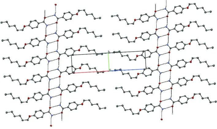
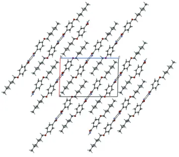
(1)° and 113.5 (1)°, respectively; Boese et al., 1999]. In the crystal structure, the amide groups are oriented head-to-head
forming N10—H102···O9 hydrogen bond at (-x, -y + 1, -z + 1) [the N···O distance is 1.941 Å] to generate a hydrogen
bond dimer. These dimers are further linked together by N10—H101···O9 hydrogen bonding at (x,y + 1, z) [N···O distance
is 3.106 Å] generating a ladder-like motif along the b axis (Table 1, Figs. 2 and 3) (Pagola & Stephens, 2009).
S2. Experimental
Attempts to crystallize 1,2-bis(4-heptylbenzylidene)hydrazine by liquid diffusion method from n-buthanol and water led
to crystals of the title compound, presumably due to slow hydrolysis by supervenient of water (Gallardo & Begnini,
1995; Pala Wilgus et al., 1995).
S3. Refinement
The H atoms were all located in a difference map, but those attached to carbon atoms were repositioned geometrically.
The H atoms were initially refined with soft restraints on the bond lengths and angles to regularize their geometry (C—H
in the range 0.93–0.98, N—H in the range 0.86–0.90 Å) and Uiso(H) (in the range 1.2–1.5 times Ueq of the parent atom),
Figure 1
Molecular structure of (I) with arom numbering and displacement ellipsoids at the 50% probablity level.
Figure 2
[image:3.610.128.482.359.570.2]Figure 3
Packing diagram of the title compound viewed along the a axis; hydrogen bond are shown as dashed lines.
4-Hexyloxybenzamide
Crystal data
C13H19NO2 Mr = 221.30 Monoclinic, P21/n Hall symbol: -P 2yn a = 12.5507 (2) Å b = 5.16441 (9) Å c = 18.9322 (3) Å β = 91.4702 (16)° V = 1226.72 (4) Å3 Z = 4
F(000) = 480 Dx = 1.198 Mg m−3
Cu Kα radiation, λ = 1.54184 Å Cell parameters from 8720 reflections θ = 3.5–70.8°
µ = 0.64 mm−1 T = 150 K
Needle-like, colourless 0.31 × 0.08 × 0.05 mm
Data collection
Oxford Diffraction Gemini E diffractometer
Radiation source: sealed x-ray tube Graphite monochromator
ω/2θ scans
Absorption correction: multi-scan
(CrysAlis PRO: Oxford Diffraction, 2006) Tmin = 0.894, Tmax = 1.000
13258 measured reflections 2355 independent reflections 2165 reflections with I > 2σ(I) Rint = 0.018
θmax = 71.0°, θmin = 4.2° h = −15→13
Refinement on F2 Least-squares matrix: full R[F2 > 2σ(F2)] = 0.037 wR(F2) = 0.095 S = 1.01 2101 reflections 145 parameters 0 restraints
Primary atom site location: structure-invariant direct methods
Hydrogen site location: inferred from neighbouring sites
H-atom parameters constrained
Method = Modified Sheldrick w = 1/[σ2(F2) + (0.05P)2 + 0.41P],
where P = [max(Fo2,0) + 2Fc2]/3 (Δ/σ)max = 0.0002608
Δρmax = 0.18 e Å−3 Δρmin = −0.18 e Å−3
Special details
Refinement. For this compound, 13258 reflections were measured and collected during the refinement. However after merging the symmetry equivalent reflections, there were only 2355 independent reflections. Further 254 more reflections were filtered, as sigma cutoff was set at 3.0 and (sin theta x 2)set to >0.01 (to eliminate reflection measured near the vicinity of beam stop) therefore reduced the number of reflection to 2101 which were used in the Refinement.
Fractional atomic coordinates and isotropic or equivalent isotropic displacement parameters (Å2)
x y z Uiso*/Ueq
O1 0.50007 (7) 0.75339 (18) 0.27977 (5) 0.0339
C2 0.40797 (9) 0.7236 (2) 0.31525 (6) 0.0276
C3 0.40594 (10) 0.5166 (2) 0.36254 (7) 0.0327
C4 0.31610 (10) 0.4686 (2) 0.40089 (6) 0.0302
C5 0.22742 (9) 0.6304 (2) 0.39492 (6) 0.0258
C6 0.23026 (10) 0.8374 (2) 0.34797 (6) 0.0283
C7 0.31893 (10) 0.8824 (2) 0.30718 (6) 0.0292
C8 0.13215 (9) 0.5718 (2) 0.43773 (6) 0.0265
O9 0.11152 (7) 0.34604 (16) 0.45547 (5) 0.0322
N10 0.07033 (9) 0.7708 (2) 0.45501 (6) 0.0326
C11 0.50247 (10) 0.9496 (3) 0.22591 (7) 0.0334
C12 0.61157 (10) 0.9498 (2) 0.19414 (7) 0.0337
C13 0.64375 (10) 0.6928 (2) 0.16146 (7) 0.0321
C14 0.74928 (10) 0.7091 (2) 0.12345 (7) 0.0314
C15 0.78664 (10) 0.4500 (3) 0.09497 (7) 0.0339
C16 0.89010 (11) 0.4699 (3) 0.05528 (7) 0.0392
H31 0.4688 0.4087 0.3672 0.0424*
H41 0.3139 0.3171 0.4318 0.0397*
H61 0.1691 0.9533 0.3429 0.0368*
H71 0.3177 1.0309 0.2744 0.0385*
H112 0.4882 1.1252 0.2475 0.0433*
H111 0.4461 0.9082 0.1885 0.0424*
H122 0.6652 1.0029 0.2319 0.0438*
H121 0.6111 1.0874 0.1576 0.0426*
H131 0.6479 0.5555 0.1986 0.0402*
H132 0.5859 0.6413 0.1263 0.0417*
H141 0.8049 0.7782 0.1575 0.0417*
H142 0.7433 0.8391 0.0847 0.0407*
H152 0.7299 0.3781 0.0628 0.0445*
H162 0.9124 0.2983 0.0389 0.0627*
H163 0.9472 0.5385 0.0870 0.0619*
H161 0.8823 0.5875 0.0142 0.0630*
H101 0.0938 0.9339 0.4498 0.0433*
H102 0.0124 0.7437 0.4808 0.0432*
Atomic displacement parameters (Å2)
U11 U22 U33 U12 U13 U23
O1 0.0292 (4) 0.0377 (5) 0.0350 (5) 0.0032 (4) 0.0051 (4) 0.0078 (4)
C2 0.0281 (6) 0.0268 (6) 0.0279 (6) −0.0015 (5) 0.0011 (4) −0.0021 (5)
C3 0.0319 (6) 0.0296 (6) 0.0365 (7) 0.0059 (5) 0.0000 (5) 0.0042 (5)
C4 0.0367 (7) 0.0238 (6) 0.0300 (6) 0.0010 (5) 0.0007 (5) 0.0046 (5)
C5 0.0312 (6) 0.0199 (5) 0.0264 (5) −0.0024 (4) 0.0004 (4) −0.0026 (4)
C6 0.0317 (6) 0.0209 (6) 0.0325 (6) 0.0024 (5) 0.0019 (5) 0.0006 (5)
C7 0.0346 (6) 0.0220 (6) 0.0311 (6) −0.0002 (5) 0.0029 (5) 0.0039 (5)
C8 0.0331 (6) 0.0216 (6) 0.0248 (5) −0.0019 (5) 0.0001 (5) −0.0006 (4)
O9 0.0390 (5) 0.0201 (4) 0.0378 (5) −0.0009 (3) 0.0095 (4) 0.0028 (3)
N10 0.0382 (6) 0.0204 (5) 0.0399 (6) −0.0006 (4) 0.0136 (5) 0.0011 (4)
C11 0.0356 (7) 0.0278 (6) 0.0373 (7) 0.0011 (5) 0.0070 (5) 0.0042 (5)
C12 0.0353 (7) 0.0279 (6) 0.0382 (7) −0.0037 (5) 0.0071 (5) 0.0001 (5)
C13 0.0316 (6) 0.0277 (6) 0.0370 (7) −0.0031 (5) 0.0030 (5) −0.0013 (5)
C14 0.0335 (6) 0.0271 (6) 0.0335 (6) −0.0027 (5) 0.0024 (5) −0.0002 (5)
C15 0.0335 (7) 0.0292 (7) 0.0391 (7) −0.0018 (5) 0.0013 (5) −0.0039 (5)
C16 0.0368 (7) 0.0393 (8) 0.0415 (7) 0.0014 (6) 0.0031 (6) −0.0071 (6)
Geometric parameters (Å, º)
O1—C2 1.3605 (14) C11—H112 1.013
O1—C11 1.4384 (15) C11—H111 1.011
C2—C3 1.3954 (17) C12—C13 1.5233 (17)
C2—C7 1.3915 (17) C12—H122 1.007
C3—C4 1.3792 (17) C12—H121 0.992
C3—H31 0.968 C13—C14 1.5258 (17)
C4—C5 1.3943 (17) C13—H131 0.998
C4—H41 0.978 C13—H132 1.007
C5—C6 1.3912 (16) C14—C15 1.5216 (17)
C5—C8 1.4926 (16) C14—H141 1.003
C6—C7 1.3904 (17) C14—H142 0.996
C6—H61 0.976 C15—C16 1.5203 (18)
C7—H71 0.986 C15—H151 0.995
C8—O9 1.2423 (14) C15—H152 0.997
C8—N10 1.3338 (15) C16—H162 0.982
N10—H101 0.898 C16—H163 0.989
N10—H102 0.898 C16—H161 0.990
O1—C2—C3 115.66 (11) C11—C12—H122 108.3
O1—C2—C7 124.71 (11) C13—C12—H122 110.2
C3—C2—C7 119.63 (11) C11—C12—H121 106.8
C2—C3—C4 120.28 (11) C13—C12—H121 109.7
C2—C3—H31 118.1 H122—C12—H121 106.9
C4—C3—H31 121.6 C12—C13—C14 112.69 (10)
C3—C4—C5 120.75 (11) C12—C13—H131 110.0
C3—C4—H41 119.7 C14—C13—H131 110.0
C5—C4—H41 119.5 C12—C13—H132 107.7
C4—C5—C6 118.62 (11) C14—C13—H132 108.8
C4—C5—C8 118.89 (10) H131—C13—H132 107.5
C6—C5—C8 122.47 (11) C13—C14—C15 113.43 (10)
C5—C6—C7 121.16 (11) C13—C14—H141 108.4
C5—C6—H61 120.1 C15—C14—H141 109.0
C7—C6—H61 118.8 C13—C14—H142 109.6
C2—C7—C6 119.50 (11) C15—C14—H142 110.5
C2—C7—H71 121.8 H141—C14—H142 105.7
C6—C7—H71 118.6 C14—C15—C16 112.97 (11)
C5—C8—O9 120.85 (10) C14—C15—H151 109.9
C5—C8—N10 117.12 (10) C16—C15—H151 109.9
O9—C8—N10 122.03 (11) C14—C15—H152 108.8
C8—N10—H101 120.1 C16—C15—H152 109.2
C8—N10—H102 119.9 H151—C15—H152 105.8
H101—N10—H102 118.5 C15—C16—H162 110.4
O1—C11—C12 108.58 (10) C15—C16—H163 109.8
O1—C11—H112 109.7 H162—C16—H163 107.9
C12—C11—H112 109.4 C15—C16—H161 111.2
O1—C11—H111 108.7 H162—C16—H161 109.2
C12—C11—H111 110.3 H163—C16—H161 108.3
H112—C11—H111 110.1
Hydrogen-bond geometry (Å, º)
D—H···A D—H H···A D···A D—H···A
N10—H101···O9i 0.90 2.14 3.0153 (18) 164
N10—H102···O9ii 0.90 2.04 2.9401 (18) 174