Robert K. Breger1 Alan L. Williams David L. Daniels Leo F. Czervionke Leighton P. Mark Victor M. Haughton Rose Anne Papke Mary Coffer
This article appears in the MayfJune 1989 issue of AJNR and the August 1989 issue of AJR.
Received May 27, 1988; accepted after revision November 21, 1988.
This work was supported by a grant from Berlex and by NIH grant #5 R01 NS22913-03.
' All authors: Department of Radiology, Medical College of Wisconsin, Froedtert Memorial Lutheran Hospital, 9200 W. Wisconsin Ave., Milwaukee, WI 53226. Address reprint requests to V. M. Haughton.
AJNR 10:633-637, MayfJune 1989 0195-6108/89/1003-0633
© American Society of Neuroradiology
Contrast Enhancement in Spinal
MR Imaging
633
We evaluated 44 patients with suspected spinal tumors or previous laminectomies with gadolinium-DTPA MR imaging in order to characterize the enhancement in normal, postoperative, and neoplastic intraspinal tissue. Using the signal intensity of CSF as an internal control, we calculated the percentage increase in signal intensity from pre- to postgadolinium studies. Tumors (astrocytoma, ependymoma, schwannoma) enhanced 70-350%; epidural scar, normal epidural venous plexus, and dorsal root ganglion enhanced up to 200%. Contrast enhancement does not per se distinguish neoplastic from normal tissue. Enhancement with gadolinium-DTPA appeared to increase the conspicuousness of intramedullary tumors but not intraosseous metastases.
We believe that gadolinium-enhanced MR imaging is a valuable adjunct to routine MR imaging in the evaluation of intraspinal neoplastic processes and may be useful in delineating normal and postoperative structures in the spinal canal.
Gadolinium (Gd)-DTPA is under evaluation by the FDA as a contrast medium tor MR imaging of the spine. Enhancement of spinal tumors has been shown previously
[1-5]. The purpose of this study was to measure the degree of contrast enhance-ment in neoplasms as compared with normal and nonneoplastic spinal tissues after Gd-DTPA administration.
Materials and Methods
Forty-two patients were studied under two different phase Ill protocols. In one protocol (group 1), 22 patients with suspected spinal tumors were studied. Of the 22 enrolled, 15 had surgical or postmortem verification of the diagnosis. Among these patients there were three astrocytomas, three ependymomas, three schwannomas, two metastases, and four cysts not associated with a tumor. One of the 15 patients had previously undergone resection of an ependymoma and was evaluated for a presumed recurrence but has not yet been reoperated. Four of the 22 patients had negative MR studies without anatomic confirmation, and three had positive studies. In a second protocol (group II), 20 patients who had previously undergone laminectomy were studied to investigate possible scar tissue or recurrent disk herniations. Eight patients with MR evidence of epidural scar at one or more levels had surgical confirmation of epidural scar (with or without herniated disk). Measurements of contrast enhancement were made in the 14 tumor tissues in group I, in epidural scar in group II, and in normal tissues in both groups.
634 BREGER ET AL. AJNR:10, May/June 1989
to nullify any change in CSF signal intensity between pre- and post-Gd-DTPA images. The contrast enhancement in each tissue was expressed as a ratio of the difference between post-and pre-Gd-DTPA signal intensities to the signal intensity in pre-Gd-DTPA scans (i.e., percent enhancement). All contrast-enhancement measurements were rounded to the nearest 1 0%, and tissues were grouped into 0-20% enhancement, 21-100%, or greater than 100%. One investiga-tor assessed contrast enhancement qualitatively by comparing the pre-and post-Gd-DTPA images (displayed at similar window width and level); and for each tumor or tissue, assigning grade 0 for no visible enhancement, 1 for moderate enhancement, and 2 for marked enhancement.
Results
In each of the spinal tumors, contrast enhancement was
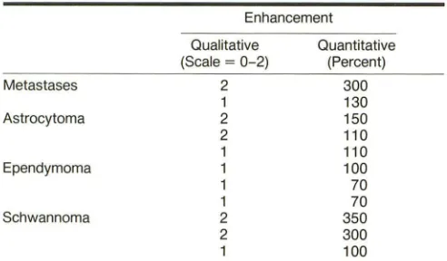
evident qualitatively and quantitatively (Table 1 ). Metastases
to the vertebrae (prostate, breast) showed grade 1 or 2
enhancement qualitatively and 130-300% contrast
enhance-ment quantitatively (Fig. 1 ). Although the metastatic tumors
enhanced, they were less conspicuous in the enhanced
im-ages. The three astrocytomas showed grade 1 or 2
enhance-ment and 11 0-150% increase in signal intensity with IV
Gd-TABLE 1: Contrast Enhancement in Spinal Tumors
Metastases
Astrocytoma
Ependymoma
Schwan noma
Enhancement
Qualitative (Scale = 0-2)
2 1 2 2 1 1 1 1 2 2 1 Quantitative (Percent)
B
300 130 150 110 110 100 70 70 350 300 100DTPA (Fig. 2). The two proved and one presumed recurrent ependymomas (Fig. 3) showed grade 1 qualitative enhance-ment and 70-1 00% increase in signal intensity. The three schwannomas showed marked or moderate enhancement (Fig. 4) and 100-350% enhancement. The intramedullary and intradural extramedullary tumors were more conspicuous in the enhanced than in the unenhanced images (800/20, 2500/ 20 or 2500/80 sequences).
Enhancement in normal tissues is summarized in Table 2.
Among normal tissues, greatest enhancement was observed in bladder urine (940%). The epidural plexus enhanced incon-sistently. Enhancement could usually be detected quantita-tively or qualitaquantita-tively in the cervical epidural plexus (Fig. 5) and sometimes in the lumbar venous plexus. The dorsal root ganglion enhanced qualitatively and quantitatively (Fig. 6). Measurements in 12 cases showed enhancement of 1 00-140%, which was qualitatively assessed as grade 1. The ganglion's contrast with respect to the surrounding fat was diminished in enhanced images. Scar enhanced to a variable
degree (Fig. 7). In eight cases with surgically verified epidural
scar tissue, enhancement averaged 80% (range, 0-240%). On qualitative assessment it appeared most commonly as grade 1 . No correlation with age of scar was attempted because of the small number of cases. Enhancement in bone marrow, muscle, and ligamentum flavum could be measured, but not usually detected by inspection of images at conven-tional windows.
Discussion
Less precision is achieved in studying MR signal intensities from the spine than from the head [6]. Because of the nonuniform signal reception in a surface coil, signal intensity measured from a region of interest in the spine is distorted by any movement of the patient. The radiofrequency pulses generated by a round head coil have a higher degree of homogeneity than those from a surface coil in which the radiofrequency signal is dependent upon the coil geometry and position. For small spinal structures such as ganglia,
[image:2.612.55.301.367.511.2] [image:2.612.55.387.543.728.2]AJNR:1 0, May/June 1989 CONTRAST ENHANCEMENT IN SPINAL MR
635
Fig. 2.-A and B, Recurrent astrocytoma (ar -rows) demonstrated with pre- (A) and post- (8) Gd-DTPA spin-echo 800/20/2 MR images. The tumor had grade 1, 110% enhancement. Autopsy confirmed tumor in the spinal cord. The cord adheres posteriorly to scar tissue, which also enhances after Gd-DTPA.
A
B
Fig. 3.-Presumed recurrent ependymoma.
A
A, MR image, 800/20/2, shows normal diameter and signal intensity in spinal cord of 56-year-old man with increasing weakness after resection of a spinal cord ependymoma.
8, Post-Gd-DTPA MR image, 800/20/2, shows enhancement in the cord (arrow) and in scar tissue dorsal to the spine. High signal in vertebral bodies represents fatty marrow replacement caused by radiation therapy.
roots, veins, and meninges, partial volume averaging is a significant source of inaccuracy in the measurements. Another variable was introduced by use of the automatic prescan. Attenuation constants were presumably not identical for
pre-B
A
B
Fig. 4.-A and 8, Schwannoma. Compared with pre-Gd-DTPA 800/20/2 MR image (A), the post-Gd-DTPA 800/20/2 MR image (8) shows 350% increase in signal intensity in the tumor (arrow).
and post-Gd-DTPA imaging. External standards to which measurements can be normalized are not practical when surface coils are used. Therefore, an internal standard (CSF) was used to normalize measurements.
[image:3.614.152.553.69.605.2] [image:3.614.52.296.344.599.2]636
BREGER ET AL. AJNR:1 0, May{June 1989TABLE 2: Contrast Enhancement in Tissues in and Near the Spine
Enhancement
Qualitative Quantitative
(Scale = 0-2) (Percent)
Bladder 2 >200
Epidural plexus 2 100-200
Epidural scar 1
Dorsal root ganglion 1
Bone marrow 0 21-100
Paraspinal muscle 0
Ligamentum flavum 0
Epidural fat 0 0-20
Spinal cord 0
Anulus fibrosus 0
Nucleus pulposus 0
Cortical bone 0
A
8
A
8
spinal tumors. Although schwannomas seemed to enhance on the average more than astrocytomas or ependymomas,
different types of tumors could not be differentiated by their degree of contrast enhancement. Some ependymomas and astrocytomas had the same degree of enhancement.
Nonneoplastic tissues also enhanced. Normal dorsal root ganglion, muscle, and marrow enhanced because they have a fenestrated capillary endothelium, that is, no blood-brain barrier. For example, normal dorsal root ganglion may en-hance to the same degree as some tumors; therefore, normal tissues, scar, and tumorous tissues cannot be identified by their degree of contrast enhancement alone. Although we found no contrast enhancement in the normal spinal cord, we did not evaluate the possibility of enhancement in the cord after trauma, surgery, or radiation therapy. Epidural scar, which enhanced inconsistently, could not be characterized accurately by its degree of enhancement. It enhanced
incon-Fig. 5.-A and 8, Enhancement in cervical spinal epidural plexus is shown in pre- (A) and post- (8) Gd-DTPA 800/20/2 MR images. Note increased signal intensity (arrows) in region of epidural plexus, dural sac, and root sleeves.
Fig. 6.-A and 8, Enhancement of spinal
[image:4.612.57.298.98.260.2] [image:4.612.57.393.272.732.2]AJNR:10, May/June 1989 CONTRAST ENHANCEMENT IN SPINAL MR 637
Fig. 7.-A and 8, Enhancement in epidural scar. Axial images at L5-S11evel without (A) and with (8) IV enhancement. In the enhanced image, the scar (arrows) is more easily distinguished from the dural sac and root sheath (arrowhead).
A
spicuously in some cases, markedly in others. The extent of scar could be judged more accurately in some cases because of enhancement. The differentiation of root sheath and scar could be made more reliably on enhanced than unenhanced images; therefore, the characteristic pattern of epidural scar was often more evident on enhanced images.
Contrast enhancement in a tumor did not inevitably increase its contrast with respect to adjacent tissues. As others have noted [4, 5], the enhancement of intramedullary (astrocytoma and ependymoma) and intradural extramedullary (schwan-noma) tumors increased their conspicuousness; enhance -ment in the intraosseous (metastatic) tumors decreased their conspicuousness because the enhanced tumors had a signal intensity similar to that of the adjacent bone marrow. There-fore, to obtain the greatest benefit of Gd-DTPA enhancement, patients must be selected thoughtfully.
The diagnostic benefit of enhancement in normal tissues needs further study. In the cervical spine, Gd-DTPA increases the signal from the large venous plexus in the neural foramina. Detection of herniated disk fragments in the neural foramen by MR imaging may be improved by enhancement, as some authors have suggested it is by CT [7].
The study emphasizes the need for a measurement of sensitivity of Gd-DTPA-enhanced MR versus unenhanced T1-or T2-weighted images. It shows that quantification of con-trast enhancement in spine imaging has little clinical utility
B
because of the variability in enhancement of normal spinal tissues and tumors and the imprecision in measurements of enhancement.
REFERENCES
1. Bydder GM, Brown J, Niendorf HP, Young IR. Enhancement of cervical intraspinal tumors in MR imaging with intravenous gadolinium DTPA. J Comput Assist Tomogr 1985;9:847-851
2. Schroth G, Thron A. Guhl L. Voigt K, Niendorf HP, Garces LR. Magnetic resonance imaging of spinal meningiomas and neurinomas. Improvement of imaging by paramagnetic contrast enhancement. J Neurosurg
1987;66:695-700
3. Stimac G, Porter B, Olson D, Gerlach R, Genton M. Gadolinium-DTPA· enhanced MR imaging of spinal neoplasms: Preliminary investigation and comparison with unenhanced spin-echo and stir sequences. AJNR
1988;9: 839-846
4. Sze G, Krol G. Zimmerman R. Deck M. Intramedullary disease of the spine: diagnosis using gadolinium-DTPA-enhanced MR imaging. AJNR 1988; 9:847-858
5. Tanaka 0, Yoshikawa H, Takahashi K. et al. Clinical experience of Gd-DTPA in MRI of brain and spinal cord tumors. J Pharmacal Ther 1987;15/ 6:2661-2669
6. Breger RK, Papke RA, Pojunas KW, Haughton VM, Williams AL, Daniels DL. Benign extraaxial tumors: contrast enhancement with Gd-DTPA. Ra-diology 1987;163:427-429
[image:5.612.224.558.79.326.2]