With 12 figures
(in Great Britain
APNOEA IN AMPHIBIANS AND REPTILES
BY G. SHELTON AND R. G. BOUTILIER
School of Biological Sciences, University of East Anglia, Norwich, NR\ yTJ
SUMMARY
1. The amphibian ancestor had two gas exchangers, one working in water and one in air. It also ventilated the aerial exchanger in an inter-mittent fashion. The functional repercussions of thie ancestry can be seen in all amphibians and reptiles.
2. During periods of apnoea, which almost all amphibians and reptiles show in their breathing patterns, there are variable fluctuations in lung and arterial POl and] PCo.- Arterial PCOj and pH show only small fluctu-ations during apnoea induced by voluntary dives, and blood lactate does not build up. More considerable acidoses and lactate accumulations occur in forced dives which the animals can readily survive, even when the dives are prolonged. Anaerobic metabolism plays an important role in their capacity to survive prolonged apnoea.
3. Amphibians ventilate their lungs with a buccal pump whose pattern of activity differs from species to species. Short periods of apnoea charac-terize the more terrestrial forms. Long dives punctuated by bursts of lung ventilation, or irregular bouts of breathing with no distinctive pattern occur in more aquatic forms. Similar differences in pattern can be seen in terrestrial and aquatic reptiles. The most striking characteristic of venti-lation is its extreme variability, even in a single individual.
4. Responses occur to decreasing oxygen tensions and increasing carbon dioxide tensions in inspired air, alveolar air, and arterial blood, the animals almost always increasing lung ventilation and decreasing the duration of apnoea. However, no simple relationship can be seen between the tensions of respiratory gases in lungs or arterial blood and the beginning or end of an apnoeic period.
5. Periods of apnoea are accompanied by vasoconstriction in the lung vasculature. This has the effect of decreasing blood flow to the lung and increasing right to left shunt in the incompletely divided heart.
INTRODUCTION
in [HCO3~] as well as in CO2, so that substantial changes in blood pH do not occu* (Rahn, 1967; Robin, Bromberg & Cross, 1969; Howell, 1970). Furthermore, gm ventilation in fishes seems to be related primarily to their O2 demands and the O2 content of the medium and not to the regulation of PCOt and pH (Janssen & Randall, 1975; Heisler, 1980) which are held constant in gill breathers by mechanisms other than adjustments of ventilation. In well-adapted air breathers, on the other hand, variations in ventilation are of fundamental importance in the control of PCOl and pH in the body fluids.
In conditions of O2 depletion in the water in which they live, many fish have devel-oped the ability to gulp air, coming to the surface for very brief periods in order to replenish the gas in the accessory air breathing structure. A few of these animals were of importance in the further evolution of the tetrapods, but many others were not, and they have remained fully adapted to life at or about the water surface. The posses-sion of two gas-exchangers, one working in water and the other in air, together with the habit of ventilating the air exchanger intermittently, has resulted in similar changes in the respiratory physiology of a wide variety of unrelated fish and amphibians. Many of the adaptations can still be seen in other intermittent ventilators, which include the more terrestrial amphibians and most of the reptiles, particularly those which are good swimmers and divers. In this article the view is taken that the history of dual gas-exchange and intermittent breathing has had functional repercussions in all amphibians and reptiles. Only in birds and mamrrtals have the gas exchangers and their associated control systems become completely emancipated from the early influence of the air-water interface.
GAS EXCHANGE
(a) Characteristics of dual (bimodal) exchange systems
tJearly constitute sites of extrapulmonary exchange, it cannot be assumed that the Bicker, more protected integument of reptiles is completely unimportant in this respect. Many workers (Belkin, 1968; Jackson, Allen & Strupp, 1976; Crawford & Schultetus, 1970; Graham, 1974; Standaert & Johansen, 1974) have reported appre-ciable loss of COa through extrapulmonary routes in reptiles, particularly aquatic forms. The lungs of reptiles do not necessarily operate at an R value equivalent to the metabolic RQ, as would be expected in a fully terrestrial animal with an impermeable integument.
(b) Fluctuations in respiratory variables
The above considerations of complex systems operating through two unevenly balanced exchangers give a view of respiratory control that is essentially rather static. This view is reinforced in the literature because values given for the major respiratory variables in arterial blood (P0 |, Pco,. pH, [HCO3~]) are often single ones, usually used to illustrate the consequences of evolutionary change from water to air. Indeed the accepted view of an effective respiratory control system would require an animal to show stability in these variables, as well as some matching between metabolic demands and the ventilation and perfusion of the gas exchanger. Almost all amphibians and reptiles ventilate their lungs intermittently to some extent, with variable periods of apnoea occurring between ventilation cycles whose depth, frequency and number during the ventilation period are also variable. Clearly, complete stability of the respiratory variables cannot exist in such intermittent breathers. It is important when assessing control systems to see how far these animals depart from the homeostatic ideal.
(i) Lung gases
The time course of changes in lung and blood gases during apnoea have, on the whole, been best described in well-adapted divers. The apnoea in these animals is prolonged and stable when compared to that in more terrestrial forms. Even so, measurements of gas tensions in lungs and blood (Lenfant et al. 1970; Toews, Shelton, & Randall, 1971; Emilio & Shelton, 1974) are not easy to carry out without disturbing the experimental animals. Breathing can be prevented in diving animals simply by making the surface inaccessible. Forced dives of this sort (Lenfant & Johansen, 1967; Jones, 1972; Emilio, 1974; Emilio & Shelton, 1980) can yield useful information but the results may not reflect those found during voluntary dives (see also Butler - this volume, 1982).
248 G. SHELTON AND R. G. BOUTILIER
Time (min) j I Out 400 °-H O t p In -400
2 (torr)
-no
Fig. 1. Continuous recordings of lung ventilation (shown as vertical deflexions produced by gas flow through a pneumotachograph) and corresponding lung gas tensions in freely diving and surfacing Xenopus laevit. The traces of lung gas tensions have been adjusted in time to take account of the delay in the extrapulmonary loop used for tension measurements. Animal in air equilibrated water and breathing air at a surface blowhole. Temp. 25 °C. (From Boutilier,
1981.)
approaching the metabolic RQ of 0-9 (Fig. za). Because ventilation in Xenopus is extremely effective in pumping freshly inspired air into the lungs (Brett & Shelton, 1979) and dead space is small, high levels of alveolar POt were seen during a breathing burst thus allowing high R values in spite of the low alveolar
PcOi-During apnoea the O2-CO2 relationship changed, following a curve towards the mixed venous (i.e. pulmonary artery) blood point (Fig. za) such as would be predicted from the known effects of low ventilation: perfusion ratios. Venous blood also changed in composition with pulmonary artery POt falling by as much as 15 torr in Xenopus (Emilio & Shelton, 1974) and other amphibians (Toews, Shelton & Randall, 1971) during voluntary dives. Venous PCOl increased very little because of its elimination through the skin and storage in blood and tissues, so that the shift in venous blood point was almost horizontal on the O2-CO4 diagram. The total effect of these changes was to reduce lung R values to 0-1-0-2.
Activity in the diving animal raised the lung PcOi values as Fig. 2 (a) shows; it also caused the dive to be curtailed so that breathing was initiated at relatively high POt levels in the lung. The relationships also changed if the dives were prolonged experi-mentally by allowing the animals to submerge voluntarily and then preventing access to the surface for about 30 min. Xenopus often exceeded this time in voluntary dives and could survive very much longer forced dives. However, in a forced dive of any duration substantial changes, with POl falling and Fcoi increasing, were seen after the animal had unsuccessfully attempted to surface and breath (Fig. zb). When a forced dive ended, the pronounced hyperventilation and release of CO2 from stores in blood and tissue caused exchange to occur at R values higher than the metabolic RQ (Fig. zb).
249
30
20
10
0-4 0-6 10 2 0 - R
(a)
50 100 Alveolar POj(torr)
150
30
r-20
o 0,°
I
<
1 0-0
o 0-2
• \
(6)
1
0-4 0-6 H O 2 0 - R
\ \ \ 1
50 100 150
[image:5.451.59.353.49.560.2]Alveolar/"ojCtorr)
Time (min)
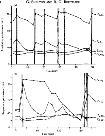
Fig. 3. Fluctuations in alveolar (PA,OV PA.OO.) and femoral artery (-P.,OH -P«,OO,) gas tensions
in freely diving Pteudemys tcripta. Periods of lung ventilation are shown by the shaded vertical bars. (From Burggren and Shelton, 1979.) (a) Dives of short duration. (6) Prolonged dive with changes in alveolar relationships. About ao % of the voluntary dives were of this type.
6; Jackson, Allen & Strupp, 1976), so that PCOl in its venous blood changes less •ran it does during apnoea in the tortoise.
(ii) Blood gases and acid base balance
A simple specification of the range of gas tensions, pH and related variables in the blood of amphibians and reptiles during apnoea is not possible. The central shunt of arterial and venous blood in the undivided ventricle makes outgoing different from incoming blood. Thus blood from the right atrium is mixed with that from the left as it flows to the pulmonary artery, and the converse occurs in blood flowing to the systemic arterial system. These two directions of mixing are known respectively as left-to-right and right-to-left shunts, and the extent to which they occur depends on a number of factors, one of the most important being the degree of lung vasoconstriction (Shelton, 1970, 1976). In addition, the blood vessels serving lungs and extrapulmonary exchangers are often connected to functionally different parts of the circulatory system. Thus a clear distinction between venous blood from metabolizing tissues and arterial blood from the gas exchanger cannot be made as it can in mammals and birds.
In Amphiuma (Toews et al. 1971), Xenopus (Emilio & Shelton, 1974), Pseudemys and Testudo (Burggren & Shelton, 1979), the largest oscillations in POl are seen in blood sampled from the pulmonary vein, followed in descending order by that from the dorsal aorta, pulmonary artery, and inferior vena cava. A similar statement cannot be made for Pc0,» since the gradients are smaller (Fig. 3 a) and the extrapul-monary exchange relatively more important. Blood returning to the heart from the body does not show significantly greater fluctuations in PCOt than blood from the lungs. Almost all measurements have been carried out on blood sampled from some part of the systemic arterial system and the following account will be confined to experiments of this type. Because there are persistent gradients between alveolar gas and pulmonary venous blood, and because such blood is mixed with left atrial blood in its passage to the systemic circulation, large tension differences exist, both for Oa and CO2, between alveolar gas and systemic arterial blood.
Closed extracorporeal loops incorporating the electrodes for measurement of gas tensions in blood are obviously advantageous in avoiding disturbance to the animal and blood loss due to sampling. Such a loop connected to the leg vessels of Xenopus (Brett, 1980) showed large and variable fluctuations in arterial POt between 80-125 t o r r during active lung ventilation and 10-15 torr at the end of a voluntary dive lasting about 30 min at 20-25 °C (Fig. 4). Just as was found in the case of lung POl, both the upper levels at which breathing stopped and the lower levels at which it started again were unpredictable and varied greatly from dive to dive.
13 100H
5 0
-100
5 0-0
20 40 60
20 40 60
Time(min)
Fig. 4. Continuous recordings of arterial PO i in freely diving Xetwput lacvis. Water Po.~I3°
torr upper trace, 88 torr lower trace. Temp. 20 °C. The vertical lines indicate timing of lung ventilations. For the upper trace recording the extracorporeal loop used for measuring Po, ran from femoral artery to vein, and for the lower trace from femoral artery back to femoral artery. (From Brett, 1980.)
between the PCOt or pH levels at which the animals were stimulated to begin or end periods of apnoea. When Xenopus was subjected to forced dives of about 30 min duration, arterial PCOl and lactate concentration increased to much higher levels (Fig. 5). A combined metabolic and respiratory acidosis caused a fall of more than 0-2 pH units, with a net reduction in plasma bicarbonate. The differences between forced and unforced dives were almost certainly all attributable to the increased levels of activity in the former. Between 1 and 4 h were necessary for Xenopus to restore PCOt and pH to normal after a forced 30 min dive, whereas recovery from even a prolonged voluntary dive was usually completed during the first breathing burst.
Fluctuations in arterial POj> similar in extent and variability to those described for
Xenopus, were found in aquatic reptiles during voluntary dives (Fig. 3 a, b). The PCOt fluctuations were some two to three times larger than in Xenopus and approached levels of 40 torr after 30 min of apnoea at equivalent temperatures (Lenfant et al. 1970; Wood & Johansen, 1974; Burggren & Shelton, 1979). In more terrestrial forms such as Testudo, PCOt fluctuated over a similar range but the range of arterial Po, was significantly smaller, due to the much shorter periods of apnoea (Burggren & Shelton, 1979). Forced dives can produce much more extreme changes. Pseudemys, in dives lasting from 4 h to 5 days at temperatures between 17 and 24 °C, survived arterial
POt values approaching zero, Pcot or" more than 100 torr, and pH of 6-6-6-8 (Robin
% ' ' •
I Out
In
253
i3
<
30
25
20
15
7-8 •a 7-7
i =
< 7-6
7-5
0 10 20 30 40 50 0 10 20 30 40 50 Time (min)
Fig. 5. Fluctuations in P(x>t, pH, and lactate concentrations (all plotted as solid circles) in blood samples taken from the femoral arteries of two Xenopui laevii during the course of prolonged voluntary dives at 25 °C. Lung ventilations (as recorded by a pneumotachograph) are shown in the upper trace and their timing represented by shaded areas on the graphs. More substantial changes produced by 30 min forced dives in eight animals (means ± s.E.M.) are shown in the graphs on the left, plotted as solid triangles. (From Boutilier, 1981.)
time with hyperventilation continuing for several hours, even though arterial Po, was higher and PCOi lower than normal quite early in the recovery period. Plasma lactate concentrations were particularly slow to return to normal. Though such experimental dives cannot be related to normal control situations they do reveal a great deal about the abilities of amphibians and reptiles to cope with, and recover from, long periods of apnoea in extreme circumstances. In particular they illustrate the considerable capacity for anaerobic metabolism that these vertebrates possess.
(c) Anaerobiosis and oxygen debt
whose end-product in these animals is mainly lactic acid, is of major importance ia the activity energetics of all but the most sluggish poikilotherms. Though activi^l and apnoea place different demands on energy producing systems (Bennett, 1978), the integral part that anaerobiosis plays in activity must also form the basis of a preadaptation to prolonged apnoea. The metabolic adaptations (Hochackha, 1980) necessary to produce energy in the absence of O2 are already well developed as part of normal metabolism in all poikilotherms.
There is little evidence to show how far anaerobic pathways are important in undisturbed, voluntary apnoea. Measurements of heat loss have shown that Rana
esculenta (Jones, 1972) and Pseudemys (Jackson, 1968; Jackson & Schmidt Nielsen,
1966) reduce metabolic rate during the early stages of enforced apnoea and utili2e Ojj stores, indicating a low level of anaerobiosis. The data for Xenopus suggest that voluntary dives are terminated before anaerobic contributions become necessary. Recovery is rapid and principally a matter of replenishing depleted oxygen stores. However, it is clear that dives can be greatly extended as part of normal behaviour if access to the surface is prevented for any reason (see also Gatten, 1981), and then anaerobiosis makes a progressively greater contribution to the total metabolism.
(d) Temperature
Changes in temperature affect not only the physical parameters associated with gas exchange and transport but also the rates of O2 consumption and CO2 production in amphibians (Whitford, 1973) and reptiles (Bennett & Dawson, 1976). In addition control systems, in particular those regulating PCOi ar>d pH, are temperature-depen-dent as discussed later in this article. Major changes in respiratory physiology are therefore to be expected at different environmental temperatures. At very low tempera-tures (around o °C) it is clear that indefinite survival without access to air is possible in some amphibians and reptiles and that, even in the latter, gas exchange with the medium is of considerable importance (Ultsch & Jackson, 1982). The lungs of amphibians make a smaller percentage contribution to the total oxygen uptake as temperatures decrease (Hutchison, Whitford & Kohl, 1968; Guimond & Hutchison, 1976). In reptiles the effects of temperature vary. In Pseudemys, for example, it has been reported that decreasing temperatures between 30 and 10 °C cause no change in respiratory minute volume (Jackson, 1978). In Chelonia midas (Kraus & Jackson, 1980) and Terrapene ornata (Glass, Hicks & Reidesel, 1979), however, minute volume decreases at lower temperatures (Fig. 8 c), as it does in the marine iguana,
Ambly-rhynchus cristatus (Ackerman & White (1980)). The varanid lizards show an even more
VENTILATION
The view of respiratory control emerging from these considerations is that it allows broad oscillations of POt and, to a lesser extent, of PCOl to occur in lungs, blood and probably in tissues, with accompanying pH fluctuations in body fluids. The extent of the oscillations depends on the animal's habit, since more terrestrial forms show shorter periods of apnoea than aquatic ones. Nevertheless it appears that the apnoeic pause is a normal part of the control system output. Anaerobic mechanisms are also part of the normal system and become increasingly important as apnoea is prolonged. The closely regulated respiratory exchange seen in birds and mammals, in which apnoea represents a deviation of the control system from its normal state, is not a useful model.
(a) Patterns of ventilation
Both the mechanics and patterns of lung ventilation vary enormously among amphibians and reptiles. The buccal pump is the basic mechanism of ventilation in all lung-breathing amphibians but the timing of the pump's activity, in relation to nasal and glottal valve operation and lung gas flow, differs from species to species, (Guimond & Hutchison, 1973, 1974; de Jongh & Gans, 1969; West & Jones, 1975; Brett & Shelton, 1979). In lizards and snakes the costal musculature is used to ventilate the lungs directly (Rosenberg, 1973). In chelonians, on the other hand, the lungs are indirectly ventilated by changes in the visceral volume caused by muscles working on, or close to, the limbs and girdles (Gaunt & Gans, 1969; Gans & Hughes, 1967). Crocodiles utilize a combination of intercostal activity and movements of the visceral mass and liver at the posterior end of the pleural cavity to change lung volume (Gans, 1976).
The timing and frequency of ventilations are determined by many factors but the basic pattern depends very greatly on the degree of aquatic or terrestrial adaptation. Urodele amphibians rely extensively, and sometimes completely (e.g. Desmognathus
fuscus - Gatz, Crawford & Piiper, 1974) on extrapulmonary mechanisms for gas
exchange. Thus in Cryptobranchus alleganiensis (Boutilier, McDonald & Toews, 1980) and Necturus maculosus maculosus (Guimond & Hutchison, 1976) lung ventila-tions were found to occur infrequently (0-6 h"1) at temperatures up to 25 °C, and the lungs made very little contribution to gas exchange. Disturbance or activity put up the ventilation frequency in both animals and lung exchange could then become important. Miller & Hutchison (1979) describe a diurnal cycle in O2 consumption and spontaneous activity in Necturus. The increased locomotor activity was of low level and supported entirely aerobically by the stimulation of lung ventilation. In Amphiuma
means means and Siren lacertina lung ventilation was also infrequent (0-25-5
venti-lations h"1), but, because the ventilation mechanisms were very effective and the lungs more highly developed in these animals, gas exchange through the lungs was greater than in Cryptobranchus or Necturus (Guimond & Hutchison, 1974).
At the other extreme, the more terrestrial anurans such as Rana pipiens (West & Jones, 1975), Rana catesbeiana (de Jongh & Gans, 1969) and Bufo marinus (Maclntyre & Toews, 1976; Boutilier & Toews, 1977) have breathing patterns characterized by very short periods of apnoea when they are in air. These animals ventilate the buccal
256
G. SHELTON AND R. G. BOUTILIER2 -5 S S (c)
H-" # •
5 in
fs«> h
(«> 111 lliliniliii
oo
30 min
Fig. 6. Breathing patterns in Xenopui laevit at 25 °C, as recorded by a pneumotachograph. Discrete breathing bursts followed by dives are shown in (a) and (6). Prolonged apnoea with single ventilations followed by rapid dives (marked by arrows) as in (c), or with no surfacing at all as in (d), can be caused by surface threat. Breathing bouts, when the animal remains at the surface with its nostrils in air and ventilates its lung at irregular intervals, are shown in
cavity by continuous movements (buccal oscillations) of its muscular floor (mean of 91 min"1 at 23 °C in Bufo). At irregular intervals the oscillations are punctuated by more powerful movements of the buccal floor associated with lung ventilations (mean of 31 min-1 in Bufo). Finally, modified lung ventilations (inflation cycles) occur occasionally (1-2 h"1 in Bufo). These comprise a succession of buccal movements which pump larger volumes of air into the lungs and inflate them to higher pressures which are then maintained for several (1-32) seconds. All the species of Rana examined are also capable of prolonged periods of apnoea during dives, whereas Bufo marinus rarely engages in diving behaviour.
Xenopus is entirely aquatic in habit and one of the best-adapted anuran divers. All
movements of the buccal pump are concerned with lung ventilation since the animal shows neither the continuous buccal oscillations nor, under normal conditions, the inflation behaviour described above. Recordings, taken over a total of 44 h in seven intact and undisturbed animals at 25 °C, gave a mean breathing frequency of 077 (s.E. 0-17) per min-1 (Boutilier, 1981). The recordings also showed an enormous variation in behaviour though two main patterns of breathing could be discerned. In one (burst breathing) long periods of diving apnoea were occasionally interrupted by brief visits to the surface, when a series of lung ventilations occurred (Fig. 6a, b). In the other pattern Xenopus remained at the surface with its nostrils in air. Lung ventilations then occurred, not in discrete bursts, but intermittently over a long period of time (a breathing bout- Fig. 6e,f). If the animal was threatened in any way, it either remained submerged (Fig. 6d) or made very brief excursions to the surface (Fig. 6c), during each of which a single lung ventilation occurred, followed by an immediate dive.
In 400-i
Ventilation || j n i
flow 0 - - V if | M
( m l / m i n )
O u t 4 0 0 -Time ,
(0-2 min intervals)
[image:13.451.41.405.74.170.2]Volume change (1 ml intervals)
Fig. 7. A single breathing burst in Xenopus laevis at 25 °C. The record of ventilation flow, determined by a pneumotachograph, shows the three main types of lung ventilation, i.e. single expirations followed by one, two or three inspirations. Integration of the flow record gives the change in lung volume as shown in the lower trace.
smaller inspirations. Occasionally single inspirations were seen, and much less frequently there were three inspiratory components in a lung ventilation. Fig. 7 shows a record of a brief burst which was unusual in that it contained all three patterns. Integration of flow records showed that the volume of gas expired was usually smaller than that inspired by a factor of 0-8-0-9, though there was a lot of variation in indi-vidual ventilations (Fig. 7). The first ventilations in a burst following apnoea usually showed relatively large inspiratory volumes; in fact the three-inspiration pattern, if it appeared at all, was almost always the first breath in a burst. Some of the decrease in volume of expired gas can be explained by the unequal exchange of O2 and CO2 in the lung. The larger decreases cannot entirely be explained in this way. Transfer of N2 from the lung, ultimately into the water surrounding the animal, must also occur because Pift in the lung increases substantially during apnoea.
The complete repertoire of breathing patterns in Xenopus is obviously extensive and the control mechanisms must be complex. Not only can the temporal pattern of lung ventilations vary but each ventilation can itself differ in composition. The tidal volume of the buccal pump can also change as Fig. 7 shows. Finally variations occur in the timing of nasal and glottal valves and the buccal pump (Brett & Shelton, 1979). The control system produces outputs of considerable variety from ventilation to ventilation.
Most species of reptile also show arrhythmic breathing patterns (Wood & Lenfant, 1976), with periods of apnoea of variable duration alternating with lung ventilations which may be single or grouped into bursts. As in amphibians, apnoea occurs at the end of inspiration with lung deflation being prevented by glottal closure rather than by prolonged activity of the inspiratory muscles. The difference between aquatic and terrestrial animals is again marked, though there are very few data on unstressed animals breathing in a normal fashion. Unrestrained turtles (Chelys fimbriata -Lenfant et al. 1970), snakes (Acrochordus javanicus - Glass and Johansen, 1976) and caiman {Caiman crocodilus - Gans & Clark, 1976) dive and remain submerged for periods of some 30 min or more at 25 °C. Other aquatic forms ventilate after shorter apnoeic pauses at 18-25 °C (Pseudemys - mean 3-8 min: Burggren, 1975; Chrysemys
picta - mean i-6min: Milsom & Jones, 1980; Pelomedusa subrufa - mean 4-3 min:
burst of lung ventilations whose number correlates roughly with the duration of t]g preceding dive or apnoea. Members of the terrestrial genus Testudo, on the otHH hand, produce single ventilations separated by much shorter apnoeic pauses. The mean duration of these pauses at 20-25 °C ranges from 0-4-1-6 min and again the apnoeic interval is the most variable component in the whole breathing pattern (Burggren, 1975; Glass et al. 1978; Benchetrit & Dejours, 1980).
(b) Control of ventilation
Very little is known of the mechanisms within the central nervous system that are responsible for coordinating the movements of ventilation in amphibians and reptiles. Lumsden (1923) suggested that the prolonged periods of apnoea after inspiration were similar to the apneustic cramps seen in mammals after midpontine transection of the brain and bilateral vagotomy. Though the similarity in the two patterns is striking, the basic concept of a continuously active oscillator of the mammalian type is not entirely appropriate. Certainly the periods of apnoea in amphibians and reptiles are not inspiratory cramps, the muscles of ventilation being relaxed with only the glottal musculature active.
The way in which lungs, and other accessory air breathing structures, are thought to have evolved lends support to the view that prolonged periods of apnoea, with the exchanger full of gas, are fundamental components of the basic breathing pattern. In fact rhythmic activity is rarely seen in amphibian breathing, the exception being the buccal oscillations of many Anura. Lung ventilating movements are much less regular, however, even when anurans are breathing in air. When they are allowed to dive very few traces of rhythmicity remain. The ventilations in a breathing burst in Xenopus, for example, vary considerably in interval, as well as in other characteristics such as the number and depth of inspirations; and the duration of the ventilation interval greatly exceeds that of the ventilation itself. It is difficult to imagine that these patterns are the product of an oscillator of the mammalian type, in which interactions between mutu-ally inhibitory groups of neurones are continuous and influenced in depth and fre-quency by mechano- and chemoreceptor feedback. A more attractive model is one in which activity, coordinating the sequence of events in a ventilation, is switched on and off by appropriate trigger signals arising from the changing conditions during apnoea or breathing. The difficulty here lies in identifying the trigger systems concerned. Though there is now a certain amount of information on respiratory responses of whole animals, or of receptor systems, to mechanical and chemical stimuli, the relationships between these responses and the detailed breathing patterns are by no means clear. The subject has been well reviewed by Wood & Lenfant (1976) and by Jackson (1978) and only the major points will be discussed here.
(i) Chemoreceptor responses
•>me lizards, for example, breathing rate was substantially decreased at high levels !Tf C02, even though tidal volumes went up (Pough, 1969; Nielsen, 1961). The ability to survive high CO2 levels in inspired air is also vastly different in different species. Xenopus cannot tolerate levels higher than 1 % CO2 for more than a few hours whereas Bufo survives indefinitely at these levels. The experiments on chelonians, which have a high tolerance of CO2, used 5-10% mixtures and produced 3- to 10-fold increases in ventilation with no long-term problems of survival at 25 °C.
Temperature changes affect the response to CO2 since they influence both metabolic rate and the levels to which arterial pH ar.dPCOj are regulated. In many amphibians and reptiles, arterial pH moves to lower and PC 0 | to higher values as the temperature of the animal rises, so that extracellular fluids maintain a fixed relative alkalinity with respect to water (Rahn, 1967; Howell & Rahn, 1976; Reeves, 1977). Though extra-pulmonary exchange of CO2 must be of considerable importance in such adjustments, especially in amphibians, increased arterial PCOt can be achieved by reducing lung ventilation relative to the rate of CO2 production (i.e. by a reduction in the ratio ^E/^COI> more usually the related ratio PrB/VOl-the air convection requirement - is specified). In Pseudemys the air convection requirement was found to be reduced at higher temperatures as Fig. 8(a) and (b) shows (Jackson et al. 1974), whether the animal was breathing air or air-CO2 mixtures. A rise in temperature was found to have very little effect on ventilation in Pseudemys, most of the fall in convection require-ment being due to the considerable increase in O2 consumption. The addition of CO2 to inspired air caused only small changes in O2 consumption, however, so the increased convection requirements shown at progressively higher CO2 levels in Fig. 8 (a), do represent substantial changes in minute volume.
Temperature effects, different from those predicted by the relative alkalinity hypothesis, have been found on pH, PC 0 | and alveolar ventilation in a number of animals, particularly at temperatures below the normal range of the active animal. In the box turtle, Terrapene ornata, minute volume and convection requirement decreased as acclimation temperature fell below 15 °C- Fig. 8(6) and (c) (Glass et al. 1979) whilst alveolar PCOl remained relatively constant. Moreover, the green turtle, Chelonia
mydas (Kraus & Jackson, 1980); and the marine iguana, Amblyrhynchus cristatus
(Ackerman & White, 1979), had lower arterial pH and higher Pc o, values at 15 °C than the relative alkalinity hypothesis would suggest. Finally, the varanid lizards, which are extremely active reptiles with a high aerobic capacity, differ over a much greater temperature range. Varanus exanthematicus maintained a relatively constant pH and showed only small changes in arterial PCOl at temperatures from 15 to 38 °C (Wood et al. 1977, 1981). In these animals the air convection requirement is not affected by temperature and acid-base balance is linked to O2 demand (Fig. Sb).
Despite the complications due to temperature, which may or may not cause the range and sensitivity of the overall response to change, CO2 increases minute volume by stimulating a higher breathing frequency, or both a higher frequency and a larger tidal volume. A fall in the duration of periods of apnoea is part of the change and in some animals is the single most important factor.
ihe response and its speed (Milsom & Jones, 1976, 1979; Fedde, Kuhlmann & Scheid, •977). Another site of sensitivity to CO2 has been demonstrated by Hitzig & Jackson (1978) in the turtle. Perfusion of the ventricles of the brain with artificial CSF showed that small changes in [HCO3~] had substantial effects on lung ventilation. Other COa receptors may exist (Jackson, 1978), perhaps in the central arterial system (Frankel
et al. 1969; Benchetrit, Armand & Dejours, 1977), but the evidence is indirect.
Changes in the O2 content of inspired gas and arterial blood can also affect ventila-tion in amphibians and reptiles, though the experimental results are often variable and generalizations difficult to make. Thus Nielson (1962), working on lizards, found that breathing frequency went down and tidal volume increased if inspired gas con-tained less than 10% Oa. Boyer (1966) found no change in breathing frequency in a snake, lizard, and alligator but claimed that tidal volume increased. Bufo marinus re-sponded to progressive hypoxia by increasing the frequency of lung ventilations (Boutilier & Toews, 1977) though as hypoxia became intense (0-2% O2) fewer normal ventilations occurred and inflation cycles, with the lungs at higher than normal volumes, became an increasingly dominant part of the breathing pattern. More con-sistent results came from aquatic species. Hypoxic mixtures caused Xenopus to in-crease minute volume progressively, leading to a twofold change at 10% O2 that was due almost entirely to reduction in duration of the apnoeic periods (Brett, 1980). Boyer (1966) found that the periods of apnoea were substantially reduced in Chelydra after breathing in low oxygen concentrations, as were the dive durations of Chelys (Lenfant
et al. 1970) and Acrochordus (Glass & Johansen, 1976). In Pelomedusa and Testudo
(Glass et al. 1978) hypoxia stimulated an increase in minute volume but affected both tidal volume and frequency.
Jackson (1973) has shown that, in Pseudemys, the magnitude of the response to hypoxic gas mixtures depends on temperature (Fig. 9), hyperventilation becoming more marked as the temperature increases. Interpretation of these results is compli-cated by the uncertain acid-base state of the animal. Hyperventilation will obviously cause a respiratory alkalosis to develop, and shortage of O2 at high temperatures may lead to a metabolic acidosis. The final stimulus to any receptor system is not clear in experiments in which only inspired gas concentrations are monitored, as Wood & Lenfant (1976) have pointed out.
•a
e
800
700
600
500
400
300
200
100
0
30°C 20°C 10°C
25 50 75 100 125 150
Inspired />Oj(torr)
Fig. 9. Pseudemys tcripta. The effect of changes in inspired oxygen concentration on ventilation (expressed as percentages of control values determined when air was inspired) at tempera-tures of 10, 20 and 30 °C. (From Jackson, 1973.)
(ii) Mechanoreceptor responses
Electrophysiological recordings from the vagus show that stretch receptors exist in the lungs of both amphibians and reptiles. In the turtle their characteristics are well documented and somewhat resemble those of mammals (Jones & Milsom, 1979). It is also clear that information about the degree of lung distension is important in the coordination of normal breathing. Lung denervation in Xenopus led to a large increase in the number of inspirations in a lung ventilation (B. Evans, personal communication) The resulting over-inflation of the lungs was often so great that the animals were unable to dive. In chelonians also, stretch receptor information is important in regulating tidal volume (Milsom & Jones, 1980; Benchetrit & Dejours, 1980). Vago-tomy increased both tidal volume and the duration of apnoeic pauses in these animals, as well as changing the pattern of response to hypercapnia.
witching lung ventilations on and off. Such is the variety in breathing pattern that, at different times, ventilations are initiated at different gas tensions in alveolar air or arterial blood and are terminated at similarly unpredictable levels. In amphibians, particularly those in which the skin is a major route for CO2 removal, it is likely that alveolar and arterial PCOt do not change substantially in the later stages of a period of voluntary apnoea (Figs. 2 and 5). This suggests that the continually falling P0 | may be the more effective stimulus to breathe though the sensitivity of the total system for monitoring oxygen may be increased at the high equilibrium' levels of CO2. In less aquatic amphibians and reptiles, however, CO2 levels continue to change throughout apnoea and may be more important in initiating ventilation. Control of tidal volume, frequency of breaths and duration of a breathing burst is equally difficult to under-stand. All the components depend in a general way on the length of the preceding apnoea and severity of hypoxia or hypercapnia produced. But breathing often con-tinues at a high level long after the Oa and CO2 tensions in lung gas and blood have reached values that are not subsequently exceeded.
This is not to suggest that the exchange system reaches a steady state, such as that seen in birds and mammals, even when the animals continue to ventilate at a high level. Oscillations in POt and PcOi undoubtedly occur throughout all patterns of breathing in amphibians and reptiles. Breathing is regulated much less precisely than in higher forms. The reason for this may be that the feedback is less precise or that the coordinating system operates with much greater error. Finally it is not possible to predict, by examining the oscillating tensions in a breathing burst, whether the pause after any ventilation will be short, so that the burst is continued, or long, as in a prolonged dive.
In all diving animals there must be important connexions between the higher centres of the brain and that part responsible for coordination of breathing. In totally undisturbed amphibians, at least, the alternating cycles of breathing and apnoea and the consequent oscillations in all the respiratory variables discussed above, are much more regular than in animals which are disturbed. It seems that the behavioural component, which determines the time of surfacing, can override information from chemo- and mechano-receptors. Though anaerobic metabolism is avoided if conditions are favourable for breathing, most amphibians and reptiles seem to be capable of delaying ventilation when conditions are unfavourable and of deriving energy from anaerobiosis. The consequence of prolonging apnoea in this way is mainly one of a greatly extended recovery time.
PERFUSION
more important factors in evolutionary design, though not the only consideration This aspect of performance could be assessed as the rate of gas transfer achieved fol each unit of energy expenditure in pumping the exchanging media and in maintaining the exchanger itself. In these terms it would be inefficient to perfuse an unventilated gas exchanger at all unless it was being used as an oxygen store. If this were the case then variable rates of perfusion could be used to control gas transfer into or out of the store.
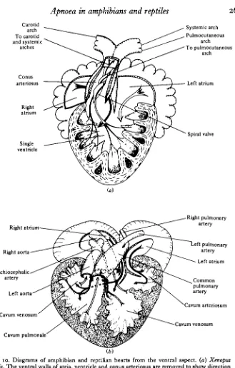
In birds and mammals, with completely divided circulations, the opportunities for controlling pulmonary blood flow are much more restricted than they are in the lower tetrapods. The anatomy of Xenopus and Pseudemys hearts is shown diagrammatically in Fig. 10(a) and (b). The completely undivided ventricle of the former makes it possible for the relative amounts of blood flowing to lungs and body to be adjusted over an infinitely variable range, the only condition being that total input and total output are the same. Cardiac output in these hearts has to be determined from measurements in all output vessels and not, as in birds and mammals, from measurements in either the aorta or the pulmonary artery. Even in the partially divided ventricle of reptiles, of which the diagram in Fig. io(b) is reasonably representative, central shunts can be equally extensive. The right and left atria empty into the partially separated cavum venosum and cavum arteriosum respectively. Blood from the cavum venosum also flows over a muscular ridge into the cavum pulmonale and it is from these two cham-bers that the output vessels arise, as Fig. 10(6) shows. Except in the varanid lizards and crocodiles there is no separation within the ventricle during either diastole or systole.
The circulation of lower tetrapods is affected considerably by periods of apnoea. Bradycardia develops progressively as apnoea becomes prolonged (Anderson, 1966), but even during the relatively short intervals between breaths there are fluctuations in heart rate. In addition vasoconstriction in some part of the pulmonary vasculature greatly restricts blood flow to the lungs. Fig. 11 shows the variation in flow in the pulmocutaneous arch of Xenopus during the ventilations of a breathing burst. Flow increased when ventilation occurred and declined gradually after ventilation ceased, even in the relatively short intervals of a breathing burst. The flow variations were obviously due to vasoactive changes in the pulmonary vessels because blood pressure fell slightly as flow increased and vice versa. Similar pulmonary flow changes, linked to lung ventilation, have been described in chelonians (White & Ross, 1966; Johansen, Lenfant & Hanson, 1970; Shelton & Burggren, 1976). Fig. 12 shows the effects in
Pseudemys both of surfacing and breathing after the prolonged apnoea of a voluntary
Apnoea in amphibians and reptiles
265
Carotid arch To carotid and systemic
arches
Conus arteriosus
Right atrium
Single ventricle
Systemic arch Pulmocutaneous
arch To pulmocutaneous
arch
Left atrium
Spiral valve
Right atrium
Right aorta
Brachiocephalic artery
Left aorta
Cavum venosum
Cavum pulmonale
Right pulmonary artery
eft pulmonary artery Left atrium
Common pulmonary artery Cavum arteriosum
[image:21.451.65.394.53.569.2]Cavum venosum
Fig. 10. Diagrams of amphibian and reptilian hearts from the ventral aspect, (a) Xenopus
laevis. The ventral walls of atria, ventricle and conus arteriosus are removed to show direction
of flow streamlines necessary to account for selective distribution of blood (from Shelton,
IO
266 G. SHELTON AND R. G. BOUTILIER
Breathing movements: buccal cavity
Blood n o w •O-l—i right pulmocutaneous I (ml/s) OH
Blood flow. left systemic (ml/s)
Blood pressure. 60 right systemic
(cm HjO) 20
Blood pressure: left pulmocutaneous
(cm
Time (min)
Fig. 11. Pressures and flows in the arterial arches of Xenopus laevis. Pressure changes in the buccal cavity during lung ventilating movements are recorded on the upper trace. The effect of lung ventilations on individual flow and pressure pulses can just be seen at the paper speed used. Changes in the pulmocutaneous arch are much greater than those in the systemic arch. (From Shelton, 1970.)
Minute flow (ml/min) 45| 40. 30| 20
t
267
Left pulmonary artery flow (ml/s)
Common pulmonary artery pressure (cm H,O)
40 301 20
10
40 Right aorta 30 pressure (cm HjO) 20'
10
[ 1 r 1 1 1 1 1 1 1 1 1 1 1
[image:23.451.53.404.63.399.2]Time (min)
vasoconstriction and hypertension mainly by means of adrenergic mechanisms and intrinsic factors in the lung.
Little is known of the sensory and central nervous mechanisms involved in the control of ventilation: perfusion relationships. White (1970, 1976) found in the alligator that deflation of the lungs caused a bradycardia that could be terminated by inflation with nitrogen. In amphibians, lung inflation was also found to be important in terminating bradycardia (Jones, 1966) and in producing pulmonary vasodilation (Emilio & Shelton, 1972). Constriction of the sphincter in the pulmonary artery of
Rana occurred when the lung was deflated, leaving the cutaneous artery unaffected.
De Saint Aubain & Wingstrand (1979) suggested that this reflex was the basis of blood-flow redistribution from lungs to skin during apnoea. However, the fact that the lungs are inflated during apnoea is not entirely consistent with hypotheses relating stretch of the lung to high heart rate and pulmonary vasodilation, and more work is needed. Chemoreceptors also seem to be important. When O2 was used to inflate
Xenopus lungs, a greater pulmonary vasodilation resulted than when air was used, and
N2 proved to be less effective than air (Emilio & Shelton, 1972). The relationships are not simple, however, and results such as those in Fig. 3(6), in which the rate of depletion of lung O2 and the POt gradients from lung to blood both vary considerably as apnoea progresses, suggest changes in lung perfusion that are incompatible with simple reflex control. Direct interactions between parts of the medulla responsible for respiratory and circulatory coordination (Burggren, 1975) may have a significant role in the production of bradycardia and selective vasoconstriction during apnoea. Anticipatory changes, such as heart-rate elevation before an animal surfaces to breathe (White, 1976), lead to the conclusion that higher centres in the brain are also involved.
Pulmonary vasoconstriction causes major readjustments in the pattern of blood flow through the heart. Evidence from experiments on a variety of amphibians and reptiles shows that, despite the undivided ventricle, blood in the systemic circulation contains more oxygen than that in the pulmonary arteries (White, 1959; Johansen & Ditadi, 1966; Tucker, 1966; Toews, Shelton & Randall, 1971; Emilio & Shelton, 1974; Burggren & Shelton, 1979). Such separation must be maintained by the laminar flow of blood in more or less uncontaminated streamlines from atria into the ventricle and from the ventricle into the arterial arches (Shelton, 1976). The flow patterns are very easily disturbed, and even in breathing animals a great deal of mixing goes on. The spatial relationships in the conus arteriosus of amphibians (Fig. 10a), and between the atria and three partially separated chambers in the ventricle of reptiles (Fig. 10 b), hardly seem conducive to undisturbed flow. An equal flow of blood from the two atria probably represents the condition for minimal mixing in either direction. Reduction of flow to the lungs will increase right-to-left shunt as the left atrium carries progressively less blood. The difference in O2 concentrations between systemic and pulmonary circulations decreases during apnoea, as the hypothesis of gradually increasing shunt demands. However, direct experimental verification of the full extent and direction of mixing during the normal alternation of ventilation and apnoea is still lacking in any of the lower tetrapods.
fipund. In varanids a complete separation of two ventricular chambers, the cavum pulmonale and the cavum arteriosum, is achieved during systole, the former pumping blood at low pressure to the lungs and the latter at high pressure to the body (Millard & Johansen, 1974; Burggren & Johansen, 1982). The myocardium is appropriately developed, that surrounding the cavum arteriosum being the more powerful. The partition, which is completed during systole by the meeting of a muscular ridge, does not persist during diastole, thus permitting shunts to develop as the ventricle fills. Berger & Heisler (1977) have shown by using microsphere tracer techniques that mixing in both left-to-right and in right-to-left directions does occur. Independent adjustment of pulmonary flow in these animals, some of which show periods of pro-longed apnoea, is therefore possible and Millard & Johansen (1974) have demonstrated an increase in pulmonary resistance during diving in Varanus niloticus. Flow and shunt patterns in the course of normal breathing behaviour have yet to be established. The crocodiles have a complete interventricular septum separating left and right ventricles. Though the connexions of the right aorta, together with the carotid and branchial vessels, are made with the left ventricle, the left aorta opens from the right ventricle alongside the pulmonary artery. The significance of this connexion was first appreciated by White (1969). He was able to show that, in air-breathing alligators, pressures in the systemic vessels greatly exceeded those in the right ventricle and pulmonary artery. As a result the valves between the right ventricle and left aorta never opened, since this large arterial trunk was in direct contact with the right aorta via both the foramen of Panizza and the dorsal aorta. The circulation performed as a completely divided one, with a high-pressure systemic circulation and a low-pressure pulmonary circulation, the blood flow to each side being equal. The myo-cardium surrounding the left ventricle is more powerfully developed than that of the right, as it is in birds and mammals. However, White (1969, 1970) also found that, when the alligator dived and bradycardia ensued, much larger pressures were produced within the right ventricle. As the dive progressed these pressures came to equal those in the systemic circuit so that the valves between the right ventricle and left aorta opened and blood flowed out to the body; a right-to-left shunt developed and flow to the lungs was reduced. Blood pressures in the pulmonary artery remained low because of contraction of the pulmonary outflow tract. It is difficult to assess the extent to which the shunt opeates because no flow measurements have yet been made. Clearly the right side of the heart becomes more and more dominant as the shunt develops and flow from lungs is reduced. The thinner myocardiumfof the right ventricle is thus pumping larger volumes of blood than the more powerfully developed left ventricle. It may be that the adjustment is made by changes in end diastolic volume and fibre length. It is also possible that differential innervation of the two ventricles could cause changes in contractility. The fact that the pattern of depolarization in the heart of chelonians changes as the animals alternate breathing with apnoea (Burggren, 1978) shows that important differences may exist in the innervation of right and left sides of the reptilian heart.
when applied to studies of gas exchange and ventilation in amphibians and reptiles, is also applicable to work on the circulatory systems. These systems are remarkably well adapted to matching ventilation and perfusion in gas exchangers that seldom operate in a steady state.
We should like to thank our colleagues Dr S. S. Brett and Dr M. G. Emilio, who were involved in much of the work and discussion that led to this article. In particular we are grateful to Dr Brett for allowing us to use some of his unpublished work.
REFERENCES
ACKERMAN, R. A. & WHITE, F. N. (1979). Cyclic carbon dioxide exchange in the turtle, Pteudemys
scripta. Physiol. Zool. 53, 378-389.
ACKERMAN, R. A. & WHITE, F. N. (1980). The effects of temperature on acid-base balance and ventila-tion of the marine iguana. Resp. Physiol. 39, 133-147.
ANDERSEN, H. T. (1966). Physiological adaptations in diving vertebrates. Phytiol. Rev. 46, 212-243. BELKIN, D. A. (1968). Aquatic respiration and underwater survival of two freshwater turtle species.
Resp. Physiol. 4, 1-14.
BENCHETRIT, G., ARMAND, J. & DEJOURS, P. (1977). Ventilatory chemoreflex drive in the tortoise
Testudo horsfieldi. Resp. Physiol. 31, 183-191.
BENCHETRIT, G., ARMAND, J. & DEJOURS, P. (1977). Ventilatory chemoreflex drive in the tortoise
Testudo horsfieldi. Resp. Physiol. 31, 183-191.
BENCHETRIT, G. & DEJOURS, P. (1980). Ventilatory carbon dioxide drive in the tortoise Testudo horsfieldi.
J. exp. Biol. 87, 229-236.
BENNETT, A. F. (1978). Activity metabolism of the lower vertebrates. A. Rev. Physiol. 40, 447-469. BENNETT, A. F. & DAWSON, W. R. (1976). Metabolism. In Biology of the Reptila, vol. 5 (ed. C. Gans
and W. R. Dawson), pp. 127-223. London, New York, San Francisco: Academic Press.
BERGER, P. J. (1972). The vagal and sympathetic innervation of the isolated pulmonary artery of a lizard and a tortoise. Comp. gen. Pharmacol. 3, 113-124.
BERCER, P. J. & HEISLER, N. (1977). Estimation of shunting, systemic and pulmonary output of the heart, and regional blood flow distribution in unanaesthetised lizards (Varanus exanthematicus) by injection of radioactively labelled microspheres. J. exp. Biol. 71, 111-122.
BOUTILIER, R. G. (1981). Gas exchange and transport during intermittent ventilation in the aquatic amphibian, Xenopus laevis. Ph.D. dissertation, University of East Anglia.
BOUTILIER, R. G., MCDONALD, D. G. & TOEWS, D. P. (1980). The effects of enforced activity on
ventilation, circulation and blood acid-base balance in the aquatic gill-less urodele, Cryptobrattehus
alleganiensis; a comparison with the semi-terrestrial anuran, Bufo marinus. J. exp. Biol. 84, 289-302.
BOUTILIER, R. G., RANDALL, D. J., SHELTON, G. & TOEWS, D. P. (1979). Acid-base relationships in the blood of the toad, Bufo marinus. I. The effects of environmental COS. J. exp. Biol. 82, 331—344.
BOUTILIER, R. G. & TOEWS, D. P. (1977). The effect of progressive hypoxia on respiration in the toad
Bufo marinus. J. exp. Biol. 68, 99-107.
BOVER, D. R. (1966). Comparative effects of hypoxia on respiratory and cardiac function in reptiles.
Physiol. Zool. 39, 307-316.
BRETT, S. S. (1980). Breathing and gas exchange in an aquatic amphibian, Xenopus laevis. Ph.D. dis-sertation, University of East Anglia.
BRETT, S. S. & SHELTON, G. (1979). Ventilatory mechanisms of the amphibian, Xenopus laevis; the role of the buccal force pump. J. exp. Biol. 80, 251-269.
BURCGREN, W. W. (1975). A quantitative analysis of ventilation tachycardia and its control in two chelonians, Pseudemys scripta and Testudo graeca. J. exp. Biol. 63, 367—380.
BURGGREN, W. W. (1977). The pulmonary circulation of the chelonian reptile: morphology, haemo-dynamics and pharmacology. J. comp. Physiol. 116, 303-323.
BURGGREN, W. W. (1978). Influence of intermittent breathing on ventricular depolarisation patterns in chelonian reptiles. J. Physiol., Lond. 278, 349—364.
BURCGREN, W. W. & JOHANSEN, K. (1982). Ventricular haemodynamics in the monitor lizard Varanus
exanthematicus: pulmonary and systematic pressure separation. J. exp. Biol. 96, 343-354.
BURGGREN, W. W. & SHELTON, G. (1979). Gas exchange and transport during intermittent breathing in chelonian reptiles. J. exp. Biol. 8a, 75-92.
CAMPBELL, G. (1971). Autonomic innervation of the pulmonary vascular bed in a toad (Bufo marinus).
KRAWFORD, E. C. & SCHULTETUS, R. R. (1970). Cutaneous gas exchange in the lizard Sauromalus obesus.
Copeia, pp. 179-180.
DAVIES, D. G. & KOPETZKY, M. T. (1976). Effect of body temperature on the ventilatory response to hypercapnia in the awake alligator. Fedn PTOC. Fedn Am. Socs exp. Biol. 35, 840.
DEJOURS, P. (1976). Water versus air as the respiratory media. In Rapiration of Amphibious Vertebrates (ed. G. M. Hughes), pp. 1-15. London, New York, San Francisco: Academic Press.
EMILIO, M. G. (1974). Gas exchanges and blood gas concentrations in the frog, Rana ridibunda. J. exp.
Biol. 60, 901-908.
EMILIO, M. G. & SHELTON, G. (1972). Factors affecting blood flow to the lungs in the amphibian,
Xenopus laevis. J. exp. Biol. 56, 67-77.
EMILIO, M. G. & SHELTON, G. (1974). Gas exchange and its effect on blood gas concentrations in the amphibian, Xenopus laevis. J. exp. Biol. 60, 567—579.
EMILIO, M. G. & SHELTON, G. (1980). Carbon dioxide exchange and its effect on pH and bicarbonate equilibria in the blood of the amphibian, Xenopus laevis. J. exp. Biol. 85, 253-262.
FEDDE, M. R., KUHLANN, W. D. & SCHEID, P. (1977). Intrapulmonary receptors in the Tegu lizard. I. Sensitivity to CO,. Resp. Physiol. 39, 35-48.
FRANKEL, H. M., SPITZER, A., BLAINE, J. & SCHOENER, E. P. (1969). Respiiatory response of turtles (Pseudemys scripta) to changes in arterial blood gas composition. Comp. Biochem. Physiol. 31,
535-S46.
GANS, C. (1976). Ventilatory mechanisms and problems in some amphibious aspiration breathers. In
Respiration of Amphibious Vertebrates (ed. G. M. Hughes), pp. 357-374. London, New York, San
Francisco: Academic Press.
GANS, C. & CLAHK, B. (1976). Studies on ventilation of Caiman crocodilus. Resp. Physiol. 36, 285-301. GANS, C. & HUGHES, G. M. (1967). The mechanism of lung ventilation in the tortoise, Testudo graeca.
J. exp. Biol. 47, 1-20.
GATTEN, R. E. (1981). Anaerobic metabolism in freely diving painted turtles (Ckrysemys picta). J. exp.
Zool. 316,
377-385-GATZ, R. N., CRAWFORD, E. C. & PIIPER, J. (1974). Metabolic and heart rate response of the pletho-dontid salamander Desmognathus fuscus to hypoxia. Resp. Physiol. ao, 43-49.
GAUNT, A. S. & GANS, C. (1969). Mechanics of respiration in the snapping turtle, Chelydra serpentina.
J. Morph. 128, 195-218.
GLASS, M., BURGOREN, W. W. & JOHANSEN, K. (1978). Ventilation in an aquatic and a terrestrial chelo-nian reptile. J. exp. Biol. 73, 165-179.
GLASS, M. L., HICKS, J. W. & RIEDESEL, M. L. (1979). Respiratory responses to long term temperature exposure in the box turtle, Terrapene ornata. J. comp. Physiol. 131, 353-359.
GLASS, M. & JOHANSEN, K. (1976). Control of breathing in Acrochordus javanicus, an aquatic snake.
Physiol. Zool. 49, 328-340.
GRAHAM, J. B. (1974). Aquatic respiration in the sea snake, Pelamis platuris. Resp. Physiol. 21, 1-7. GUIMOND, R. W. & HUTCHISON, V. H. (1973). Trimodal gas exchange in the large aquatic salamander
Siren lacertma. Comp. Biochem. Physiol. 46, 249-268.
GUIMOND, R. W. & HUTCHISON, V. H. (1974). Aerial and aquatic respiration in the congo eel
Amphiuma means means. Resp. Physiol. 20, 147-159.
GUIMOND, R. W. & HUTCHISON, V. H. (1976). Gas exchange of the giant salamanders of North America. In Respiration of Amphibious Vertebrates (ed. G. M. Hughes), pp. 313-338. London, New York, San Francisco: Academic Press.
HEISLER, N. (1980). Regulation of the acid-base status in fishes. In Environmental Physiology of Fishes (ed. M. A. Ali), pp. 123-162. New York: Plenum.
HEMMINGSON, A. M. (i960). Energy metabolism as related to body sifce and respiratory surfaces, and its evolution. Rep. Steno Mem. Hosp. Nord. Insulinlab. 9, 1-110.
HITZIG, B. M. & JACKSON, D. C. (1978). Central chemical control of ventilation in the unanaesthetised turtle. Am. J. Physiol. 335, R257-R264.
HOCHACHKA, P. W. (1980). Living Without Oxygen. Cambridge, Mass., London: Harvard University Press.
HOWELL, B. J. (1970). Acid-base balance in transition from water breathing to air breathing. Fedn Proc.
Fedn Am. Socs exp. Biol. 39, 1130-1134.
HOWELL, B. J. &-RAHN, H. (1976). Regulation of acid-base balance in reptiles. In Biology of the Reptilia vol. 5 (ed. C. Gans and W. R. Dawson), pp. 335-363. London, New York, San Francisco: Academic Press.
HUGHES, G. M. (1967). Evolution between water and air. In Development of the Lung (ed. A. V. S. de Reuck and P. Porter), pp. 64-84. London: Churchill.
HUTCHISON, V. H., WHITFORD, W. G.. & KOHL, M. (1968). Relation of lx>dy size and surface area to gas exchange in anurans. Physiol. Zool. 41, 65-85.
ISHII, K., HONDA, K. & ISHII, K. (1966). The function of the carotid labyrinth in the toad. TohokuJ.
ISHII, K. & ISHII, K. (1973). Fiber composition and derivation of afferent and efferent nerve fibers i f the carotid nerve innervating the carotid labyrinth of the toad. TohokuJ. exp. Med. 109, 323-337. JACKSON, D. C. (1968). Metabolic depression and oxygen depletion in the diving turtle. J. appl. Phytiol.
M,
5O3-5O9-JACKSON, D. C. (1973). Ventilatory response to hypoxia in turtles at various temperatures. Rap. Pkysiol. 18, 178-187.
JACKSON, D. C. (1976). Non-pulmonary CO! loss during diving in the turtle, Pseudemyt scripta. Comp.
Biochem. Physiol. 55 A, 237-241.
JACKSON, D. C. (1978). Respiratory control in air breathing ectotherms. In Research Topics in Physiology:
Regulation of Ventilation and Gas Exchange (ed. D. G. Davies and C. D. Barnes), pp. 93-130.
London, New York, San Francisco: Academic Press.
JACKSON, D. C , ALLEN, J. & STRUPP, P. K. (1976). The contribution of non-pulmonary surfaces to
CO, loss in 6 species of turtles at 20 °C. Conip. Biochem. Physiol. 55 A, 243-246.
JACKSON, D. C , PALMER, S. E. & MEADOW, W. L. (1974). The effect of temperature and carbon dioxide breathing on ventilation and acid-base status of turtles. Resp. Physiol. 20, 131-146.
JACKSON, D. C. & SCHMIDT-NIELSEN, K. (1966). Heat production during diving in the freshwater turtle,
Pseudemys scripta. J. cell Physiol. 67, 225-232.
JACKSON, D. C. & SILVERBLATT, H. (1974). Respiration and acid-base status of turtles following experi-mental dives. Am. J. Phyiiol. 226, 903—909.
JANSSEN, R. G. & RANDALL, D. J. (1975). The effect of changes in pH and PCo, in blood and water on
breathing in rainbow trout, Salmo gairdneri. Resp. Physiol. 25, 235-245.
JOHANSEN, K. & DITADI, A. S. F. (1966). Double circulation in the giant toad, Bufo paracnemis. Physiol.
Zool. 39, 140-150.
JOHANSEN, K., LENFANT, C. & HANSON, D. (1970). Phylogenetic development of pulmonary circulation.
Fedn Proc. Fedn Am. Socs exp. Biol. 29, 1135-1140.
JONES, D. R. (1966). Factors affecting the recovery from diving bradycardia in the frog. J. exp. Biol. 44.
397-411-JONES, D. R. (1972). Anaerobiosis and the oxygen debt in an anuran amphibian, Rana esculenta.J. comp.
Physiol. 77, 356-382.
JONES, D. R. & MILSOM, W. K. (1979). Functional characteristics of slowly adapting pulmonary stretch receptors in the turtle (Chrysemys picta). J. Physiol., Land. 291, 37-49.
DE JONCH, H. J. & GANS, C. (1969). On the mechanism of respiration in the bullfrog, Rana catesbeiana: a reassessment. J. Morph. 137, 259—290.
KRAUS, D. R. & JACKSON, D. C. (1980). Temperature effects on ventilation and acid-base balance of the green turtle. Am. J. Physiol. 239, R254-R258.
KROGH, A. (1904). On the cutaneous and pulmonary respiration of the frog. Scdnd. Arch. Physiol. ig, 328-419.
LENFANT, C. & JOHANSEN, K. (1967). Respiratory adaptations in selected amphibians. Resp. Physiol. 2, 247-260.
LENFANT, C , JOHANSEN, K., PETERSEN, J. A. & SCHMIDT-NIELSEN, K. (1970). Respiration in the fresh water turtle, Chelys fimbriata. Resp. Physiol. 8, 261-275.
LUMSDEN, T. (1923). The regulation of respiration in the tortoise. J. Physiol., Lond. 58, 259-266. MACINTYRE, D. H. & TOEWS, D. P. (1976). The mechanics of lung ventilation and the effects of
hyper-capnia on respiration in Bufo marinus. Can.J. Zool. 54, 1364-1374.
MILLARD, R. W. & JOHANSEN, K. (1974). Ventricular outflow dynamics in the lizard, Varanus niloticus: responses to hypoxia, hypercarbia and diving. J. exp. Biol. 60, 871-880.
MILLER, K. & HUTCHISON, V. H. (1979). Activity metabolism in the mudpuppy, Necturus maculosus.
Physiol. Zool. 52, 22-37.
MILSOM, W. K. & JONES, D. R. (1976). Are reptilian pulmonary receptors mechano- or chemo-sensitive?
Nature, Lond. 261, 327-328.
MILSOM, W. K. & JONES, D. R. (1979). Pulmonary receptor chemosensitivity and the ventilatory response to inhaled CO, in the turtle. Resp. Physiol. 37, 101-107.
MILSOM, W. K. & JONES, D. R. (1980). The role of vagal afferent information and hypercapnia in control of the breathing pattern in Chelonia. J. exp. Biol. 87, 53-63.
MILSOM, W. K., LANGILLE, B. L. & JONES, D. R. (1977). Vagal control of pulmonary vascular resis-tance in the turtle (Chrysemys scripta). Can. J. Zool. 55, 350—367.
NIELSEN, B. (1961). On the regulation of the respiration in reptiles. I. The effect of temperature and CO, on the respiration of lizards (Lacerta). J. exp. Biol. 38, 301-314.
NIELSON, B. (1962). On the regulation of respiration in reptiles. II. The effect of hypoxia with and without moderate hypercapnia on the respiration and metabolism of lizards. J. exp. Biol. 39, 107-117. PIIPER, J. & SCHEID, P. (1977). Comparative physiology of respiration: functional analysis of gas exchange
BOUGH, F. H. (1969). Physiological aspects of the burrowing of sand lizards (Uma, Iguanidae) and other lizards. Comp. Biochem. Physiol. 31, 869-884.
TIAHN, H. (1966). Aquatic gas exchange: theory. Respir. Physiol. I, 1-12.
RAHN, H. (1967). Gas transport from the external environment to the cell. In Development of the
Lung. (ed. A. V. S. de Reuck and R. Porter), pp. 3-29. London: Churchill.
RAHN, H. & HOWELL, B. J. (1976). Bimodal gas exchange. In Respiration of Amphibious Vertebrates (ed. G. M. Hughes), pp. 271-285. London, New York, San Francisco: Academic Press.
REEVES, R. B. (1977). The interaction of body temperature and acid-base balance in ectothermic vertebrates. A. Rev. Physiol. 39, 559-586.
ROBIN, E. D., BROMBERG, P. A. & CROSS, C. E. (1969). Some aspects of the evolution of vertebrate acid-base regulation. YaleJ. Biol. Med. 41, 448-467.
ROBIN, E. D., VESTER, J. W., MURDAUOH, H. V. & MILLEN, J. E. (1964). Prolonged anaerobiosis in a vertebrate: anaerobic metabolism in the freshwater turtle. J. cell comp. Physiol. 63, 287-297. ROSENBERG, H. I. (1973). Functional anatomy of pulmonary ventilation in the garter snake, Thavmophis
elegans.J. Morph. 140, 171-184.
DE SAINT-AUBAIN, M. L. & WINGSTRAND, K. G. (1979). A sphincter in the pulmonary artery of the frog Rana temporaria and its influence on blood flow in skin and lungs. Ada zool., Stock. 60, 163-172. SHELTON, G. (1970). The effect of lung ventilation on blood flow to the lungs and body of the amphibian,
Xenopus laevis. Resp. Physiol. 9, 183-196.
SHELTON, G. (1976). Gas exchange, pulmonary blood supply, and the partially divided amphibian heart. In Perspectives in Experimental Biology (ed. P. Spencer Davies), pp. 247-259. Oxford, New York: Pergamon.
SHELTON, G. & BURGGREN, W. W. (1976). Cardiovascular dynamics of the Chelonia during apnoea and lung ventilation. J. exp. Biol. 64, 323-343.
SMYTHE, D. H. (1939). The central and reflex control of respiration in the frog. J. Physiol., Land. 95,
305-3*7-STANDAERT, T. & JOHANSEN, K. (1974). Cutaneous gas exchange in snakes. J. comp. Physiol. 89, 313-320. TOEWS, D. P., SHELTON, G. & BOUTILIER, R. G. (1982). The amphibian carotid labyrinth: some
anatomical and physiological relationships. Can. J. Zool. (In the Press.)
TOEWS, D. P., SHELTON, G. & RANDALL, D. J. (1971). Gas tensions in the lungs and major blood vessels of the urodele amphibian, Amphiuma tridactylum. J. exp. Biol. 55, 47-61.
TUCKER, V. A. (1966). Oxygen transport by the circulatory system of the green iguana (Iguana iguana) at different body temperatures. J. exp. Biol. 44, 77-92.
ULTSCH, G. R. & JACKSON, D. C. (1982). Long-term submergence at 3 °C of the turtle, Chrysemys
picta bellii, in normoxic and severely hypoxic water. J. exp. Biol. 96, 11-28.
WEST, J. B. (1980). Pulmonary Gas Exchange, 2 vols. New York, London, San Francisco: Academic Press.
WEST, N. H. & JONES, D. R. (1975). Breathing movements in the frog Rana pipiens. I. The mechanical events associated with lung and buccal ventilation. Can. J. Zool. 53, 332-344.
WHITE, F. N. (1959). Circulation in the reptilian heart. {Squamata). Anat. Rec. 135, 129-134. WHITE, F. N. (1969). Redistribution of cardiac output in the diving alligator. Copeia, pp. 567-570. WHITE, F. N. (1970). Central vascular shunts and their control in reptiles. Fedn Proc. Fedn Am. Socs
exp. Biol. 29, 1149-1153.
WHITE, F. N. (1976). Circulation. In Biology of the Reptilia, vol. 5. (ed. C. Gans and W. R. Dawson), pp. 275-334. London, New York, San Francisco: Academic Press.
WHITE, F. N. & Ross, G. (1966). Circulatory changes during experimental diving in the turtle. Am. J.
Physiol. a n , 15-18.
WHITFORD, W. G. (1973). The effects of temperature on respiration in the Amphibia. Am. Zool. 13,
SOS-S'2-WOOD, S. C , GLASS, M. L. & JOHANSEN, K. (1977). Effects of temperature on respiration and acid-base balance in a monitor lizard. J. comp. Physiol. 116, 287-296.
WOOD, S. C. & JOHANSEN, K. (1974). Respiratory adaptations to diving in the Nile monitor lizard,
Varanus niloticus. J. comp. Physiol. 89, 145-158.
WOOD, S. C , JOHANSEN, K., GLASS, M. L. & HOYT, R. W. (1981). Acid-base regulation during heating
and cooling the lizard. Varanus exanthematicus. J. appl. Physiol. 50, 779-783.
WOOD, S. C. & LENFANT, C. J. M. (1976). Respiration: mechanics, control and gas exchange. In
Biology of the Reptilia, vol. 5 (ed. C. Gans and W. R. Dawson), pp. 225-274. London, New York,