MR in the Initial Diagnosis of Multiple Sclerosis
Mechli W. Tas, Frederik Barkhol, Marianne A. A. van Walderveen, Chris H. Polman, Otto R. Hommes, and Jacob Valk
PURPOSE: To determine whether gadolinium can improve the sensitivity and specificity of MR
imaging for the initial diagnosis of multiple sclerosis. METHODS: Patients (n557) with neurologic symptoms suggesting multiple sclerosis were studied prospectively. MR imaging consisted of T2-weighted and gadolinium-enhanced T1-weighted spin-echo images. Lumbar puncture was performed for cerebrospinal fluid analysis in 34 patients. RESULTS: After imaging, 17 patients (35%) had clinically definite multiple sclerosis. Cerebrospinal fluid examination had a sensitivity of 69% and specificity of 38%. Using liberal criteria, the sensitivity of T2-weighted MR imaging was 94% and the specificity 55%; using more strict criteria, the specificity increased to 65% with a sensitivity of 88%. Gadopentetate dimeglumine enhancement increased the specificity further to 80% with a loss of sensitivity (59%). CONCLUSION: Gadolinium enhancement increases the specificity of MR imaging in the early diagnosis of multiple sclerosis.
Index terms: Sclerosis, multiple; Magnetic resonance, comparative studies; Magnetic resonance,
contrast enhancement
AJNR Am J Neuroradiol16:259 –264, February 1995
Multiple sclerosis is the most common neu-rologic disorder in young adults, starting at an age between 20 and 45 years. A clinical diag-nosis of multiple sclerosis requires fulfillment of two fundamental criteria, that is, demonstration of dissociation in place and demonstration of dissociation in time (1). At present the most widely use diagnostic criteria are those by Poser et al (2), allowing in addition to the Schumacher criteria the use of paraclinical tests (cerebrospi-nal fluid [CSF], oligoclo(cerebrospi-nal banding or increased intrathecal IgG synthesis, evoked potentials, computed tomography, and magnetic reso-nance [MR] imaging) (2).
Demonstration of dissociation in place can be fulfilled by clinical evidence of two separate
le-sions in the central nervous system (CNS) or clinical evidence of one lesion in the CNS and paraclinical evidence of involvement of another separate lesion in the CNS (2). MR imaging is the most sensitive paraclinical test in demon-strating the dissociation in place (3, 4). Dem-onstration of dissociation in time requires two attacks lasting at least 24 hours, involving dif-ferent parts of the CNS, separated by a period of at least 1 month, for clinically definite multiple sclerosis (2).
One of the earliest events in the development of multiple sclerosis lesions is characterized by inflammation with, in the early stage, disruption of the blood-brain barrier, leading to extravasa-tion of gadolinium-diethylenetriaminepentaace-tic acid (Gd-DTPA; gadopentetate dimeglu-mine) in the CNS, giving rise to a higher signal intensity on T1-weighted MR images (5). Thus in the context of multiple sclerosis lesions ga-dopentetate dimeglumine gives the opportunity to differentiate between new, active (enhanc-ing) lesions and old, inactive (nonenhanc(enhanc-ing) lesions (6, 7). When in one patient both enhanc-ing and nonenhancenhanc-ing lesions can be demon-strated, these lesions must differ in age by at least a few weeks, because lesions enhance for
Received June 3, 1994; accepted after revision October 25. This work was supported by grant 28-1453 from the Praeventie Fonds. From the Departments of Diagnostic Radiology (M.W.T., F.B., M.A.A.V.W., J.V.) and Neurology (C.P.), Free University Hospital, Amster-dam, The Netherlands; and Institute of Neurology, Catholic University Hospital, Nijmegen, The Netherlands (O.R.H.).
Address reprint requests to Mechli W. Tas, MD, Department of Diag-nostic Radiology, Free University Hospital, De Boelelaan 1117, 1007 MB Amsterdam, The Netherlands.
AJNR 16:259–264, Feb 1995 0195-6108/95/1602–0259
qAmerican Society of Neuroradiology
about 1 month, giving radiologists the opportu-nity to demonstrate clinically silent dissociation in time (8 –11).
In most patients with isolated neurologic symptoms suggesting multiple sclerosis, sev-eral white matter lesions will show on MR scans (4). There is a reasonable chance that there will be both enhancing and nonenhancing lesions on MR scan at the time of first presentation enabling simultaneous radiologic demonstra-tion of dissociademonstra-tion in both place and time, permitting a very early diagnosis of multiple sclerosis (12).
The purpose of this study is to determine whether an MR demonstration of dissociation in both time and place can predict the develop-ment of clinically definite multiple sclerosis, thereby increasing the specificity of MR imaging in the early diagnosis of multiple sclerosis.
Methods
This prospective study involved patients presenting for the first time with monophasic neurologic symptoms of the kind seen in multiple sclerosis, such as optic neuritis, somatosensory symptoms, or motor deficit, not attribut-able to other diseases. The patients were referred by neu-rologists and ophthalmologists from the area around Am-sterdam. Clinical information was obtained by the referring physicians. Preferably, patients were scanned within 4 weeks after onset of symptoms, but occasionally patients with signs or symptoms of longer duration were included. The present material includes all patients (n 5 57) referred before November 1992. The presenting symp-toms were classified according to the functional systems (pyramidal, cerebellar, brain stem, somatosensory, blad-der, or bowel dysfunction; visual, mental, and multiple symptoms) (13). CSF was analyzed for the presence of oligoclonal banding or increased IgG level. Accurate follow-up history and physical examinations were per-formed by the referring qualified physicians to determine whether the patients spontaneously presented with new symptoms, allowing a positive diagnosis of clinically def-inite multiple sclerosis according to the Poser criteria (2). The diagnosis of conversion to clinically definite multiple sclerosis was made by standard clinical and paraclinical tests excluding MR imaging. Because all patients had monophasic disease and were usually unaware of the chance of developing a potentially disabling disease such as multiple sclerosis, it was judged unethical to reinvesti-gate patients who did not present spontaneously with new symptoms.
MR imaging was performed on a 0.6-T machine with a standard head coil. From a midsagittal scout image, double oblique axial series were planned. T2-weighted spin-echo images (2755/60,120/2 [repetition time/echo
time/excitations]) were obtained. Nineteen sections with a section thickness of 5 mm (1.25-mm gap) and an in-plane resolution of 1.031.3 mm were obtained. Gadopentetate dimeglumine was administered intravenously at a dose of 0.2 mmol/kg with the patient remaining in the same posi-tion in the head coil. Nineteen T1-weighted spin-echo im-ages (450/28/4) were obtained, starting 5 to 10 minutes after the injection. The MR scans were evaluated by two of the authors in conference (M.W.T. and F.B.), who were unaware of the clinical follow-up.
The T2-weighted images were scored according to the Paty and Fazekas criteria (3, 14). The Paty criteria define a scan as definite abnormal when four or more lesions are present, or at least three lesions, if one is located periven-tricularly (3). The Fazekas criteria require at least three lesions and two of the following features: (a) lesions 6 mm or larger; (b) a lesion abutting the bodies of the lateral ventricles; and (c) a lesion located infratentorially (14, 15). The gadolinium-enhanced images were scored ab-normal when one or more areas of increased signal inten-sity (enhancement) were observed, related to the areas of abnormality on the T2-weighted images, provided that not all lesions enhanced (Fig. 1).
The value of CSF and MR findings with regard to clinical follow-up is expressed as sensitivity (true-positive/[true-positive 1 false-negative]), specificity (true-negative/ [true-negative1false-positive]) and accuracy ([true-pos-itive 1 true-negative]/[true-positive 1 false-positive 1 true-negative 1 false-negative]). True-positive is defined as abnormal paraclinical and conversion to clinically def-inite multiple sclerosis (2), false-positive as positive para-clinical in absence of conversion to para-clinically definite mul-tiple sclerosis, false-negative as normal paraclinical but conversion to clinically definite multiple sclerosis, and true-negative as normal paraclinical and no conversion to clinically definite multiple sclerosis.
Results
The ages of the patients (34 female and 23 male) ranged from 12 to 64 years (mean, 33.2; SD, 10.4). The presenting symptoms scored according to the functional symptoms were 1 pyramidal, 10 brain stem, 8 somatosensory, 28 visual, and 10 multiple symptoms (13). The median duration of symptoms was 3.5 weeks.
Of the 34 patients who underwent CSF anal-ysis at initial evaluation 22 (65%) had abnormal CSF findings, still not allowing a diagnosis of multiple sclerosis according to the Poser criteria (2). Of these patients, 9 converted to clinical definite multiple sclerosis, 1 had abnormal CSF findings with no T2-weighted abnormalities, and 4 of 13 patients in whom clinically definite multiple sclerosis eventually developed were not identified by CSF examination but were cor-rectly identified with T2-weighted MR imaging (Table 2).
MR imaging initially revealed T2-weighted abnormalities according to the Paty criteria (3) in 34 (60%) of 57 patients, thus fulfilling the criteria for dissociation in place, and allowing a diagnosis of clinically probable multiple scle-rosis. Based on the Paty criteria, MR imaging
correctly identified all but one patient who even-tually developed clinical definite multiple scle-rosis. However a substantial number of patients without clinical definite multiple sclerosis (yet) also fulfilled the Paty criteria. In the initial group of 57 patients, 28 were negative according to the Fazekas criteria (14, 15) and 29 positive. In 15 of these 29 patients clinical definite multiple sclerosis developed, clinical definite multiple sclerosis developing was in 2 patients was not identified by the Fazekas criteria (Table 2).
Within the group of patients with T2 abnor-malities, 18 patients (59%) showed gadolinium enhancement on the T1-weighted images, thus fulfilling the radiologic criteria for dissociation in time. Of these 18 patients, clinical definite mul-tiple sclerosis became definite in 10. Of the 39 patients without gadolinium enhancement,
clin-Fig 1. MR images of previously healthy 32-year-old man who presented with diplo-pia.
[image:3.612.56.378.100.309.2]A, An abducens paresis was found. No other neurologic abnormalities were found. CSF analysis showed an increased IgG level. Time to diagnosis of clinically definite multi-ple sclerosis was 6 months. Multimulti-ple white matter lesions can be seen on the T2-weighted image B, one of which enhances with gadopentetate dimeglumine on the T1-weighted image.
TABLE 1: Patients (n59) with diagnoses other than multiple sclerosis
Sex/Age, y Presenting Symptom* CSF Exam
Unenhanced MR
Gd-enhanced MR Diagnosis
Paty Faz
M/32 Somatosensory 1 1 1 2 Lyme disease
F/47 Visual 2 2 2 2 Lyme disease
M/45 Multiple ND 2 2 2 Vascular disease
F/43 Pyramidal 2 1 1 2 Vascular disease
F/32 Visual ND 2 2 2 Menigeoma
F/22 Multiple 1 2 2 2 Normocytic anemia
F/57 Visual ND 2 2 2 Pituitary tumor
F/43 Somatosensory 2 2 2 2 Cervical hernia
F/47 Visual ND 2 2 2 Ophthalmologic disease
Note.—Paty indicates Paty criteria (3); Faz, Fazekas criteria (14, 15); ND, not done;1, abnormal; and2, normal.
[image:3.612.57.553.586.716.2]ically definite multiple sclerosis became definite in 7 (Table 2).
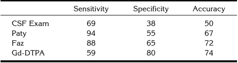
The diagnostic value of CSF examination, T2-weighted imaging (Paty and Fazekas crite-ria), and gadolinium-enhanced imaging are presented in Table 3. The Paty criteria secure of high sensitivity, with a relatively low specificity. The Fazekas criteria show a lower sensitivity, with a high specificity and accuracy. Gado-linium enhancement secures a high specificity and accuracy but a loss of sensitivity.
Discussion
Most studies on the reliability of MR criteria in the diagnosis of multiple sclerosis have been retrospective studies (16 –22). Because of the selection bias of well-established patients (with a long disease duration) and “super controls” (healthy subjects without neurologic diseases), these studies are of less value in establishing the true value of MR imaging in the initial diagnosis of multiple sclerosis. This prospective study in patients with a first episode of clinical symp-toms suggesting multiple sclerosis involves both T2-weighted MR imaging for evaluation of
the Paty and Fazekas criteria (3, 14, 15) and gadolinium-enhanced T1-weighted MR imaging in the early diagnosis of multiple sclerosis.
Multiple sclerosis can be diagnosed with cer-tainty only histologically. For clinical and re-search purposes it is desirable to diagnoses multiple sclerosis during lifetime as early as possible in the course of the disease. For that purpose, criteria have been developed to make a clinical diagnosis of multiple sclerosis (1, 2); these, however, do not provide a correct diag-nosis in all cases. Clinical misdiagdiag-nosis occurs in 9% to 12% of patients; 4% to 5% of patients are at first not diagnosed as having multiple sclerosis (23). Although it is impossible to achieve a sensitivity of 100% with paraclinical tests (lack of standard of reference), our results confirm those of previous studies that multiple T2 abnormalities (ie, MR demonstration of dis-sociation in place) are the most sensitive para-clinical indications (sensitivity, 94%) (3, 4).
In the present study MR imaging measures (Paty and Fazekas criteria) show a higher sen-sitivity and specificity than CSF examination. The Fazekas criteria (14, 15) provide both a relatively high sensitivity (88%) and higher specificity (65%) than the Paty criteria (3). The fact that Offenbacher et al (15) recently re-ported much higher values for sensitivity and specificity illustrates the limited applicability of findings from retrospective studies in a prospec-tive diagnostic setting.
Using gadolinium enhancement it is possible to increase the specificity of MR imaging in mul-tiple sclerosis even further to 80%. The pres-ence of the radiologic combination of both en-hancing and nonenen-hancing lesions seems to be much more specific for multiple sclerosis than the simple demonstration of dissociation in place. This could be because if there are both enhancing and nonenhancing lesions, they probably differ in age, thus indicating dissocia-tion not only in place but also in time and ful-filling both clinical prerequisites. Gadolinium-enhancement is, however, by no means completely restricted to multiple sclerosis, ex-plaining why a specificity of 100% cannot be achieved. It occurs in multiple sclerosis variants (24 –26), vasculitis (27), neurosarcoidosis (28), and most infections. However, demonstration of contrast enhancement on MR probably rules out vascular changes of normal aging, migraine, Alzheimer disease, and Binswanger disease. As with the Poser criteria, good clinical
judg-TABLE 2: Relation between CSF findings, T2-weighted MR abnormalities, gadolinium-enhanced MR, and follow-up findings
Conversion to Clinically Definite Multiple Sclerosis?
Yes No
CSF1 9 13
CSF2 4 8
Paty1 16 18
Paty2 1 22
Faz1 15 14
Faz2 2 26
Gd-DTPA1 10 8
Gd-DTPA2 7 32
[image:4.612.57.298.125.264.2]Note.—Paty indicates Paty criteria (3); Faz, Fazekas criteria (14, 15); Gd-DTPA, gadolinium-enhanced imaging;1, abnormal findings; and2, normal findings.
TABLE 3: The diagnostic value (%) of CSF examination, T2-weighted imaging, and gadolinium-enhanced imaging
Sensitivity Specificity Accuracy
CSF Exam 69 38 50
Paty 94 55 67
Faz 88 65 72
Gd-DTPA 59 80 74
[image:4.612.57.298.662.726.2]ment is needed to rule out diseases other than multiple sclerosis when contrast enhancement is demonstrated.
One could question whether gadopentetate dimeglumine should be routinely used in addi-tion to T2-weighted MR imaging of the brain in the initial diagnosis of multiple sclerosis. It probably depends on the clinical setting whether more value will be added to the sensi-tivity or specificity. When, for example, a first-degree family member of a patient with multiple sclerosis want to have multiple sclerosis rule out, a high sensitivity is needed; in that situa-tion, a T2-weighted scan seems sufficient. When, on the other hand, it is important in a patient with multiple white matter lesions to dif-ferentiate between incidental white matter le-sions of aging and multiple sclerosis (eg, when considering treatment with b-interferon), high specificity is needed, to prevent inappropriate administration of drugs. It should be kept in mind that the “conversion” rate to clinical defi-nite multiple sclerosis until now has been only 35% because of the relatively short follow-up time (5 to 28 months), and it is likely that the conversion rate will increase with a longer fol-low-up time. We did not attempt to calculate predictive values, because they are determined by the prevalence of multiple sclerosis (as yet unknown) in this particular patient population.
In conclusion, the presence of multiple ab-normalities on T2-weighted MR imaging pro-vides very sensitive paraclinical evidence of clinically silent dissociation in place. This pro-spective study indicates that gadolinium enhancement provides paraclinical evidence for the demonstration of clinically silent dissocia-tion in time and increases the specificity of MR imaging in multiple sclerosis. These findings are important especially in view of the fact that now more-effective treatments for multiple sclerosis, such as b-interferon (29 –31), seem to come available for clinical use, thus making a early specific diagnosis of multiple sclerosis most im-portant. Gadolinium-enhanced MR imaging seems to provide the clinician with the ideal tool for making an early, justified diagnosis of mul-tiple sclerosis and effectively “buying time” for the patient.
Acknowledgments
We thank all neurologists and ophthalmologists for re-ferring their patients to our department and for their
col-laboration in the evaluation of the assessment of the clin-ical follow-up. We also thank Ton Schweigmann for his excellent technical support and Luc Truyen for his critical remarks.
References
1. Schumacher GA, Beebe G, Kibler RE, et al. Problems of experi-mental trials of therapy in multiple sclerosis: report by the panel on evaluation of experimental trials of therapy in multiple sclero-sis.Ann NY Acad Sci1965;122:552–568
2. Poser CM, Paty DW, Scheinberg L, et al. New diagnostic criteria for multiple sclerosis: guidelines for research protocols.Ann Neurol 1983;13:227–231
3. Paty DW, Oger JJF, Kastrukoff LF, et al. Magnetic resonance imaging in the diagnosis of multiple sclerosis (MS): a prospective study of comparison with clinical evaluation, evoked potential, oligoclonal banding, and CT.Neurology1988;38:180 –185 4. Lee KH, Hashimoto S, Hooge JP, et al. Magnetic resonance
im-aging of the head in the diagnosis of multiple sclerosis: a prospec-tive 2-year follow-up with comparison of clinical evaluation, evoked potential, oligoclonal banding, and CT.Neurology1991; 41:405– 414
5. Kermode AG, Thompson AJ, Tofts PS, et al. Breakdown of the blood-brain barrier precedes symptoms and other MRI signs of new lesions in multiple sclerosis. Pathogenic and clinical implica-tions.Brain1990;133:1477–1489
6. Grossman RI, Gonzalez-Scarano F, Atlas SW, et al. Multiple scle-rosis: gadolinium enhancement in MR imaging.Radiology1986; 161:721–725
7. Miller DH, Rudge P, Johnson G, et al. Serial gadolinium enhanced magnetic resonance imaging in multiple sclerosis.Brain1988; 111:927–939
8. Barkhof F, Valk J, Hommes OR, et al. Gadolinium enhancement of multiple sclerosis lesions on long TR images at 0.6 T.AJNR Am J Neuroradiol1992;13:1257–1259
9. Harris JO, Frank JA, Patronas N, et al. Serial gadolinium-en-hanced magnetic resonance imaging scans in patients with early, relapsing-remitting multiple sclerosis: implications for clinical tri-als and natural history.Ann Neurol1991;29:548 –555
10. Thompson AJ, Kermode AG, Wicks D, et al. Major differences in the dynamics of primary and secondary progressive multiple scle-rosis.Ann Neurol1991;29:53– 62
11. Thompson AJ, Miller D, Youl B, et al. Serial gadolinium enhanced MRI in relapsing remitting multiple sclerosis of varying disease duration.Neurology1992;42:60 – 63
12. Heun R, Kappos, Bittkau S, Sta¨dt D, Rohrbach E, Schuknecht B. Magnetic resonance imaging and the early diagnosis of multiple sclerosis.Lancet1988;II:1202–1203
13. Kurtzke JF. Rating neurological impairment in multiple sclerosis: an expanded disability status scale (EDSS).Neurology1983;33: 1444 –1452
14. Fazekas F, Offenbacher H, Fuchs, et al. Criteria for an increased specificity of MRI interpretation in elderly subjects with suspected multiple sclerosis.Neurology1988;38:1822–1825
15. Offenbacker H, Fazekas, Schmidt R, et al. Assessment of MRI criteria for a diagnosis of MS.Neurology1993;43:905–909 16. Scotti G, Scialfa G, Biondi A, Landoni L, Caputo D, Cazzullo CL.
Magnetic resonance in multiple sclerosis.Neuroradiology1986; 28:319 –323
18. Ormerod IEC, Miller DH, McDonald WI, et al. The role of NMR imaging in the assessment of multiple sclerosis and isolated neu-rological symptoms.Brain1987;110:1579 –1616
19. Mushlin AI, Detsky AS, Phelps CE, et al. The accuracy of mag-netic resonance imaging in patients with suspected multiple scle-rosis.JAMA1993;269:3146 –3151
20. Yetkin FZ, Haughton VM, Papke RA, Fischer ME, Rao SM. Multiple sclerosis: specificity of MR for diagnosis.Radiology 1991;178: 447– 451
21. Young IR, Hall AS, Pallis CA, Legg NJ, Bydder GM, Steiner RE. Nuclear magnetic resonance imaging of the brain in multiple sclerosis.Lancet1981;II:1063–1066
22. Runge VM, Price AC, Kirshner HS, Allen JH, Partain CL, James AE. Magnetic resonance imaging of multiple sclerosis: a study of pulse-technique efficacy.AJNR Am J Neuroradiol1984;5:691– 702
23. Herndon RM, Brooks B. Misdiagnosis of multiple sclerosis.Semin Neurol1985;5:94 –98
24. Eblen F, Poremba, Grodd W, Opitz H, Roggendorf W, Dichgans J, Mylinoclastic diffuse sclerosis (Schilder’s disease): cliniconeuro-radiologic correlations.Neurology1991;41:589 –591
25. Barkhof F, Scheltens P, Valk J, Waalewijn C, Uitdehaag BMJ, Polman CH. Serial quantitive MR assessment of optic neuritis in a case of neuromyelitis optica, using gadolinium-“enhanced” STIR imaging.Neuroradiology1991;33:70 –71
26. Mendez MF, Pogacar S. Malignant monophasic sclerosis of “Mar-burg’s disease.”Neurology1988;38:1153–1155
27. Kazui S, Naritomei H, Imakita S, Yamada N, Ogawa M, Sawada T. Sequential gadolinium-DTPA enhanced MRI studies in neuro-Be-hcet’s disease.Neuroradiology1991;33:136 –139
28. Smith SA, Meisler DM, Weinstein MA. High signal periventricular lesions in patients with sarcoidosis: neurosarcoidosis or multiple sclerosis.AJNR Am J Neuroradiol1989;10:485– 490
29. Arnason BGW. Interferon beta in multiple sclerosis. Neurology 1993;43:641– 643
30. The IFBN Multiple Sclerosis Study Group. Interferon beta-1b is effective in relapsing-remitting multiple sclerosis, I: clinical results of a multicenter, randomized, double-blind, placebo-controlled trial.Neurology1993;43:655– 661