fe'tf 13 figures in Great Britain
THE EFFECTS OF SODIUM-TRANSPORT
INHIBITORS AND COOLING ON MEMBRANE POTENTIALS
IN COCKROACH CENTRAL NERVOUS CONNECTIVES
BY Y. PICHON AND J. E. TREHERNE*
Laboratoire de Neurobiologie Celkilaire de C.N.R.S., Gif-sw-Yvette 91190, France
(Received 20 December 1973)
SUMMARY
1. Cooling caused axonal depolarization in desheathed and urea-treated connectives, but induced hyperpolarizing responses (measured with micro-electrodes and with the sucrose-gap) in intact preparations. Hyperpolarizing responses were also recorded with extracellularly-located microelectrodes in intact connectives.
2. Strophanthidin (0-2 mM/1) caused axonal depolarization in desheathed preparations, ethacrynic acid being without appreciable effect. Ethacrynic acid (0-2 mM/1) induced apparent hyperpolarizations in intact connectives and abolished or reduced the effects of cooling.
3. It is concluded that the axonal sodium pump is pharmacologically sep-arable from that associated with the perineurial and/or the glial membranes: the former being inhibited by cardiac glycosides, the latter by ethacrynic acid. 4. The results are discussed in relation to extra-axonal sodium regulation and the possible involvement of an electrogenic sodium pump associated with the perineurial or glial membranes.
INTRODUCTION
The electrical activity of excitable cells results from the presence of steep ionic gradients across their membranes, being generally dependent on a high external concentration of sodium ions relative to those of potassium. In contrast to most vertebrates and marine invertebrates, insect species frequently possess haemolymph containing exceptionally low concentrations of sodium and relatively high concen-trations of potassium ions (cf. Shaw & Stobbart, 1963). The available evidence indicates, however, that insect axons are essentially similar to those of other higher invertebrates in that the inward current of the action potential is largely carried by sodium ions and is, consequently, dependent upon a relatively high external con-centration of this cation (cf. Narahashi, 1963; Pichon, 1074).
The existence of an extra-axonal sodium regulation is particularly evident in phytophagous insects such as the stick insect, Carausius morosus, or the hawk moth, Manduca sexta, in which the sodium requirement for axonal function is approximately
204 Y. P I C H O N AND J. E. TREHERNE
an order higher than the relatively low concentration of this cation in the haemolymfH (Treherne & Maddrell, 1967 a, b; Weidler & Diecke, 1969; Pichon, Sattelle & Lane, 1972). Extra-axonal sodium regulation is also apparent in the cockroach, Periplaneta americana, an insect with a relatively high sodium concentration of 157 mM/1 in the haemolymph (cf. Treherne, 1961). This has been inferred from the larger action potentials observed in intact as compared with desheathed connectives (Pichon & Boistel, 1967) and from an equivalent decline in amplitude of the action potentials recorded in connectives in which the blood-brain barrier system was disrupted by brief exposure to hypertonic urea solutions (Treherne, Schofield & Lane, 1973).
It has been proposed that a high extra-axonal sodium concentration would be maintained, by Donnan forces (Treherne, 1962) due to the anionic groups associated with the extracellular acid mucopolysaccharides (Ashhurst, 1961). The activity coefficient of sodium ions associated with such a fixed charge system would, however, be low and would not effectively elevate the concentration of the free cation in the fluid bathing the axon surfaces (Treherne & Maddrell, 19676). It has been suggested, therefore, that a transport of sodium ions from the haemolymph, or bathing medium, via the perineurium and possibly the glial elements, would be necessary to effectively raise the extra-axonal sodium level (Treherne, 1967; Treherne & Maddrell, 19676; Pichon & Boistel, 1967; Treherne, 1974).
The existence of such a sodium transporting system could account for some recent observations on the rate of recovery of action potentials in sodium-depleted cockroach connectives (Schofield & Treherne, in preparation). In preparations in which exposure to sodium-free conditions produced a measurable decline in the amplitude of the action potentials it was found that the normally rapid recovery, obtained on return to normal saline, was slowed down in the presence of dilute dinitrophenol (5 x io~s M) or ethacrynic acid (io~* M). NO recovery of the action potentials was observed when sodium-depleted connectives were exposed to lithium saline, despite the observation that lithium ions substitute for those of sodium in maintaining axonal function in desheathed preparations. Rapid recovery was, how-ever, observed in lithium saline in preparations in which the blood-brain barrier was disrupted by brief exposure to hypertonic urea solution.
A simple explanation of the above observations would be that an appreciable proportion of the sodium ions were transported to the axonal surfaces by a mech-anism which is sensitive to dinitrophenol and ethacrynic acid, but does not accept lithium ions.
Extracellular fluid Bathing Pcrincurium
medium and glia Axon
+ 20
'Sheath' or 'extracellular' potential
Apparent axonal resting potential
True axonal resting potential
- 2 0
- 4 0
6 0
[image:3.451.57.388.47.271.2]-- 8 0 L
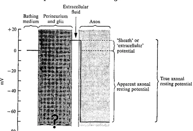
Fig. i. Schematic representation of the apparent potential profile across the various structural compartments of a cockroach connective. The heavy continuous lines show the potentials measured in previous microelectrode studies; the broken ones represent the possible potential gradients across the glial and perineurial elements, for simplicity and on the basis of ultra-structural evidence (Lane & Treherne, 1970) these are represented as linked compartments. According to this interpretation the 'extracellular' or 'sheath' potential could result either from the differential passive ionic permeabilities of the outer perineurial and of the inner perineurial and/or glial membranes or from an inwardly directed electrogenic sodium pump situated on either of these membranes.
examination shows the extracellular material to be still present (Treherne et al. 1970).
At the present time we have, however, very little concrete evidence as to the nature of the 'extracellular' or 'sheath' potential (Fig. 1). It could be conceived that this potential arises: (1) from a differential sensitivity of the inwardly perineurial and/or glial membranes to potassium ions with respect to the outer perineurial surface; (2) from a lower potassium concentration in the extracellular fluid as compared to the bathing medium, possibly related to the activity of a potassium pump; or (3) from the activity of an electrogenic sodium pump located either on the outer peri-neurial surface or on the inwardly-directed periperi-neurial or glial membranes.
The present investigation was devised to elucidate the mechanism of production of the 'extracellular' or 'sheath' potential using agents which are known to block active transport either physiologically (cooling) or pharmacologically (strophanthidin and ethacrynic acid). The experiments demonstrate mechanisms which are likely to be involved in the regulation of the ionic composition of the extracellular fluid in intact cockroach connectives.
METHODS
Intracellular and extracellular recordings were made in the penultimate connectives of the abdominal nerve cord of the cockroach, Periplaneta americana. The isolated nerve cords were mounted in a small perspex chamber similar to that described by
206 Y. PlCHON AND J. E. TREHERNE
were isolated from each other by petroleum jelly seals; the two left-hand compd ments contained normal saline solution and were connected to a Farnell pulse-generating system through a photon-coupled isolating unit via platinum wires. The middle compartment contained the experimental solution flowing at a regulated rate. It was connected to the indifferent electrode via an isotonic KCl-Agar bridge. The fourth compartment was filled by a continuously flowing isotonic mannitol solution. The right-hand compartment was filled with normal saline or isotonic KC1 solution and was connected to a high-impedance negative-capacitance amplifier via another isotonic KCl-Agar bridge. The glass microelectrodes were filled with 3-0 M-KC1 solution (their resistance ranging between 5 and 20 MQ) and were connected to a second high-impedance negative-capacitance amplifier. Both amplifiers were con-nected to a Tektronix 602 oscilloscope and to a PED Servotrace SEFRAM pen recorder.
The preparation was laid across the five compartments so that the connectives between the fourth and the fifth ganglia were continuously irrigated by the test solution. The nerve cord was supported at this level on a piece of elastomer resin to facilitate the penetration of the microelectrodes into the connectives. Extracellular recordings were made between the right-hand and the middle compartment across the 'mannitol gap'. The d.c. and action potentials were recorded between the tip of the microelectrode, in intracellular or extracellular positions, and the indifferent
electrode.
A two-way stopcock was used to change solutions. In preliminary experiments cooling was achieved by allowing the solution to flow through a length of catheter tubing contained in a Dewar flask filled with a mixture of sea water and ice. With this system cooling below 10 °C was rarely achieved within the chamber and was not easily reproducible. Cooling was routinely achieved by short applications of a 'Quik-Freeze' spray (MS240, Miller Stephenson Chemical Company) to the tubing con-necting the reservoir to the stopcock. Rapid cold pulses to temperatures as low as 6-o °C could thus be obtained. Using this system it was also possible to cool the preparation at any time during perfusion with test solution or the application of drugs. The strophanthidin in these experiments was produced by Sigma and etha-crynic acid by Merck, Sharp and Dohme.
The normal saline contained 213-3 mitf/1 Na+, 3-1 mM/1 K+, 10 mM/1 Ca2+, 233-1 mM/1 Q - , o-i mM/1 PO4H2- and 2-1 mM/1 C03H~. A relatively high calcium
con-centration was employed to take account of the observation of Pichon & Boistel (1967) and Wilson (1973) which indicate that the effective extra-axonal calcium level probably exceeds the i-8 mM/1 in the saline of Yamasaki & Narahashi (1959). The high potassium solution which was used to test the accessibility of the extra-axonal fluid to externally applied ions (i.e. the effectiveness of the blood-brain barrier) con-tained 217 mM/1 KC1.
RESULTS Effects of reduced temperature on intact connectives
30
25
°C 20
15
10
- 6 5 r
2 min
I 1
Intraccllular recording Extracellular recording
mV
- 7 0 L
mV 5 0
[image:5.451.54.386.68.377.2]--100 L 1 msec
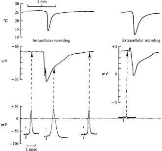
Fig. a. The effect of reduced temperature (indicated by the continuous recordings at the top of the illustration) on the resting and action potentials and on the 'extracellular' potential. The intracellular recordings were made with the tip of the microelectrode located within a giant axon; the extracellular ones were achieved after withdrawing the tip of the microelectrode into an extracellular position.
response to reduced temperature, when the tip of the microelectrode was withdrawn into an extracellular position adjacent to the surface of the giant axon. The latter result suggests that the hyperpolarizing response does not arise at the level of the axonal membrane.
Sucrose-gap recordings, which were performed to preclude any possible pinholing effects produced by microelectrodes, confirmed the above observations. As with microelectrodes, distinct hyperpolarizations were induced by cold-pulses under sucrose-gap conditions (Fig. 3). For temperatures below about 19 °C, the extent of these hyperpolarizations showed an approximately linear relation with the extent of the temperature reduction (Fig. 4). For smaller excursions, the recorded potential was rather insensitive to temperature.
208 Y . PlCHON AND J . E . TREHERNE
30 25 20 15 10 5
5mV
[image:6.451.48.378.43.369.2]2min
Fig. 3. The effects of successive cold-pulses on the d.c. potential recorded in an intact connective using the 'aucrose-gap' technique.
Effects of low temperature on desheathed connectives
In contrast to the situation in intact connectives, low-temperature pulses induced depolarizing responses in desheathed preparations (Fig. 6). An apparently linear relation exists between the depolarization and the extent of the temperature reduction (Fig. 7). No initial insensitive phase was observed, as with the hyperpolarizing res-ponses observed in intact connectives.
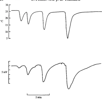
The above results suggest an explanation of the complex responses to cold-pulses recorded in some intact preparations (Fig. 5). It seems reasonable to suppose that the slight depolarizations induced by small temperature changes arise at the level of the axonal membranes, such responses being predicted from the initial region of insensitivity of the extracellular potential to small temperature reductions (Fig. 7). According to this interpretation the hyperpolarizing responses, which would only become apparent with larger temperature reductions (Fig. 4) result from extra-axonal effects, presumably at the level of the perineurial and/or glial membranes.
Effects of urea-treatment on cold responses
7r
o
1
4I 3
25 20 15 10
Temperature (°C)
Fig. 4. The relation between temperature and the extent of the cold-induced hyper-polarizations recorded in an intact connective using the 'sucrose-gap' technique.
30 25 °C 20 15 10
5mV
2 min
Fig. 5. Complex responses to successive cold-pulses recorded in an intact connective using the 'sucrose-gap' technique.
urea-treatment (Fig. 8). The change in the polarity of the cold response produced by urea-treatment was correlated with alterations in the potassium-induced d.c. potential changes (Fig. 9). The initial potassium-induced potential change, illustrated in Fig. 9, can be interpreted as a depolarization of the outwardly directed perineurial mem-brane, which is associated with a restricted intercellular access of potassium ions ^ the axonal surfaces resulting from the presence of perineurial tight junctions
2 1 0 Y . PlCHON AND J. E . TREHERNE
25
20
15
10
5mV
[image:8.451.90.328.36.288.2]2 min
Fig. 6. ' Sucroae-gap' recording showing the effects of cold-pulses on d.c. potentials in a desheathed connective.
(Treherne et al. 1970). The smaller and slower depolarizing response to high-potassium saline, obtained after urea-treatment, has been identified as the axonal depolarization resulting from potassium entry allowed by the disruption of the peripheral blood-brain barrier associated with the perineurium (Treherne, Schofield & Lane, 1973).
Effects of strophanthidin
As in a previous investigation with ouabain (P. K. Schofield & J. E. Treherne, in preparation) this cardiac glycoside was found to exert effects at the axonal level. This is illustrated for a desheathed preparation in Fig. 10 in which it can be seen that 0 2 mld/l strophanthidin produced a depolarization with a reduction in the amplitude of the response to a cold-pulse. This cardiac glycoside could not be used, therefore, to distinguish between the axonal and extra-axonal potential changes in-duced by low-temperature pulses.
Strophanthidin produced no appreciable effect in intact preparations, presumably because it could not penetrate the blood-brain barrier to reach the axonal surfaces or, alternatively, because it acted simultaneously on both inner perineurial and axonal membranes. The lack of an effect on the amplitude of the action potentials does not, however, accord with the latter hypothesis.
Effects of ethacrynic acid
20 15 Temperature (°C)
[image:9.451.123.313.81.356.2]10
Fig. 7. TTie relation between temperature and the extent of the cold-induced depolariza-tions recorded in a desheathed connective. ' Sucroae-gap' recording.
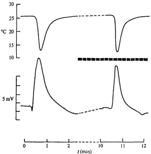
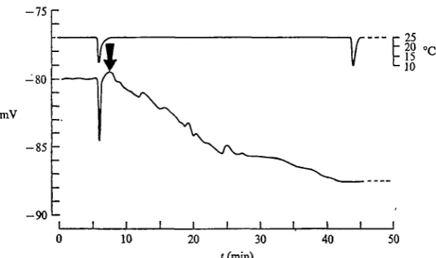
Application of 0 2 mM/1 ethacrynic acid to intact connectives, on the other hand, was found to produce a hyperpolarization under sucrose-gap conditions (Fig. 12) or with intracellularly-located microelectrodes (Fig. 13). Short exposure to ethacrynic acid resulted in a decrease in the cold-induced hyperpolarization (Fig. 12), more prolonged exposure abolishing the response to cold shocks (Fig. 13).
In two experiments (one using intracellular microelectrodes and the other the sucrose-gap technique) no appreciable hyperpolarization was recorded following exposure of intact connectives to 0-2 mM/1 ethacrynic acid. In these preparations the cold-induced hyperpolarizations were retained at full amplitude, and it is concluded that at this critical concentration there was an insufficient access of ethacrynic acid to the site of action.
DISCUSSION
Two types of electrical responses to cooling have been described: a hyperpolariza-tion, in intact connectives, and a depolarization in desheathed preparations and in those in which the blood-brain barrier was disrupted by exposure to hypertonic urea solution.
The axonal depolarization induced by cooling of desheathed preparations con-the earlier observations of Wilson (1973), who showed that con-the extent of con-the
2 1 2 Y . P l C H O N AND J . E . TREHERNE
Initial response Response after urea treatment
- 9 0 n
mV 7 0
-- 7 5 -
L-- 9 5
- 1 0 0
2 rain
Fig. 8. The effects of urea treatment on the electrical responses to cooling recorded from a giant axon with an intracellularly located microelectrode. T h e first recording shows the effects of reduced temperature on the intact connective, the second, that produced following 15 sec exposure to hypertonic urea solution.
depolarization was greater than would be predicted from thermodynamic changes resulting from temperature reduction. Wilson also observed that exposure to ouabain reduced the depolarization almost to that which would be predicted on thermo-dynamic grounds. This, and other evidence, led Wilson to discount the effects of cold-induced changes on the passive permeability properties of the axon membrane. Depolarization appeared, in fact, to result largely from the accumulation of potassium ions at the axonal surfaces, rather than from any direct effects on a hypothetical electrogenic pump situated on the axon membrane. The accumulation of potassium ions was postulated to result from the inhibition of uptake by a linked sodium-potassium pump situated on the membranes adjacent to the extra-axonal fluid. The restricted dimensions of the extra-axonal fluid layer (ca. 200 A) formed by the closely applied glial membranes (cf. Smith & Treherne, 1963; Treherne et al. 1970) would tend to exaggerate this effect.
Initial responses Responses following urea treatment 25
20
15
10
5mV
J
2 min
Fig. 9. The effects of a 15 sec exposure to hypertonic urea solution on the d.c. potentials recorded using the 'sucrose-gap' technique. The change in the polarity of the response to cooling (A) was accompanied by a reduction in the extra-neuronal potential change induced by exposure to saline in which the sodium was replaced by 214 mM/1 K+ (B). The black bars indicate periods of exposure to high potassium saline.
The fact that the cold hyperpolarization (measured in axons in intact connectives) is converted to an equivalent depolarization (in urea-treated or desheathed pre-parations) implies that the glial and/or perineurial hyperpolarization must be larger than the axonal depolarization in intact connectives. The cold-induced hyper-polarizations measured with the microelectrode tip in an extracellular position were, however, generally only of similar magnitude to the axonal depolarizations. This could be due to attenuation caused by a poor electrical seal being made along the relatively long path of the electrode through the tissues.
The hyperpolarizing effects of ethacrynic acid on intact connectives suggest that the cold-induced hyperpolarizations could result from the inhibition of extra-cellular cation regulation. This supposition is supported by the demonstrated abolition of cold responses following exposure of intact connectives to ethacrynic acid. As dilute ethacrynic acid produced no appreciable axonal effect it can reasonably be concluded that this compound exerts its effects at the level of the glial and/or
214 Y. PlCHON AND J. E . TREHERNE 30
25 °C 20 15 10
10 mV
16 17 18
Fig. io. The effects of strophanthidin (o-a min/1) on the d.c potential and the response to cooling in a desheathed connective. The broken horizontal black line indicates the period of exposure to strophanthidin. 'Sucrose-gap' recording.
30
25
°C 20
15
10
215
5mV
10 11 12
[image:13.451.75.318.57.306.2]/(min)
Fig. II. The effects of ethacrynic acid (o-a mM/1) on the d . c potential and the response to cooling in a desheathed connective. The broken horizontal black line indicates the period of exposure to ethacrynic acid. 'Sucrose-gap' recording.
30
25
°C 20
15
10
5mV
14 16 30
/ (min)
[image:13.451.46.345.357.547.2]2 l 6 Y . PlCHON AND J . E . TREHERNE
- 7 5
-80
mV - 8 5
• 2 5
20 15 10
-90 L
0 10 20 30 40 50
[image:14.451.70.376.65.246.2]/ (rain)
Fig. 13. Microelectrode recording showing the effects of ethacrynic acid (0-2 miw/l) on the potentials recorded from within a giant axon in an intact connective. The arrow indicates the time at which the preparation was exposed to ethacrynic acid. A cold-induced hyper-polarization was not evoked following exposure to ethacrynic acid.
approximately 5-9 mM/1 (i.e. from 3-1 to 9-0 HIM/I K+). By analogy with other glial membranes (Nicholls & Kuffler, 1964; Kuffler, Nicholls & Orkand, 1966), which behave effectively as accurate potassium electrodes, it can be calculated that an equivalent change in potassium concentration could induce a depolarization of the inner perineurial and/or the glial membranes of as much as 27 mV. With such a system it seems clear that strongly hyperpolarizing responses would be recorded with microelectrodes situated within a giant axon in an intact connective.
In the absence of quantitative information concerning the electrochemical properties of the inner perineurial and glial membranes it is, however, difficult to eliminate the possibility that cold- and ethacrynic-induced hyperpolarizations also resulted from the inhibition of an electrogenic component in sodium transport into the extracellular fluid. As mentioned earlier, it has been tentatively suggested that the positive 'sheath' or 'extracellular' potential (Pichon & Boistel, 1967) results from an electrogenic sodium pump situated on the inner perineurial or glial membranes (Treherne & Moreton, 1970; Treherne & Pichon, 1972). However, the existence of a pronounced effect of cold and ethacrynic acid, in inhibiting such a postulated electrogenic pump, would imply that the potassium-sensitivity of the inner perineurial and glial mem-branes approximate to that of the axonal memmem-branes, which thus effectively cancel each other so as to reveal the inhibition of the electrogenic pump.
^Bntributes to the ouabain-sensitive fraction of radio-sodium efflux in cockroach nerve cords (Treherne, 1966).
The extra-axonal responses recorded in this investigation indicate that, unlike the axonal membranes, the perineurial and/or glial membranes are sensitive to ethacrynic acid. This effect is consistent with the hypothesis that the perineurial and glial elements are involved in the transport of sodium ions to the fluid bathing the axonal surfaces in the insect central nervous system (cf. Treherne, 1967; Treherne & Mad-drell, 19676; Treherne, 1974). The present observations also accord with the effect of ethacrynic acid and dilute DNP in slowing the apparent rate of movement of sodium ions to the extra-axonal fluid in intact sodium-depleted connectives when returned to normal saline (P. K. Schofield & J. E. Treherne, in preparation). They can, in addition, be correlated with the observed inability of lithium ions to induce recovery of the action potentials in sodium-depleted preparations, despite the fact that a rapid recovery could be obtained with lithium saline in connectives in which the blood-brain barrier was disrupted by brief exposure to hypertonic urea solution. The latter observations could be interpreted as evidence for the existence of perineurial/glial transport to the extra-axonal fluid in intact connectives.
This research has been partly supported by a grant from the D.G.R.S., T.M.72 70295.
REFERENCES
ASHHURST, D. E. (1961). An acid mucopolysaccharide in cockroach ganglia. Nature, Land. 191, 1*24-5.
HAUSB, L. L., PATTILLO, R. A., SANCES, A. & MATTINGLY, R. F. (1970). Cell surface coatings and
membrane potentials of malignant and non-malignant cells. Science 169, 601-3.
KUFFLBH, S. W., NICHOLLS, J. G. & ORKAND, R. (1966). Physiological properties of glial cells in the central nervous system of amphibia. J. Neuropkysiol. 29, 768—87.
LANB, N. J. & TREHBRNB, J. E. (1970). Uptake of peroxidase by the cockroach central nervous system. Tissue & Cell a, 413-25.
LEBLANC, G. & ERLIJ, D. (1969). Effects of ethacrynic acid on sodium fluxes in frog sartorius muscle. Siochim. Biophys. Acta 173, 149-51.
LUBOWITZ, H. & WHITTAM, R. (1969). Ion movements in human red cells independent of the sodium pump. J. Physiol. aoa, m - 3 1 .
NARAHASHI, T. (1963). The properties of insect axons. In Advances in Insect Physiology (ed. J. W. L. Beament, J. E. Treherne & V. B. Wigglesworth), vol. I, pp. 175-256. London & New York:
Academic Press.
NICHOLLS, J. G. & KUFFLKR, S. W. (1964). Extracellular space as a pathway for exchange between blood and neurones in the central nervous system of the leech: ionic composition of glial cells and neurones, Neurophysiol. 37, 645-71.
PICHON, Y. (1974). Axonal conduction in insects. In Insect Neurobiology (ed. J. E. Treherne), pp. 7 3 -117. Amsterdam and London: North-Holland.
PICHON, Y. & BOISTKL, J. (1966). Application aux fibres geantes de blattes {Periplaneta americana L. et Blabera craniifer Burm.), d'une technique permettant l'introduction d'une microe'lectrode dans le tissu nerveux, sans resection prealable de la gaine. J. Physiol. Paris 58, 592.
PICHON, Y. & BOISTBL, J. (1967). Microelectrode study of the resting and action potentials of the cockroach giant axon with special reference to the role played by the nerve sheath. J. exp. Biol. 47.
357-73-PICHON, Y., SATTELLE, D. B. & LANE, N. J. (1972). Conduction processes in the nerve of the moth, Manduca sexta, in relation to haemolymph ionic composition and central nervous system ultra-structure. J. exp. Biol. 56, 717-34.
SHAW, J. & STOBBART, R. U. (1963). Osmotic and ionic regulation in insects. In Advances in Insect Physiology (ed. J. W. L. Beament, J. E. Treherne and V. B. Wigglesworth), vol. 1, pp. 315—99. London and New York: Academic Press.
2 l 8 Y. PlCHON AND J. E . TREHERNE
TREHERNE, J. E. (1961). Sodium and potassium fluxes in the abdominal nerve cord of the cockroach
Periplaneta americana.J. exp. Biol. 38, 315—22.
TREHERNE, J. E. (1062). The distribution and exchange of some ions and molecules in the central nervous system of Periplaneta americana. J. exp. Biol. 39, 193-217.
TREHERNE, j . E. (1066). The effect of ouabain on the efflux of sodium ions in the nerve cord of two insect species (Periplaneta americana and Carausius morosui). J. exp. Biol. 44, 355-62.
TREHERNE, J. E. (1967). Axonal function and ionic regulation in insect central nervous tissues. In Insects and Physiology (ed. J. W. L. Beament and J. E. Treherne). Edinburgh and London: Oliver & Boyd.
TREHERNE, J. E. (1974). The environment and function of insect nerve cells. In Insect Neurobiology (ed. J. E. Treherne), pp. 187-244. Amsterdam and London: North-Holland.
TREHERNE, J. E., LANE, N. J., MORETON, R. B. & PICHON, Y. (1970). A quantitative study of potassium
movements in the central nervous system of Periplaneta americana. J. exp. Biol. 53, 109-36. TREHERNE, J. E. & MADDRELL, S. H. P. (1967a). Membrane potentials in the central nervous system of a
phytophagous insect (Carausius morosus). J. exp. Biol. 46, 413-21,
TREHERNE, J. E. & MADDRBLL, S. H. P. (19676). Axonal function and ionic regulation in the central nervous system of a phytophagous insect (Carausius morosus). J. exp. Biol. 47, 235-47.
TRBHERNE, J. E. & MORETON, R. B. (1970). The environment and function of invertebrate nerve cells. Int. Rev. Cytol. a8, 45-88.
TRBHERNE, J. E. & PICHON, Y. (1972). The insect blood-brain barrier. In Advances in Insect Physiology (ed. J. E. Treherne, M. J. Berridge & V. B. Wiggles worth), vol. ix, pp. 257-308. London and New York: Academic Press.
TREHERNE, J. E., SCHOFIELD, P. K. & LANE, N. J. (1973). Experimental disruption of the blood-brain
barrier system in an insect (Periplaneta americana). J. exp. Biol. 59, 711-23.
WETDLER, D. J. & DIECKE, F. P. J. (1969). The role of cations in conduction of the central nervous system of the herbivorous insect Carausius morosus. Z. vergl. Pkysiol. 64, 372-99.
WHITTEMBURY, G. & FISHMAN, J. (1969). Relation between Na-extrusion and transtubular absorption in the perfused toad kidney: the effect of K, ouabain and ethacrynic acid. PflUgers Arch. 307, 138-53. WILSON, M. C. L. (1973). Cold acclimation in insect nerves. Ph.D. thesis, Cambridge University. YAMASAKI, T. & NARAHASHI, T. (1959). The effects of potassium and sodium ions on the resting and