Transport of amino acids into and out of cells is an essential part of intra- and extracellular amino acid homeostasis. These solutes play fundamental roles in a multitude of functions, including protein synthesis, hormone metabolism, catalytic
functions, nerve transmission, regulation of cell growth, production of metabolic energy, synthesis of purines and pyrimidines, nitrogen metabolism and biosynthesis of urea. A variety of amino acid transporters with overlapping substrate JEB0715
In mammalian cells, the uptake of amino acids is mediated by specialized, energy-dependent and passive transporters with overlapping substrate specificities. Most energy-dependent transporters are coupled either to the cotransport of Na+or Cl−or to the countertransport of K+.
Passive transporters are either facilitated transporters or channels. As a prelude to the molecular characterization of the different classes of transporters, we have isolated transporter cDNAs by expression-cloning with Xenopus
laevis oocytes and we have characterized the cloned
transporters functionally by uptake studies into oocytes using radiolabelled substrates and by electrophysiology to determine substrate-evoked currents. Mammalian transporters investigated include the dibasic and neutral amino acid transport protein D2/NBAT (system b0+) and
the Na+- and K+-dependent neuronal and epithelial
high-affinity glutamate transporter EAAC1 (system X−AG). A
detailed characterization of these proteins has provided new information on transport characteristics and mechanisms for coupling to different inorganic ions. This work has furthermore advanced our understanding of the roles these transporters play in amino acid homeostasis and in various pathologies. For example, in the central nervous system, glutamate transporters are critically important in maintaining the extracellular glutamate concentration below neurotoxic levels, and defects of the human D2 gene have been shown to account for the formation of kidney stones in patients with cystinuria.
Using similar approaches, we are investigating the molecular characteristics of K+-coupled amino acid
transporters in the larval lepidopteran insect midgut. In the larval midgut, K+ is actively secreted into the lumen
through the concerted action of an apical H+V-ATPase and
an apical K+/2H+antiporter, thereby providing the driving
force for absorption of amino acids. In vivo, the uptake occurs at extremely high pH (pH 10) and is driven by a large potential difference (approximately −200 mV). Studies with brush-border membrane vesicles have shown that there are several transport systems in the larval intestine with distinct amino acid and cation specificities. In addition to K+, Na+ can also be coupled to amino acid
uptake at lower pH, but the Na+/K+ratio of the hemolymph
is so low that K+is probably the major coupling ion in vivo.
The neutral amino acid transport system of larval midgut has been studied most extensively. Apart from its cation selectivity, it appears to be related to the amino acid transport system B previously characterized in vertebrate epithelial cells. Both systems have a broad substrate range which excludes 2-(methylamino)-isobutyric acid, an amino acid analog accepted by the mammalian Na+-coupled
system A. In order to gain insights into the K+-coupling
mechanism and into amino acid and K+ homeostasis in
insects, current studies are designed to delineate the molecular characteristics of these insect transporters. Recent data showed that injection of mRNA prepared from the midgut of Manduca sexta into Xenopus laevis oocytes induced a 1.5- to 2.5-fold stimulation of the Na+-dependent
uptake of both leucine and phenylalanine (0.2 mmol l−1,
pH 8). The molecular cloning of these transporters is now in progress. Knowledge of their unique molecular properties could be exploited in the future to control disease vectors and insect pests.
Key words: Amino acid transport, intestine, kidney, brain, lepidopteran insects, neurotransmitter, glutamate, GABA, cystinuria.
Summary
Introduction
MOLECULAR CHARACTERISTICS OF MAMMALIAN AND INSECT AMINO ACID
TRANSPORTERS: IMPLICATIONS FOR AMINO ACID HOMEOSTASIS
MICHELA CASTAGNA1, CHAIRAT SHAYAKUL1, DAVIDE TROTTI1, V. FRANCA SACCHI2, WILLIAM R. HARVEY3 ANDMATTHIAS A. HEDIGER1,*
1Renal Division, Department of Medicine, Brigham and Women’s Hospital and Harvard Medical School, and
Department of Biological Chemistry and Molecular Pharmacology, Harvard Medical School, Boston, MA, 02115, USA, 2Istituto di Fisiologia Generale e Chimica Biologica, Facolta di Farmacia, Via Trentacoste 2, 20134 Milano,
Italy and 3Department of Biology, Temple University, Philadelphia, PA 19122, USA
specificities ensure an adequate supply of these important solutes.
In mammalian cells, concentrative accumulation of amino acids can be achieved by coupling the uptake to the cotransport of Na+. The required inwardly directed Na+electrochemical ion
gradient is maintained by the plasma membrane Na+/K+
-ATPase. Certain epithelial amino acid transporters have also been reported to be H+-coupled (Thwaites et al. 1993), by
analogy with the intestinal brush-border H+-coupled
oligopeptide transporter (Fei et al. 1994). The required inwardly directed H+ electrochemical ion gradient is maintained by the
concerted action of the basolateral Na+/K+-ATPase and the
brush-border Na+/H+exchanger. By contrast, in the midgut of
lepidopteran larvae, amino acid transporters are energized by coupling to the cotransport of K+. The inwardly directed K+
electrogenic ion gradient is maintained by the concerted actions of the apical H+V-ATPase (Harvey and Wieczorek, 1997) and
the apical K+/H+exchanger (Lepier et al. 1994).
Concentrative uptake of amino acids is also driven in some cases either by the outwardly directed concentration gradient of other amino acids by exchange mechanisms or simply by the membrane potential. For example, in the kidney proximal tubule S3 segment, the amino acid cystine appears to be absorbed via system bo+in exchange for intracellular amino
acids. The cationic amino acid transport system y+ mediates
the entry of positively charged amino acids down their electrochemical gradient. This system also allows certain neutral amino acids to be transported in a Na+-dependent
manner, thereby mimicking dibasic amino acids.
The purpose of this review is to summarize our knowledge of the molecular and physiological characteristics of amino acid transporters in mammals and insects. The review is divided into the following sections: (1) classification and physiological relevance of amino acid transport systems; (2) recent insights into the physiology of amino acid transporters based on molecular studies; (3) K+-coupled amino acid
transporters in insect midgut; and (4) conclusion.
Classification and physiological relevance of amino acid transport systems
Kinetic studies and inhibition experiments of transporters using a variety of preparations led to the identification of distinct amino acid transport systems. In addition to nutrient roles, transporters of acidic amino acids and γ-aminobutyric acid (GABA) play important roles in neurotransmission in the central nervous system (CNS). Other transporters are involved in cellular activation and tumorigenesis (Freidman et al. 1994; Singh and Siegal, 1995). Neutral and acidic amino acids are important organic osmolytes in osmotically stressed cells, and regulation of amino acid uptake systems (e.g. systems A or X−AG) in response to hypertonic stress can contribute to cell
volume recovery (Sone et al. 1993; Chen and Kempson, 1995; Petronini et al. 1992; Chesney, 1985).
The following different systems for the uptake of neutral amino acids appear to be present in epithelial cells: systems A,
ASC, B, B0 and L (see below). Systems B, B0 and L are
thought to be expressed in apical membranes and to be involved in transepithelial absorption, whereas systems A and ASC are expressed in basolateral membranes, where they absorb amino acids from the blood for the metabolic requirement of enterocytes (Stevens et al. 1984; Tate et al. 1996). The characteristics of different amino acid transport systems are summarized below.
System A
This system, which mainly transports small aliphatic amino acids, is widely expressed in mammalian cells, including myocytes and hepatocytes. In the intestine, system A is localized to basolateral membranes where it absorbs amino acids from the blood for the metabolic requirement of enterocytes (Stevens et al. 1984; Tate et al. 1996). System A is Na+-coupled, tolerates Li+and is pH sensitive (Christensen
et al. 1965). It recognizes N-methyl amino acids, and
(N-methylamino)-α-isobutyric acid (MeAIB) is a characteristic substrate.
System A is regulated by amino acid deprivation, hormones, growth factors and hyperosmotic stress (Soler et al. 1993; Chen and Kempson, 1995). Both glucagon and insulin modulate the expression of system A (Shotwell et al. 1983; Kilberg et al. 1985). Insulin stimulates system A activity in both liver and skeletal muscle, and glucagon also stimulates it synergistically in hepatocytes (Le Cam and Freychet, 1978; Fehlmann et al. 1979; Kelley et al. 1980). In addition to being a hormone of glucose deficiency, glucagon plays important roles during periods of protein abundance, facilitating the conversion of amino acids to glucose and stimulating urea nitrogen production (Boden et al. 1990). In vitro studies on the nutrient-dependence of pancreatic glucagon and insulin release revealed different effects of individual amino acids (Ebert and Creutzfeldt, 1987; Pipeleers et al. 1985a,b). For example, alanine stimulates primarily glucagon release, leucine stimulates release of insulin but not glucagon, and arginine (a non-substrate of system A) stimulates the release of both glucagon and insulin. These responses appear to correlate with the different metabolic roles of amino acids in liver and muscle and their involvement in gluconeogenesis and ureagenesis. For example, alanine plays a key role in transamination reactions and in furnishing carbon atoms for hepatic gluconeogenesis.
Glucagon and insulin induce system A (or at least one subtype of this system) by a process that is dependent on protein synthesis (Crettaz et al. 1983; Pariza et al. 1976; Lin
et al. 1994; Cariappa and Kilberg, 1992). Micro-injection of
System ASC
This system provides cells with the essential amino acids alanine and threonine, and the non-essential amino acids serine and cysteine. Cysteine is a precursor for the synthesis of glutathione (GSH), which protects cells against oxidative stress. System ASC is easily distinguishable from system A because (1) it does not recognize MeAIB, and (2) neutral amino acid uptake is relatively pH-insensitive. This transporter also accepts acidic amino acids at reduced extracellular pH (Tate et al. 1996; Vadgama and Christensen, 1984; Makowske and Christensen, 1982).
Systems B, B0and B0+
These closely related Na+-dependent systems mediate the
absorption of aliphatic, branched-chain and aromatic amino acids. B0+ also accepts dibasic amino acids (Van Winkle,
1988). Systems B and B0have a broader specificity for neutral
amino acids than systems A and ASC. They are present in intestinal and renal epithelial brush-border membranes (Stevens et al. 1984, 1992; Maenz and Patience, 1992; Lynch
and McGivan, 1987; Doyle and McGivan, 1992).
System B0+was first characterized in mouse blastocysts but
has also been observed in Xenopus oocytes, fibroblasts, porcine endothelial cells and hepatoma cells. It has been proposed that systems B, B0 and B0+ are variations of an
evolutionarily ancient transporter (Kilberg et al. 1993). Alternatively, these systems may constitute different manifestations of the same transporter. It is not clear at present how these transport systems relate to the epithelial brush-border H+-coupled amino acid transporters recently described
by Thwaites et al. (1993).
System B0+ is both Na+- and Cl−-coupled (Van Winkle,
1985b) and may correspond to the β-alanine carrier (Munck and Munck, 1994).
System b0+
The mouse blastocyst transport system b0+ mediates Na+
-independent, high affinity transport of neutral and dibasic amino acids (Van Winkle, 1993). It is expressed in kidney and intestinal epithelia (Stevens et al. 1984; Mirchett et al. 1982).
System N
This system is Na+-coupled and specific for neutral amino
acids. It has a more restricted tissue distribution than systems A, ASC, B, B0and B0+. It is expressed in liver and muscle. In
liver, system N is involved in the transport of glutamine, asparagine and histidine and it plays an important role in glutamine metabolism (Kilberg et al. 1980).
System GLY
System GLY is specific for glycine and sarcosine and is found in liver, erythrocytes and brain (Eavenson and Christensen, 1967). The functional importance of system GLY in peripheral tissues has not yet been defined. In brain, glycine is an inhibitory neurotransmitter (Kanner, 1989, 1994), and system GLY is expressed in glial cells. This is consistent with the view that system GLY is involved in neurotransmission (see below).
System β
This system is specific for β-amino acids and taurine (Miyamoto et al. 1990). Given its high abundance in brain, it is thought to play a role in neurotransmission. It was also reported to be an important osmolyte transporter in kidney (Uchida et al. 1992; Yamauchi et al. 1992).
The imino system
This system, specific for proline, was described in brush-border membranes of intestinal enterocytes (Stevens and Wright, 1985). The system accounts for 60 % of the Na+
-dependent uptake of proline in brush-border membranes and is specific for imino acids and MeAIB. Most other neutral amino acids are not recognized. At present, it is not clear whether the imino carrier is related to the proline transporters of the type PROT from brain.
System L
Branched-chain and aromatic amino acids that are poorly metabolized in several types of cells are transported by the widespread Na+-independent system L. This system has been
further subdivided into two subtypes, which appear at different ages in primary cultures of hepatocytes and which have different affinities for amino acids (Weissbach et al. 1982). Whether a single transport system phenotypically displays the two affinities remains to be determined. Interestingly, the activity of system L is selectively decreased in B-lymphocytes of patients affected by chronic lymphatic leukemia (Segel et
al. 1984).
In the brain, system L is the major transport system of the blood–brain barrier and of glial cells (Pardridge and Oldendorf, 1977; Brookes, 1988; Su et al. 1995). The bicyclic amino acid 2-aminobicyclo(2,2,1)heptane-2-carboxylic acid (BCH) is a characteristic substrate of system L (Christensen et al. 1969).
System X−AGand related acidic amino acid transport systems
The electrogenic Na+-dependent acidic amino acid transport
system X−AG has been found in both epithelial cells and
neurons (Dall’Asta et al. 1983). Studies of glutamate uptake in epithelial cells, neurons and glial cells revealed the existence of several high- and low-affinity, Na+- and K+-dependent
acidic transporters. High-affinity (K0.5=1–50µmol l−1) and
low-affinity (K0.5>100µmol l−1) glutamate transporters were
identified in kidney, intestine and brain (Christensen, 1990; Cox et al. 1977; Fonnum, 1984; Fukuara and Turner, 1985; Lerner, 1987; Nicholls and Attwell, 1990; Schousboe, 1981; Somohano and Lopez-Colome, 1995; Wingrove and Kimmich, 1988).
System y+
Cationic amino acids are taken up by cells via system y+.
This system also accepts some neutral amino acids in the presence of Na+, resulting in electrogenic transport. Lysine is
an essential amino acid in humans and must therefore be taken up by all cells. Lysine and its derivatives also play important roles in fatty acid oxidation and polyamine synthesis. Arginine uptake is particularly important because it is required for nitric oxide (NO) synthesis (see below).
System x−c
This system is involved in Na+-independent, but Cl−
-dependent, cystine/glutamate exchange (Bannai, 1984). Thus far, system x−chas been described in fibroblasts, macrophages,
endothelial cells, glial cells and hepatocytes. This system is known to protect cells against oxidative stress. Cysteine uptake is the rate-limiting step for the synthesis of intracellular GSH, an endogenous antioxidant and radical scavenger (Orrenius et
al. 1983; Meister, 1988; Verity, 1994). Under normal
conditions, system ASC supplies cells with cysteine for GSH synthesis. However, during oxidative stress, extracellular cysteine is rapidly converted into cystine, which is not recognized by system ASC. Some cells are able to induce system x−c in response to oxidative stress (Ishii et al. 1992).
This system can transport cystine into cells, where it is
converted to cysteine for GSH synthesis. In the CNS, this system is expressed in glia to supply neurons that do not have the ability to induce system x−c(Sagara et al. 1993).
Like neurons, T-lymphocytes in normal conditions show low levels of intracellular cysteine, owing to the low plasma concentration of this amino acid and because these cells lack system x−c. However, in response to interaction with
macrophages or fibroblasts, there is an increased recovery of cysteine by T-lymphocytes that appears to determine the induction of pathways for T-cell activation (Mihm and Dröge, 1990; Dröge et al. 1991; Mihm et al. 1991). Thus, like glial cells, macrophages supply T-cells with cysteine.
Recent insights into the physiology of amino acid transporters based on molecular studies
[image:4.595.43.559.425.768.2]A deeper understanding of the characteristics and the physiological roles of amino acid transport systems required their molecular identification. Owing to the difficulties of isolating functional transporters from plasma membranes, an important tool for transporter identification has been expression-cloning using Xenopus laevis oocytes (Table 1). Analysis of the primary sequences of the different transporters identified to date has led to their classification into the following families of evolutionarily related proteins.
Table 1. Summary of amino acid transporters
Name of the Corresponding
cloned transporter Tissue distribution transport system Specificity
CAT-1 Widespread (except liver) y+ Lysine, histidine, arginine
CAT-2A Widespread (including liver)
CAT-2B Widespread (activated in T-cells)
EAAC1 Intestine, kidney, brain X−AG Glutamate, aspartate
GLAST Brain, heart, lung, skeletal muscle, Glutamate transporter placenta
GLT-1 Brain, liver Glutamate transporter
EAAT4 Brain (cerebellum), placenta Glutamate transporter
ASCT1, ASCT2 Widespread ASC Small aliphatic amino acids
GAT1,2,3,4 Brain GABA transporter
GLYT-1a, 1b, GLYT-2 Brain ‘System GLY-like’ Glycine
PROT Brain Proline
TAUT-R, M, D Brain β-like Taurine
Transporter related proteins:
D2/rBAT/NBAT Intestine, kidney b0+ Neutral and dibasic amino acids
4F2 hc Widespread (activated in T-cells) y+L Neutral amino acids: Na+-dependent,
Dibasic amino acids: Na+-independent
Not cloned
? Widespread b0+ Neutral and dibasic amino acids
? Hepatocytes, fibroblast x−c Glutamate/cystine
? Widespread A Small aliphatic amino acids
? Intestinal epithelial cells B Most neutral amino acids
? Widespread B0+ Neutral and dibasic amino acids
? Intestine imino Proline
? Widespread L Branched chains/aromatic amino acids
The cationic amino acid transporter family (CAT family)
Initially, two mouse cDNA clones encoding proteins involved in the transport of cationic amino acids were isolated: mCAT-1 and mCAT-2 (mouse cationic amino acid transporters 1 and 2) (Kim et al. 1991; Wang et al. 1991; Closs
et al. 1993a). When expressed in oocytes, these proteins
showed properties similar to the amino acid transport system y+. The CAT proteins catalyze the Na+-independent uptake of
arginine, lysine and ornithine and the Na+-dependent uptake of
some neutral amino acids, as reported for system y+ (White,
1985). The distribution of CAT-1 in most tissues, except liver, matches that of system y+. The CAT-2 isoform (also called
CAT-2A) was subsequently cloned and corresponds to the low-affinity transporter expressed in hepatocytes (Closs et al. 1993a). In addition, CAT-2B has been identified as an alternative splice product of CAT-2A with different kinetic properties (Closs et al. 1993b). In mouse, 2A and CAT-2B differ by an internal domain of 41 amino acids and are expressed in cells derived from different tissues. In macrophages and T-cells, CAT-2B expression requires cellular activation (Boyd and Crawford, 1992). CAT-2B may provide cells with cationic amino acids as part of the host immune response (see below). Hydropathy analysis showed that the CAT proteins are integral membrane proteins with 12–14 putative membrane-spanning domains (Kim et al. 1991; Wang
et al. 1991; Closs et al. 1993a,b).
The importance of the identification of these transporters and the characterization of their regulation lies in the physiological role of cationic amino acids. In humans, arginine is a non-essential amino acid which is synthesized in kidney and liver and is released to be absorbed by other cells via CAT transporters. Arginine is also the immediate precursor for the synthesis of NO (nitric oxide), a metabolite with many physiological effects (Moncada and Higgs, 1993). As a vasodilator, it plays an important role in the regulation of blood flow and blood pressure (Moncada, 1994). NO is thought to be a regulator of immune responses by both the autocrine and paracrine pathways (Albina and Henry, 1991; Boyd and Crawford, 1992). In the peripheral nervous system, it is involved in neurotransmission, acting as a mediator of cell–cell signalling. NO is released from adrenergic and non-cholinergic nerves in the peripheral nervous system (Moncada, 1994). In the CNS, NO is an important retrograde messenger that modulates long-term potentiation (Musleh et al. 1993). Arginine is also a precursor of the synthesis of creatine, an important high-energy phosphate source for muscle contraction (Balsom et al. 1994). Lysine, in contrast, is precursor of the synthesis of trimethyllysine and carnitine, which supply long-chain fatty acids through β-oxidation (Bieber, 1988). Ornithine is required for the synthesis of polyamines, which play an important role in cell and tissue growth (Janne et al. 1991).
So far, little is known about the transcriptional regulation of the expression of amino acid transporters in general and what factors control the tissue-specific expression of the alternative
splice products CAT-2A and CAT-2B. Moreover, the analysis of the transcription products of the CAT-2 gene revealed the presence of at least three promoters that control the use of different 5′ untranslated regions (Finley et al. 1995). This mechanism allows different cells to customize the expression of this unique gene, depending on the cellular needs. As indicated above, expression of the isoform CAT-2 appears to play an important role in the activation of T-lymphocytes. The activation process is characterized by expression of the Tea (T-cell early activated) gene product, which was later identified as CAT-2B. CAT-2B is normally absent in resting lymphocytes (Kavanaugh et al. 1994). Activation is also paralleled by induction of the 4F2 heavy chain (4F2hc) cell surface antigen, a type II membrane glycoprotein that, as will be discussed later, activates a cationic and neutral amino acid transporter with properties reminiscent of system y+L(Devés
et al. 1992; Fei et al. 1995). Thus, one of the consequences of
activation is accumulation of intracellular arginine, and it is likely that this correlates with the higher requirement of activated cells for NO as a regulator of the immune response (Boyd and Crawford, 1992; Parmaceck et al. 1989; Devés et
al. 1992; Kilberg and Häussinger, 1992).
The interactions of these transporters with other proteins may also be involved in their regulation. An interesting observation is the co-regulation of CAT-2 and 4F2hc in lymphocytes, because 4F2hc is thought to be a regulatory protein (see below). Recently, immunohistochemical analysis using different tissues revealed clustering of mCAT-1 protein within the membrane cytoskeleton (Woodard et al. 1994), consistent with the possibility that functional expression of mCAT-1 requires interaction with other plasma membrane proteins.
Knowledge of the regulation, synthesis and oligomeric state of dibasic amino acid transporters may, furthermore, provide new insights into amino acid transport disorders such lysinuric protein intolerance and cystinuria (Rajantie et al. 1980, 1981; Crawhall et al. 1967; Whelan and Scriver, 1968; Simell, 1989).
The glutamate transporter family (EAAC family, system X−AG)
Molecular cloning of Na+- and K+-dependent glutamate
transporters
Four high-affinity glutamate transporters, EAAC1, GLT-1, GLAST and EAAT4, have recently been cloned using different approaches. Kanai and Hediger (1992) isolated a cDNA encoding the neuronal and epithelial high-affinity glutamate transporter EAAC1 using a Xenopus laevis oocyte expression-cloning approach (Hediger and Rhoads, 1994). Kanner and coworkers (Danbolt et al. 1992) purified a 73 kDa glycoprotein (GLT-1) from crude synaptosome fraction P2 which, when
during the isolation of UDPgalactose:ceramide galactosyltransferase. Part of the protein was sequenced by Edman degradation and, on the basis of this sequence, degenerative oligonucleotides were designed to the C terminus of the protein. These were used as a probe to screen a rat brain cDNA library, and a cDNA clone was subsequently isolated which encoded a 573-residue protein. Expression in Xenopus
laevis oocytes led to the demonstration that GLAST is the
high-affinity glutamate transporter. Using polymerase chain reaction (PCR) amplification of cDNA with degenerate oligonucleotide primers, Amara and colleagues subsequently isolated a cDNA from a human motor cortex which encodes the glutamate transporter subtype EAAT4 (Fairman et al. 1995).
Importance of extracellular glutamate homeostasis during excitatory neurotransmission
L-Glutamate is considered to be the major mediator of
excitatory neurotransmission in the mammalian central nervous system (Ottersen and Storm-Mathisen, 1984) (see Fig. 1). Almost all the glutamate is located intracellularly, and nerve terminals are known to have the highest concentration (approximately 10 mmol l−1) (Storm-Mathisen et al. 1992;
Ottersen et al. 1992). Glutamate is involved in most aspects of normal brain function including cognition, memory and learning. The glutamate concentration in the synaptic cleft determines the extent of glutamate receptor stimulation. Too much extracellular glutamate is, furthermore, toxic to neurons owing to activation of glutamate receptors, leading to neuronal damage or death. Thus, extracellular glutamate concentration must be kept low for at least two reasons: (1) to ensure a high signal-to-noise ratio during neurotransmission, and (2) to prevent neuronal cell death due to excessive activation of glutamate receptors. Understanding the transport mechanisms controlling extracellular glutamate levels is therefore of critical importance.
2Na 2Na+ +
Neuron
Neuron
Glia
[image:6.595.60.546.326.585.2]≈100 mmol l
[Glu ]− ≈1mmol l
[Glu ]− ≈10 mmol l
CSF
Glu−
Glu− K+
K+
H+
Glu− H+
H+ K
+ H+
Glu− Glu−
Glu−
PKC
Glu−
Glu−
Gln +
+ −
− −
− α-Ketoglutarate
EPSP
Glu− Na+ AA
Ca
2+Ca
2+Voltage-sensitive Ca channel 2+ Ca2+
Glu− Oxygen
free radicals
?
GLT-1
EAAC1
2Na+EAAC1
−1−1
−1
(Postsynaptic cell) (Presynaptic
terminal)
Fig. 1. Glutamate transporters at glutamatergic synapses. The excitatory neurotransmitter L-glutamate (Glu−) is stored in synaptic vesicles at presynaptic terminals and released into the synaptic cleft to act on glutamate receptors. The α-Amino-3-hydroxy-5-methyl-4-isoxazolepropionic acid (AMPA) receptors mediate fast excitatory postsynaptic potentials (EPSPs) whereas the N-methyl-D-aspartate (NMDA) receptors possess a cation channel which is permeable to Ca2+. High-affinity glutamate transporters play important roles in removing released glutamate from the
synaptic cleft. These transporters are also crucial for maintaining the extracellular glutamate concentration of the cerebrospinal fluid (CSF) below neurotoxic levels. The high accumulative power of glutamate transporters is provided by coupling the transport to the co- or countertransport of Na+, K+and OH−or H+. The figure shows the glial glutamate transporters GLT-1 and GLAST (not shown) and the neuronal
Functional and kinetic properties of the cloned high-affinity glutamate transporters
Coupling stoichiometry of EAAC1. Kanai et al. (1995)
performed a detailed ion-coupling analysis of glutamate transport induced by EAAC1 expressed in Xenopus oocytes. The stoichiometry was studied by comparing the charge flux, the H+ (or OH− ) flux and the initial rates of 22Na+ and
[14C]glutamate uptake. Two-electrode voltage-clamp analysis
of glutamate-evoked currents showed a first-order dependence of the currents on extracellular glutamate concentration, indicating that one glutamate molecule is translocated with each transport cycle. Flux measurements using 22Na+ and
[14C]glutamate gave a constant Na+:glutamate coupling ratio
of 2:1. It was therefore concluded that EAAC1-mediated glutamate uptake is coupled to the cotransport of 2 Na+. To test
whether transport mediated by EAAC1 is coupled to the cotransport of H+(Bouvier et al. 1992) (or the countertransport
of OH−), the intracellular pH was measured using pH-sensitive microelectrodes impaled into oocytes. The studies revealed that EAAC1-mediated transport is associated with intracellular acidification. The H+ (or OH−) to charge coupling ratio,
estimated from the rate of decrease of the intracellular pH and the depolarization, was 1:1.
Transport mediated by EAAC1 was also shown to involve countertransport of K+(Barbour et al. 1988, 1991; Sarantis et
al. 1990). Nussberger et al. (1996) reported the K+coupling
stoichiometry for EAAC1 based on studies involving capillary zone electrophoresis (CZE) to monitor solute transport into oocytes. This technique allowed the simultaneous detection of changes of intracellular Na+ and K+ levels in response to
EAAC1-mediated Na+ cotransport and K+ countertransport.
Na+and K+were monitored by CZE using indirect on-column
ultraviolet detection. This detection mode takes advantage of the displacement of an organic ultraviolet-absorbing compound, such as imidazole, by the inorganic ion. On the basis of this analysis, EAAC1-mediated uptake of glutamate resulted in an increase in intracellular [Na+] and a decrease in
intracellular [K+], consistent with Na+ cotransport and K+
countertransport. The exact amount of Na+and K+transported
in response to glutamate application was estimated using Rb+
as an internal standard and using Na+ and K+ calibration
solutions. These data gave an overall stoichiometry for EAAC1-mediated glutamate transport of 1 glutamate to 2 Na+
to 1 H+(or OH−) to 1 K+. Thus, the uptake of each glutamate
molecule is coupled to the cotransport of two Na+, the
cotransport of 1 H+(or the countertransport of 1 OH−) and the
countertransport of 1 K+, resulting in the net translocation of
one positive charge.
Electrogenic properties of glutamate transporters. On the
basis of the above stoichiometry, glutamate transporters are electrogenic. Thus, when expressed in Xenopus oocytes or cultured mammalian cells, L-glutamate and D- and L-aspartate induce inward currents (Arriza et al. 1994; Fairman et al. 1995; Kanai and Hediger, 1992; Kanai et al. 1994, 1995; Klöckner
et al. 1993; Wadiche et al. 1995b). The detailed analysis of the
pre-steady-state and steady-state currents displayed by these
transporters in response to step changes in the membrane potential provided important information on their dynamics (i.e. the kinetics of the transitions between the empty transporters and the transporter–substrate complexes) and on their structures (i.e. whether the substrate binding sites are within or outside the membrane electric field) (Loo et al. 1993; Mager et al. 1993; Wadiche et al. 1995b). In response to sudden changes of the membrane potential, two components of electric currents were usually observed in oocytes expressing the transporters, transient pre-steady-state currents and steady-state currents. With respect to glutamate transporters such as EAAC1, the accurate determination of pre-steady-state currents has been difficult because (1) they are much smaller than those of the Na+/glucose cotransporter and
the GABA transporter, and (2) useful inhibitors that ‘freeze’ the transporter–inhibitor complex in a non-transporting form are not available for EAAC1 and GLAST (Kanai et al. 1994, 1995; Loo et al. 1993; Mager et al. 1993; Wadiche et al. 1995b). Recently, Kavanaugh and coworkers used kainate, a non-transported inhibitor of GLT-1, to isolate pre-steady-state currents for human GLT-1 (Wadiche et al. 1995b). On the basis of the Na+-dependence of the pre-steady-state currents,
these investigators concluded that the currents reflected the voltage-dependent binding and unbinding of Na+ near the
extracellular surface of the transporter. The pre-steady-state currents of GLT-1 did not appear to reflect conformational changes of the transporter molecule (Wadiche et al. 1995b). This supports the idea that the empty carrier is electroneutral (Heinz et al. 1988).
Given the electroneutral behavior of the empty carrier within the membrane, the fully loaded carrier, which carries one glutamate anion, two Na+ cations and possibly one H+, has
overall one or two positive charges, and its translocation is therefore electrogenic. Consistent with this, the current–voltage relationships of glutamate-induced steady-state currents of EAAC1, GLT-1 and GLAST exhibited strong voltage-dependences (Fairman et al. 1995; Kanai et al. 1995; Klöckner et al. 1993; Wadiche et al. 1995b). At saturating glutamate concentration, the glutamate-induced currents of EAAC1 increased with hyperpolarization of the membrane (Kanai et al. 1994). Therefore, a specific voltage-dependent step must be rate-limiting for the glutamate translocation process. This step was referred to as the voltage-dependent charge translocation step of the fully loaded carrier (Kanai et
al. 1994, 1995).
The countertransport of K+ appears to facilitate the
relocation of the empty carrier so that the relocation step is faster than the charge translocation step (Kanai et al. 1994, 1995). The role of H+or OH−in the transport process is not
yet clear. H+or OH−is either involved in the relocation or in
the charge translocation step.
approximately −20 mV. The substrate-induced current in EAAT4 had two components: a transporter current, reflecting translocation of substrates across the membrane, and a Cl− current. The latter component had the characteristics of a substrate-gated Cl− channel. The anion selectivity of the channel was NO3− > I− > Br− > Cl− > F−. Cl− was only
translocated in the presence of glutamate or related substrates. Cl− movement was not thermodynamically coupled to substrate transport. Cl−is therefore not necessary for substrate translocation. The results of Amara and coworkers strongly indicate that EAAT4 itself functions as a Cl−channel (Fairman
et al. 1995). Amara and colleagues also observed that the other
glutamate transporter isoforms, in particular GLAST, exhibited current reversals to various degrees when the membrane potential was shifted to more positive potentials (Wadiche et
al. 1995a). Thus, glutamate transporters appear to possess
structures which can function as Cl−channels.
Regulation of glutamate transport activity. There is
increasing evidence to suggest that glutamate transporters are tightly regulated, i.e. through protein-kinase-C-mediated phosphorylation (Casado et al. 1993), arachidonic acid (Trotti
et al. 1995; Volterra et al. 1992; Zerangue et al. 1995) and
oxygen free radicals (Volterra et al. 1994). It has recently been demonstrated that peroxynitrite and hydrogen peroxide (two endogenously occurring oxidants) affect glutamate transport by directly targeting a putative oxidant-sensitive site which is shared by the different glutamate transporter isoforms (Trotti
et al. 1996). Disulfide bonds and sulfhydryl groups were
furthermore shown to play an important role in regulating glutamate transport activity. A novel modulatory site which is sensitive to the redox environment was described for the three rat glutamate transporter recombinant proteins EAAC1, GLT-1 and GLAST. This site was shown to be necessary for changes in the uptake activity in response to changes of the extracellular redox environment when recombinant proteins were functionally reconstituted in artificial membranes or expressed in glial cell cultures (D. Trotti, B. Lodi-Rizzini, D. Rossi, O. Haugeto, G. Racagni, N. C. Danbolt and A. Volterra, in preparation). Similar results were obtained for EAAC1 expressed in Xenopus oocytes (D. Trotti and M. A. Hediger, in preparation).
Distribution of glutamate transporter subtypes. Several
studies have addressed the tissue and cellular distribution at the protein and mRNA levels of the different transporter subtypes (Lehre et al. 1995; Rothstein et al. 1994) by using both immunocytochemistry and in situ hybridization. To summarize, EAAC1 has been found in the neurons of brain and in the epithelia of intestine, kidney, liver and heart. GLT-1 is expressed in astrocytes throughout the CNS. GLAST is expressed in astrocytes, in particular in the cerebellum, and in heart, lung, skeletal muscle and placenta. EAAT4 is expressed in the cerebellar Purkinje cells and in the placenta.
System ASC
Recently, two new members of the high-affinity glutamate transporter family have been described: ASCT1 and ASCT2
(Shafqat et al. 1993; Arriza et al. 1993; Tate et al. 1996). They were isolated from human hippocampal and mouse testis cDNA libraries, respectively, and show moderate amino acid sequence similarity to mammalian glutamate transporters (40–44 % identity). Consistent with this, ASCT1 and ASCT2 have properties similar to system ASC. In addition to alanine, serine and cysteine, ASCT1 and ASCT2 also transport glutamate with low affinity at neutral pH and with increased affinity at reduced pH. This feature has already been described for system ASC in several cell lines (Vadgama and Christensen, 1984; Makowske and Christensen, 1982). It is interesting because it points to similar characteristics in substrate binding sites. Consistent with this, hydropathy analysis and sequence comparison not only revealed similar patterns of hydrophobicity between glutamate and ASC transporters but also the presence of a common extended hydrophobic and highly conserved stretch near the C-terminal portion of these proteins. It is conceivable that the substrate binding site in ASCT1, ASCT2 and the glutamate transporters lies in this hydrophobic stretch.
The ASCT1 and ASCT2 isoforms each have distinct characteristics. Whereas ASCT1-mediated transport is electrogenic, as reported for glutamate transporters, ASCT2-mediated transport is electroneutral, as described for system ASC (Bussolati et al. 1992). In addition, ASCT2 is reported to accept glutamine and asparagine with high affinity, and methionine, leucine and glycine with low affinity, whereas ASCT1 does not recognize these substrates. These aspects suggest that ASCT1 is more closely related to the glutamate transporters, whereas ASCT2 is more closely related to system ASC.
System ASC has been suggested to exist as different isoforms in different tissues (Christensen, 1990). It will be interesting to determine whether this is due to expression of ASCT1, ASCT2 and possibly other related transporters.
The GABA transporter family
GABA (γ-aminobutyric acid) is the predominant inhibitory neurotransmitter in brain. Recent data show that temporal-lobe epilepsy is characterized in part by a loss of glutamate-stimulated GABA release due to a reduction in the number of GABA transporters (During et al. 1995). Four different Na+
-and Cl−-dependent GABA transporters have been cloned thus far from mouse, rat and human brain (Guastella et al. 1990; Nelson et al. 1990; Borden et al. 1992; López-Corcuera et al. 1992). Their primary sequences are characterized by a high level of homology (Liu et al. 1993). Recently, homologs have also been isolated from the electric lobe of Torpedo (Guimbal
et al. 1995) and the embryo of lepidopteran Manduca sexta
non-selective blockers of neuronal GABA uptake. GAT4 also transports β-alanine and taurine (Liu et al. 1993).
GAT2 and GAT3 are low-affinity GABA transporters with a primarily glial distribution in brain. GAT3 also accepts β -alanine and taurine, whereas GAT2 has a relatively low affinity for these substrates. GAT2 and GAT3 are also expressed in liver and kidney. Cerebral expression of GAT3 is developmentally regulated, and its mRNA is more abundant in neonatal than in adult mouse brain.
Following the identification of cDNAs encoding several GABA transporter isoforms, screening brain cDNA libraries or PCR-based cloning approaches led to the isolation of clones corresponding to transporters for additional neurotransmitters such as norepinephrine, dopamine, serotonin, proline and glycine (Pacholczyk et al. 1991; Giros et al. 1991; Kilty et al. 1991; Shimada et al. 1991; Hoffman et al. 1991; Fremeau et
al. 1992; Shafqat et al. 1995; Liu et al. 1992, 1993). These
transporters share significant homology with the GABA transporters and all are Na+- and Cl−-coupled.
The proline and glycine transporters of the type ‘PROT’ and ‘GLYT’, respectively, were also found to be part of the GABA transporter family. Two PROT isoforms have been isolated from rat and human brain (Fremeau et al. 1992; Shafqat et al. 1995). The following three GLYT isoforms have been identified: GLYT-1a and GLYT-1b, which are the product of alternative splicing, and GLYT-2, which is encoded by a different gene (Liu et al. 1992, 1993). Whereas glycine acts as an inhibitory neurotransmitter in the caudal mammalian brain (Kanner, 1989, 1994), the function of proline in the CNS is not yet clearly understood, and specific high-affinity proline receptors have not been identified. Nevertheless, northern analysis showed a distinct cerebral distribution for PROT mRNA, suggesting a specific role for this transporter in the CNS (Shafqat et al. 1995). In addition, neurotoxic effects of proline have been reported (Nadler et al. 1988). It has been suggested that the function of high-affinity L-proline transport is to supply mitochondria of nerve terminals with proline as a precursor of Krebs cycle intermediates. Proline forms glutamate via oxidation processes and glutamate is converted
to α-ketoglutarate by glutamate dehydrogenase (Shafqat et al. 1995). Future studies of PROT and GLYT should be fruitful in determining their roles in the physiology and pathology of nerve transmission.
The properties of the PROT and GLYT transporters are distinct from those of the system A, ASC and ‘imino’ transporters, which are also involved in Na+-dependent
transport of glycine and proline, in addition to transport of other neutral amino acids. The latter transporters show relatively broad selectivities and affinities, whereas the PROT and GLYT transporters have high affinities for their specific substrates. The systems A and ASC transporters are widely distributed, and the imino system is thought to be mainly expressed in the intestinal brush border, whereas PROT and GLYT are primarily expressed in the cerebrum. Conceivably, the PROT and GLYT cDNA probes will lead to the molecular identification of additional general neutral amino acid transporters.
Additional cDNAs of the GAT family have been isolated that encode taurine transporters (TAUT) and a betaine transporter (BGT-1). Taurine and betaine transporters have been identified in both brain and kidney (Uchida et al. 1992; Yamauchi et al. 1992). While the neuronal roles of these amino acids have yet to be elucidated, the abundance of taurine in brain and the general importance of taurine and betaine as non-perturbing osmolytes suggest specific functions of the TAUT and BGT-1 transporters in the CNS (Uchida et al. 1992; Yamauchi et al. 1992).
Hydropathy analysis of GABA family members show that they are proteins with 12 membrane-spanning domains and consensus sites for N-glycosylation localized in the extracellular loop between transmembrane helices III and IV. The first 30 amino acids of this loop are well-conserved and are thought to be involved in substrate binding. Site-directed mutagenesis showed that tryptophan residue 222 is strictly involved in substrate binding, whereas tryptophan 230 appears to be involved in appropriate sorting of the transporter into the plasma membrane (Jursky et al. 1994). These neurotransmitter transporters also have protein kinase C consensus phosphorylation sequences in putative intracellular loops which are highly conserved in all members of the family (Shafqat et al. 1995). It was also suggested that arginine 69, which is conserved in all members of the GABA family, is involved in Cl−coupling (Jursky et al. 1994).
D2/NBAT, 4F2, b0+, y+Land L
Among the cDNAs cloned so far, it is important to note that several actually encode activators or regulators of amino acid transporters. Examples are D2/NBAT and 4F2 heavy chain (4F2hc). D2/NBAT was cloned from kidney and intestine of rat and rabbit by expression-cloning, screening cDNA libraries for the Na+-independent transport of cationic and neutral
amino acids (Wells and Hediger, 1992; Bertran et al. 1992b; Tate et al. 1992). The characteristics of the transport induced by D2/NBAT are very similar to those of system b0+, the Na+
showed that NBAT (85 kDa) is also associated with a smaller protein (50 kDa) and that the association involves one or more disulfide bonds, and it was suggested that the 50 kDa protein could play a role in amino acid transport. D2/NBAT and 4F2hc all have a conserved cysteine residue at position 111 (rat D2 nomenclature), which may be involved in dimerization through the formation of disulfide bonds.
Expression of 4F2hc in Xenopus oocytes induced amino acid transport with characteristics different from that induced by D2. 4F2hc induced Na+-independent uptake of cationic amino
acids, but not cystine, and Na+-dependent uptake of neutral
amino acids (Wells and Hediger, 1992; Bertran et al. 1992a), properties reminiscent of system y+L(Van Winkle, 1993; Fei
et al. 1995; Devés et al. 1992).
The mechanisms by which D2/NBAT and 4F2hc activate distinct amino acid transport systems is currently unknown. When expressed in oocytes, these proteins dramatically (at least 100-fold) upregulate the endogenous b0+- or y+L-like
amino acid transport systems (Wells and Hediger, 1992; Bröer
et al. 1995). One hypothesis for activation is that the D2/NBAT
and 4F2hc type II membrane proteins form heterodimers with endogenous oocyte transporters which may correspond to the 50 and 41 kDa light chains, respectively. A recent study also revealed that 4F2hc is involved in virus-mediated cell fusion and fusion of monocytes, and that this process involves activation of the integrin system (Ohgimoto et al. 1995). Additional studies will be required to elucidate the remarkable ability of these type II membrane glycoproteins to upregulate specific transport systems.
A defect of the cystine transporter responsible for renal cystine reabsorption results in cystinuria, an autosomal recessive disorder of renal amino acid transport and a common hereditary cause of nephrolithiasis. The low solubility of cystine in the urine favors the formation of cystine-containing kidney stones. Earlier studies with human isolated cortical tubules, brush-border membrane vesicles and kidney cortex slices have suggested that cystine uptake in kidney occurs via two saturable transport systems (Segal and Thier, 1995). One system has a high affinity for cystine and was thought be located in proximal tubules S3 segments. The other system corresponds to a low-affinity, high-capacity cystine transporter and appears to be expressed in proximal tubule S1 segments. The former system is Na+-independent and inhibited by dibasic
amino acids. It is defective in patients with type I cystinuria. The latter system is kidney-specific and may be defective in patients with type III cystinuria. The dependence of this system on Na+has not been established. With the exception of neutral
amino acids, D2/NBAT-induced transport in oocytes exhibited the same substrate specificity as the type I cystinuria transporter, and in situ hybridization of rat kidney demonstrated that D2 is specifically expressed in proximal tubule S3 segments (Kanai et al. 1992). Immunocytochemistry revealed that D2/NBAT is expressed in the brush-border membrane of S3 cells (Furriols et al. 1993). Therefore, the human D2/NBAT gene was investigated in patients with type I cystinuria. Recent studies established that mutations in the human D2/NBAT gene are a common cause of the formation of kidney stones in patients with type I cystinuria (Fig. 2) (Calonge et al. 1994; Miyamoto et al. 1995). A frequently occurring mutation is Met467Thr. Oocyte expression studies showed that this mutation decreases the transport rate of cystine by 80 % compared with wild-type human D2/NBAT. Other mutations identified are Thr341Ala and Gln268Lys. These gave 46.1 and 38.5 % reductions in the rate of cystine transport, respectively. Taken together, the results demonstrate that these mutations affect important regions of D2/NBAT that are required to activate the kidney b0+-like cystine transporter.
The exact mechanism of cystine reabsorption in kidney is not yet clearly understood. It is likely that proteins other than D2/NBAT are also involved in type I cystinuria. Electrophysiological studies demonstrated that the D2/NBAT-induced transporter in oocytes is an amino acid exchanger, suggesting that cystine is absorbed in exchange for intracellular amino acids (Y. Kanai and M. A. Hediger, unpublished data; Busch et al. 1994). Thus, in type III cystinuria, impaired reabsorption of cystine in proximal tubule S1 segments by the NH
COOH 685
Homology to the 4F2 heavy-chain cell surface antigen Homology to
-amylases and -glucosidases
α α Cystinuria:
D2H/NBAT membrane glycoprotein
M467T
E268K T341A
2
Plasma
[image:10.595.54.285.96.386.2]membrane
low-affinity transporter would be expected to result in accumulation of cystine in the lumen of S3 segments and, through D2/NBAT/bo+-mediated amino acid exchange, in
urinary excretion of dibasic and neutral amino acids. The exchange mechanism provides an explanation for the lysinuria observed in patients with type III cystinuria. The transporter involved in type III cystinuria has not yet been cloned.
Recent studies indicated that the y+L transport induced by
4F2hc is similar to system-L-mediated transport (Bröer et al. 1995). However, there is disagreement as to the level of inhibition of system y+Lby BCH, a characteristic substrate of
system L (Fei et al. 1995; Bröer et al. 1995). Thus, the relationship between the systems y+L and L requires further
studies. Interestingly, system L was also reported to catalyze amino acid exchange under certain conditions (Christensen, 1990), by analogy with b0+-mediated amino acid exchange.
However, whether the 4F2hc-induced transporter is an amino acid exchanger has not yet been demonstrated. The cDNAs encoding systems b0+, y+Land L have not yet been isolated.
Systems A
As alluded to above, system A is a ubiquitous, highly regulated, Na+-coupled amino acid transport system that plays
a key role in providing cells with neutral amino acids as nutrients and for protein synthesis. However, a cDNA encoding this important amino acid transport system has not yet been isolated.
System x−c
Despite the importance of this system in protecting cells from oxidative stress, system x−chas not yet been cloned.
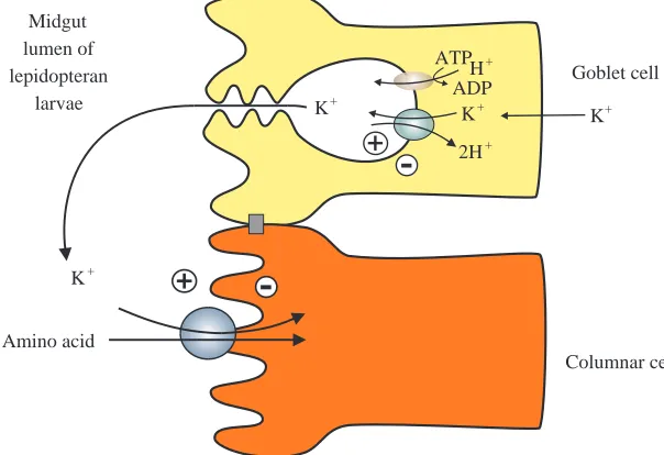
K+-coupled amino acid transporters in insect midgut
Amino acid transporters in insects have been studied in the adult stage of the orthopteran Blatera gigantea and in the larval stage of the coleopteran Leptinotarsa decemlineata and the
lepidopterans Philosamia cynthia and Manduca sexta. While more work is necessary to provide a general description of these processes, interesting characteristics of amino acid transporters in the midgut of coleopteran and lepidopteran insects have recently been reported. Although in Coleoptera it was not possible to demonstrate the presence of ion-coupled amino acid transport (Reuveni et al. 1993), orthopteran transport of amino acids was shown to be Na+-coupled; the Na+ electrochemical
ion gradient is maintained by a Na+/K+-ATPase, analogous to
mammalian cells. In contrast, Lepidoptera revealed transport features that were completely different from those found in mammals. The hemolymph of lepidopteran larvae has a very low ratio of [Na+] to [K+] and relatively high Mg2+and Ca2+
concentrations, in marked contrast to mammalian blood (Giordana et al. 1982; Dow et al. 1984).
The lepidopteran midgut is mainly composed of two types of cells: goblet cells and columnar cells (Fig. 3) (Anderson and Harvey, 1966; Cioffi, 1979). Goblet cells are involved in net secretion of K+. An apical electrogenic H+ V-ATPase in
parallel with an apical K+/2H+ antiporter (Wieczorek et al.
1991) are responsible for active secretion of K+ from the
hemolymph into the midgut lumen (Fig. 3). This K+ pump
produces a high transapical electrochemical potential difference for K+ of approximately −200 mV, which is
composed almost completely of the voltage component (Giordana et al. 1982; Dow and Peacock, 1989), as well as a large pH difference between the hemolymph (pH 6.8) and the lumen (pH 10.5) because K+ reaches the lumen accompanied
by carbonate, resulting in luminal alkalization (Dow, 1984). The columnar cells have a well-developed brush border and accumulate amino acids, coupled to the cotransport of K+
(Giordana et al. 1982, 1989).
Using brush-border membrane vesicles (BBMVs) prepared from different species of lepidopteran larva, such as
Philosamia cynthia, Manduca sexta, Bombyx mori and Pieris brassicae, it has been possible to distinguish several distinct
amino acid transport systems with overlapping specificities
Amino acid
Columnar cell Goblet cell Midgut
lumen of lepidopteran
larvae K+
K+
2H+ H+ ATP
ADP
[image:11.595.256.558.555.762.2]K+ K+
Fig. 3. K+-coupled amino acid transporters in
(Hanozet et al. 1989; Harvey and Wieczorek, 1997). With minor exceptions, general characteristics are conserved within the Lepidoptera. There is evidence for the presence of separate neutral and cationic K+-coupled amino acid transporters.
These transporters are characterized by a particularly strong dependence on the transmembrane potential difference (Parthasarathy and Harvey, 1994). In Philosamia cynthia, there is evidence for a separate proline transporter, but proline is a poor inhibitor of other neutral amino acid transporters (Giordana et al. 1989). A proline, glycine:K+symporter has
furthermore been recently described in Manduca sexta (Bader
et al. 1995). Glutamate transport is K+-coupled but appears
not to use an independent transporter in Manduca sexta (Xie
et al. 1994). A cationic K+ cotransporter has been found in
Philosamia cynthia (Giordana et al. 1985), and a K+/arginine
cotransporter, system R+, operates at near neutral pH has been
described in Manduca sexta (Liu and Harvey, 1996a). Lysine uptake probably occurs via a B-like system (see below), since leucine inhibits lysine uptake (Liu and Harvey 1996b). The neutral amino acid transporter has been studied most extensively. Inhibition, counterflow and kinetic experiments using BBMVs (Giordana et al. 1982, 1989, 1994; Hennigan
et al. 1993a,b) revealed a broad substrate specificity with most
of the neutral amino acids sharing the same transporter. This feature is reminiscent of the vertebrate epithelial amino acid transport system B. As noted above, members of system B have not been cloned yet.
The larval neutral amino acid transporters in Philosamia
cynthia, Manduca sexta, Bombyx mori and Pieris brassicae
show different dependencies on pH, but an increased uptake was generally reported at physiological alkaline pH (Sacchi et
al. 1990; Hennigan et al. 1993b). Neutral amino acid transport
is not strictly K+-dependent in vitro. Studies using BBMVs
revealed that Na+and Li+can also serve as coupling ions under
certain conditions (e.g. reduced pH). However, as noted above, the Na+ concentration is very low in relation to the K+
concentration in vivo and Li+ is absent. Although the
lepidopteran midgut has a low Na+ concentration, a large
inwardly directed electrochemical Na+gradient exists but it is
composed almost entirely of the voltage component (Giordana
et al. 1982; Dow and Peacock, 1989). Kinetic analysis revealed
that the transporter shows a higher affinity for Na+than for K+,
but that the maximal transport rate is higher in the presence of K+(Hanozet et al. 1992; Sacchi et al. 1994). The unlikelihood
of separate Na+- and K+-coupled transporters is further
demonstrated by the reduced K+-coupled leucine uptake at
increased external Na+concentration (Sacchi et al. 1994).
The following kinetic mechanism for the neutral amino acid transport process has been proposed. First, the carrier randomly binds K+and leucine with a 1:1 stoichiometry. Either the fully
loaded carrier or the carrier containing leucine alone then translocates and releases the amino acid on the opposite side of the membrane (Parenti et al. 1992).
To obtain molecular information on this K+-coupled
transporter, we have initiated expression-cloning with mRNA derived from Manduca sexta midgut and expressed in Xenopus
oocytes. Injection of mRNA prepared from the larval midgut induced Na+-dependent uptake of leucine and phenylalanine.
At pH 8.0, the uptake increased 1.5- to 2.5-fold above that of water-injected control oocytes. Similar results have been obtained with mRNA from Philosamia cynthia (Sacchi et al. 1995). Further characterization of this transporter is now in progress. Knowledge of its unique properties should yield new insights into the mechanisms of ion-coupled solute transport.
Ultimately, this study may also lead to the molecular identification of the mammalian amino acid transport system B. As mentioned above, the cDNA encoding a Na+- and Cl−
-coupled GABA transporter has been isolated from a Manduca
sexta embryo cDNA library (Mbungu et al. 1995). The primary
sequence showed moderate homology (72–77 % similarity) to mammalian GABA transporters. The clone has been expressed in Xenopus oocytes, and GABA uptake showed strong Na+
-dependence (Mbungu et al. 1995). In contrast to the mammalian homologue, Br−could substitute for Cl−.
Knowledge of the molecular characteristics of the insect neutral amino acid transporters may also lead to the design of vectors to control insect pests. It was reported that the Bacillus
thuringiensisδ-endotoxin CryIA(a) partially inhibited leucine uptake in BBMVs prepared from Bombyx mori larvae and phenylalanine uptake in Lymantria dispar midgut. This inhibition can be explained by a direct interaction of the endotoxin with the transporter complex (Wolfersberger, 1991; Parenti et al. 1995). The receptor that binds δ-endotoxin CryIA(c) in the midgut brush border of Manduca sexta larvae has been identified and was found to correspond to aminopeptidase N (Knight et al. 1994; Sangadala et al. 1994). This enzyme has been implicated in Na+-dependent neutral
amino acid transport by system B0 in bovine renal BBMVs
(Plakidou-Dymack et al. 1993).
Conclusions
The cloning and functional expression in Xenopus oocytes of vertebrate and invertebrate amino acid transporters has provided new information into the transport characteristics, regulation and physiological roles of amino acids transporters. These studies have also significantly advanced our understanding of the pathophysiology of transporters in neurotransmission, intestinal nutrient absorption, nephrolithiasis, diabetes, cancer, etc. Although cDNA clones corresponding to several classically characterized amino acid transport systems have been isolated, much work remains to be done to complete their molecular characterization. Several important amino acids transporters, such as systems A, B, L, x−c and the imino system, await characterization at the
molecular level. In addition, the cDNAs encoding the transporters activated by D2/NBAT and 4F2-hc (i.e. b0+, y+L,
etc.) have also not been isolated.
References
ALBINA, J. E. ANDHENRY, W. J. (1991). Suppression of lymphocyte
proliferation through the nitric oxide synthesizing pathway. J. surg. Res. 50, 403–409.
ANDERSON, E. ANDHARVEY, W. R. (1996). Active transport by the Cecropia midgut. II. Fine structure of the midgut epithelium. J. Cell Biol. 31, 107–134.
ARRIZA, J. L., FAIRMAN, W. A., WADICHE, J. I., MURDOCH, G. H.,
KAVANAUGH, M. P. AND AMARA, S. G. (1994). Functional comparisons of three glutamate transporter subtypes cloned from human motor cortex. J. Neurosci. 14, 5559–5569.
ARRIZA, J. L., KAVANAUGH, M. P., FAIRMAN, W. A., WU, Y. N.,
MURDOCH, G. H., NORTH, R. A. ANDAMARA, S. G. (1993). Cloning and expression of a human neutral amino acid transporter with structural similarity to the glutamate transporter gene family. J. biol. Chem. 268, 15329–15332.
BADER, A. L., PARTHASARATHY, R. AND HARVEY, W. R. (1995). A novel proline, glycine:K+ symporter in midgut brush-border
membrane vesicles from larval Manduca sexta. J. exp. Biol. 198, 2599–2607.
BALSOM, P. D., SODERLUND, K. ANDEKBLOM, B. (1994). Creatine in humans with special reference to creatine supplementation. Sports Med. 18, 268–280.
BANNAI, S. (1984). Transport of cysteine in mammal cells. Biochim.
biophys. Acta 779, 289–306.
BARBOUR, B., BREW, H. AND ATTWELL, D. (1988). Electrogenic
glutamate uptake in glial cells is activated by intracellular potassium. Nature 335, 433–435.
BARBOUR, B., BREW, H. ANDATTWELL, D. (1991). Electrogenic uptake of glutamate and aspartate into glial cells isolated from the salamander (Ambystoma) retina. J. Physiol., Lond. 436, 169–193. BERTRAN, J., MAGAGNIN, S., WERNER, A., MARKOVICH, D., BIBER, J.,
TESTAR, X., ZORZANO, A., KUHN, L. C., PALACIN, M. ANDMURER, H. (1992a). Stimulation of system y+-like amino acid transport by
the heavy chain of human 4F2 surface antigen in Xenopus laevis oocytes. Proc. natn. Acad. Sci. U.S.A. 89, 5606–5610.
BERTRAN, J., WERNER, A., MOORE, M. L., STANGE, G., MARKOVICH, D., BIBER, J., ZORZANO, A., PALACIN, M. ANDMURER, H. (1992b).
Expression cloning of cDNA from rabbit kidney cortex that induces a single transport system for cystine and dibasic and neutral amino acids. Proc. natn. Acad. Sci. U.S.A. 89, 5601–5605.
BIEBER, L. L. (1988). Carnitine. A. Rev. Biochem. 57, 261–283.
BODEN, G., TAPPY, L., JADALI, F., HOELDTKE, R. D., REVANI, I. AND
OWEN, O. E. (1990). Role of glucagon in disposal of an amino acid
load. Am. J. Physiol. 259, E225–E232.
BONADONNA, R. C., SACCOMANI, M. P., COBELLI, C. ANDDEFRONZO,
R. A. (1993). Effect of insulin on system A amino acid transport in human skeletal muscle. J. clin. Invest. 91, 514–521.
BORDEN, L. A., SMITH, K. E., HARTIG, P. R., BRANCHEK, T. A. AND
WEISHANK, R. L. (1992). Molecular heterogeneity of the γ
-aminobutyric acid (GABA) transport system. J. biol. Chem. 267, 21098–21104.
BOUVIER, M., SZATKOWSKI, M., AMATO, A. ANDATTWELL, D. (1992). The glial cell glutamate uptake carrier countertransports pH-changing anions. Nature 360, 471–474.
BOYD, C. A. AND CRAWFORD, D. H. (1992). Activation of cationic
amino acid transport through system y+correlates with expression
of the T-cell early antigen gene in human lymphocytes. Eur. J. Physiol. 422, 87–89.
BRÖER, S., BRÖER, A. ANDHAMPRECHT, B. (1995). The 4F2 hc surface
antigen is necessary for expression of system L-like neutral amino
acid-transport activity in C6-BU-1 rat glioma cells: evidence from expression studies in Xenopus laevis oocytes. Biochem. J. 312, 863–870.
BROOKES, N. (1988). Neutral amino acid transport in astrocytes:
characterization of Na+-dependent and Na+-independent
components of alpha-aminoisobutyric acid uptake. J. Neurochem.
51, 1913–1918.
BUSCH, A. E., HERZER, T., WALDEGGER, S., SCHMIDT, F., PALACIN, M., BIBER, J., MARKOVICH, D., MURER, H. AND LANG, F. (1994).
Opposite directed currents induced by the transport of dibasic and neutral amino acids in Xenopus oocytes expressing the protein rBAT. J. biol. Chem. 269, 25581–25586.
BUSSOLATI, O., LARIS, P. C., ROTOLI, B. M., DALL’ASTA, V. AND
GAZZOLA, G. C. (1992). Transport system ASC for neutral amino acids. An electroneutral sodium/amino acid cotransport sensitive to membrane potential J. biol. Chem. 267, 8330–8335.
CALONGE, M. J., GASPARINI, P., CHILLARON, J., CHILLO, M., GALLUCCI, M., ROUSAUD, F., ZELANTE, L., TESTAR, X., DALLAPICCOLA, B., DI
SILVERIO, F., BARCELO, P., ESTIVILL, X., ZORZANO, A., NUNES, V.
ANDPALACIN, M. (1994). Cystinuria caused by mutations in rBAT, a gene involved in the transport of cystine. Nature Genetics 6, 420–425.
CARIAPPA, R. ANDKILBERG, M. S. (1992). Plasma membrane domain localization and transcytosis of the glucagon-induced hepatic system A carrier. Am. J. Physiol. 263, E1021–E1028.
CASADO, M., BENDAHAN, A., ZAFRA, F., DANBOLT, N. C., ARAGON, C., GIMENEZ, C. ANDKANNER, B. I. (1993). Phosphorylation and
modulation of brain glutamate transporters by protein kinase C. J. biol. Chem. 268, 27313–27317.
CHEN, J. G. ANDKEMPSON, S. A. (1995). Osmoregulation of neutral amino acid transport. Proc. Soc. exp. Biol. Med. 210, 1–6. CHESNEY, R. W. (1985). Taurine: its biological role and clinical
implications. Adv. Pediatr. 32, 1–42.
CHRISTENSEN, H. N. (1990). Role of amino acid transport and counter
transport in nutrition and metabolism. Physiol. Rev. 70, 43–77. CHRISTENSEN, H. N., HANDLOGTEN, M. E., LAM, I., TAGER, H. S. AND
ZAND, R. (1969). A bicyclic amino acid to improve discrimination among transport systems. J. biol. Chem. 244, 1510–1520. CHRISTENSEN, H. N., OXENDER, D. L., LIANG, M. ANDVATZ, K. A.
(1965). The use of N-methylation to direct the route of mediated transport of amino acids. J. biol. Chem. 240, 3609–3616. CIOFFI, M. (1979). The morphology and fine structure of the larval
midgut of the moth (Manduca sexta) in relation to active ion transport. Tissue & Cell 11, 467–479.
CLOSS, E. I., ALBRITTON, L. M., KIM, J. W. ANDCUNNINGHAM, J. M.
(1993a). Identification of a low affinity, high capacity transporter of cationic amino acids in mouse liver. J. biol. Chem. 268, 7538–7544.
CLOSS, E. I., LYONS, C., KELLY, C. ANDCUNNINGHAM, J. M. (1993b). Characterization of the third member of the MCAT family of cationic amino acid transporters. J. biol. Chem. 268, 20796–20800. COX, D. W., HEADLEY, M. H. ANDWATKINS, J. C. (1977). Actions of L- and D-homocysteate in rat CNS: a correlation between low-affinity uptake and the time courses of excitation by microelectrophoretically applied L-glutamate analogues. J. Neurochem. 29, 579–588.
CRAWHALL, J. C., SCOWEN, E. F., THOMSON, C. J. ANDWATTS, R. W.
(1967). The renal clearance of amino acids in cystinuria. J. clin. Invest. 46, 1162–1171.