Visual pigments (generically, rhodopsins) are intrinsic membrane proteins, opsins, coupled with retinoid chromophores. They are functionally distinguished by the wavelength of maximal absorbance (λmax). There is good
reason to believe that amino acid residues in the opsin-binding pocket surrounding the chromophore interact with its unsaturated polyene chain to determine the λmax of that
particular rhodopsin. However, the molecular details are far from settled (Yokoyama, 1995; Yokoyama and Yokoyama, 1996).
The presence in an eye of two or more rhodopsins tuned to different regions of the spectrum can enable an animal to discriminate relevant visual stimuli on the basis of wavelength. Behavioral, electrophysiological and biochemical studies indicate that wavelength discrimination in insects is commonly based on a set of three rhodopsins tuned to absorb maximally in the ultraviolet, blue and green regions of the spectrum (Menzel and Backhaus, 1991; Peitsch et al. 1992). At least in Hymenoptera, this capacity fits the definition of true trichromatic color vision (Goldsmith, 1990; Menzel and Backhaus, 1991; Peitsch et al. 1992). Some insects also have additional red receptors (Bernard and Remington, 1991; Peitsch et al. 1992).
Vertebrate, cephalopod and arthropod opsins occupy distinct branches of a phylogenetic tree that may have
separated as early as the Paleozoic period (Goldsmith, 1990; Gärtner and Towner, 1995). The sequences of nearly 50 assorted vertebrate opsins are now available (Yokoyama and Yokoyama, 1996). In contrast, only a few insect (and other invertebrate) opsins are known (for references and sequence comparisons, see Smith et al. 1993; Carulli et al. 1994; Chang et al. 1995; Gärtner and Towner, 1995). The scant comparative evidence suggests that the molecular basis of spectral tuning (apart from very general considerations) may be dissimilar in insect and vertebrate rhodopsins (Britt et al. 1993; Chang et al. 1995; Gärtner and Towner, 1995). Thus, opsin amino acid sequences provide the primary data for attacking several related problems: the molecular basis of spectral tuning, the phylogeny of visual pigments and the evolution of color vision. The primary structures of insect visual pigments promise, in addition, a valuable comparative perspective provided by the molecular products of parallel evolution.
It is not surprising that most of our information on insect opsins comes from flies. Four opsin-encoding genes from Drosophila melanogaster have been known for some time (Pollock and Benzer, 1988). Each is expressed in specific photoreceptor cells of the compound eye, the dorsal ocelli and the larval photosensory organ. The opsins they encode fall into two groups on the basis of amino acid sequence and rhodopsin JEB1083
Three distinct opsin-encoding cDNAs, designated MANOP1, MANOP2 and MANOP3, were isolated from the retina of the sphingid moth Manduca sexta. MANOP1 codes for a protein with 377 amino acid residues. It is similar in sequence to members of a phylogenetic group of long-wavelength-sensitive arthropod photopigments, most closely resembling the opsins of ants, a praying mantis, a locust and the honeybee. MANOP2 and MANOP3 opsins have 377 and 384 residues respectively. They belong to a related group of insect visual pigments that include the ultraviolet-sensitive rhodopsins of flies as well as other
insect rhodopsins that are also thought to absorb at short wavelengths. The retina of Manduca sexta contains three rhodopsins, P520, P450 and P357, with absorbance peaks, respectively, at green, blue and ultraviolet wavelengths. There is evidence that MANOP1 encodes the opsin of P520. We suggest that MANOP2 encodes P357 and that MANOP3, representing a class of blue-sensitive insect photopigments, encodes P450.
Key words: Lepidoptera, Sphingidae, Manduca sexta, compound eye, rhodopsin, opsin cDNA.
Summary
Introduction
THREE OPSIN-ENCODING cDNAS FROM THE COMPOUND EYE OF MANDUCA
SEXTA
MICHAEL R. CHASE, RUTH R. BENNETT ANDRICHARD H. WHITE*
Department of Biology, University of Massachusetts Boston, 100 Morrissey Boulevard, Boston, MA 02125-3393, USA
Accepted 10 July 1997
absorb in the blue-green region of the visible spectrum. Rh3 and Rh4 encode ultraviolet-sensitive rhodopsins. Two more distinctive Drosophila melanogaster opsin sequences, Rh5 (Chou et al. 1996) and Rh6 (Huber et al. 1997), have just been discovered. Efforts to characterize opsins from insect taxa other than Diptera have yielded sequences from a mantid species (Towner and Gärtner, 1994), the locust Schistocerca
gregaria (Gärtner and Towner, 1995; Towner et al. 1997), two
species of ant, Cataglyphis bombycina and Camponotus
abdominalis (Popp et al. 1996), and the honeybee Apis mellifera (Chang, 1995; Chang et al. 1996; Bellingham et al.
1997). However, the corresponding rhodopsins have not been directly identified from the absorbance spectra of expressed gene products or by in situ localization.
To explore systematically the lineage and function of insect visual pigments requires the continued collection of opsin sequences from insect orders other than Diptera. It is particularly desirable to characterize the full complement of opsins from compound eyes that deploy the three photopigments of typical λmaxfor wavelength discrimination.
The sphingid moth Manduca sexta has such a representative trichromatic visual system based on characterized rhodopsins, P520, P450 and P357, absorbing in the green, blue and ultraviolet regions respectively (White et al. 1983a,b; Bennett and Brown, 1985) and it supports wavelength-dependent behavior (White et al. 1994; Cutler et
al. 1995). The present paper reports the amino acid sequences
of three opsins deduced from retinal cDNA and addresses the problem of assigning them among the three identified rhodopsins.
Materials and methods
Isolation and cloning of opsin-encoding cDNA Manduca sexta were reared on a carotenoid-rich artificial
diet under conditions described previously (Bennett and White, 1989; Chase et al. 1996). Approximately 200 retinas from compound eyes were frozen quickly in liquid nitrogen and stored at −80 °C. Total RNA was isolated using guanidinium thiocyanate extraction and pelleted twice by successive centrifugation through 5.7 mol l−1 CsCl (MacDonald et al.
1987) in order to remove traces of ommachrome screening pigment and/or other factors that may interfere with reverse transcription (Smith et al. 1993).
Total RNA was reverse-transcribed using a primer (RT-1, Table 1) targeted to poly(A) in order to select mRNA. Approximately 200 ng of RNA and 100 pmol of the primer were added to 50µl of 5×reverse transcription buffer (Gibco BRL, Rockville, Maryland, USA). The solution was heated to 65 °C and allowed to cool to room temperature for 10 min. Reverse Transcriptase RNase Minus (100 units; M-MLV, Gibco) and l mmol l−1each of dATP, dCTP, dGTP and dTTP
were then added, and the reverse transcription reaction was run at 42 °C for 1 h. The reaction was terminated by heating to 70 °C for 10 min.
polymerase chain reaction (PCR) using suitable primers. PCR reactions were typically cycled 35 times at 94 °C for 1 min, 48 °C for 1 min and 72 °C for 1 min. Fragments were separated on agarose gels, excised, cloned into t-vectors (Novagen, Madison, WI), and sequenced with the Taq fmol system (Promega, Madison, WI). cDNA molecules encoding one of the opsins eventually identified, MANOP3, were sequenced on an ABI model 373 sequencer using a dye-terminator sequencing kit (Perkin Elmer Applied Biosystems, Foster City, CA). In order to verify the nucleotide sequences of cDNA fragments, 2–4 clones from independent PCR reactions were sequenced on the forward and reverse strands.
For the initial approach to isolating opsin-encoding 3′cDNA fragments, RT-1 was paired with a degenerate sense-strand primer (OPS-FD, Table 1) designed to a conserved amino acid sequence, (E\D)QAKKMN, from loop 4–5 of known arthropod opsins (Hariyama et al. 1993; Smith et al. 1993; Towner and Gärtner, 1994; Chang et al. 1995). This strategy yielded fragments of two unique opsin-encoding cDNAs, designated MANOP1 and MANOP2 (Chase et al. 1996). To amplify upstream regions, antisense primers were designed to unique sequences in MANOP1 (MSN-1, Table 1) and MANOP2 (UVAS-1, Table 1) approximately 150 base pairs downstream from the OPS-FD site. These opsin-specific primers were coupled with a sense degenerate primer (MSH-1, Table 1) designed to a conserved sequence, GNG(MLV)V(IVM)YW, in helix I of insect opsins.
[image:2.609.309.560.567.742.2]This approach for capturing upstream regions worked only for MANOP1. MANOP1 was then extended into the 5′ untranslated region (UTR) using a 5′ RACE (rapid amplification of cDNA ends) strategy (Frohman et al. 1988): cDNA fragments were tailed with dCTP using terminal transferase (Gibco BRL) and reverse transcription reactions were run using a 5′anchor primer (GIBCO, Table 1) provided with the Gibco kit and a specific antisense primer (LWN-1, Table 1) downstream from the MSH-1 site. The 3′ region of
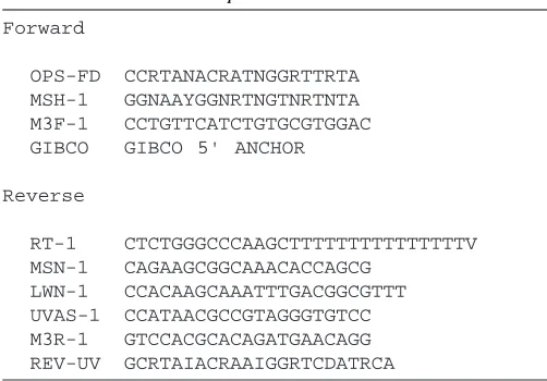
Table 1. Forward and reverse primers used for isolating
opsin cDNAs
Forward
OPS-FD CCRTANACRATNGGRTTRTA MSH-1 GGNAAYGGNRTNGTNRTNTA M3F-1 CCTGTTCATCTGTGCGTGGAC GIBCO GIBCO 5' ANCHOR
Reverse
RT-1 CTCTGGGCCCAAGCTTTTTTTTTTTTTTTV MSN-1 CAGAAGCGGCAAACACCAGCG
MANOP1
M D P G P G L A A L Q A W A A K S P A Y G A A N Q T V V D K V P 1 CGGCAATGGA TCCCGGGCCC GGTTTGGCGG CCCTGCAAGC ATGGGCGGCC AAGTCGCCAG CTTACGGCGC TGCAAATCAA ACTGTCGTCG ACAAAGTGCC P D M M H M I D P H W Y Q F P P M N P L W H A L L G F T I G V L G 101 TCCAGACATG ATGCACATGA TCGACCCTCA CTGGTATCAA TTTCCGCCTA TGAACCCACT TTGGCATGCG CTGTTAGGAT TTACAATTGG TGTTCTGGGA F V S I S G N G M V I Y I F M S T K S L K T P S N L L V V N L A F 201 TTTGTTTCGA TATCGGGCAA CGGCATGGTC ATCTACATCT TTATGTCAAC TAAGAGTCTT AAAACGCCGT CAAATTTGCT TGTGGTCAAT CTTGCTTTTT S D F L M M C A M S P A M V V N C Y Y E T W V W G P F A C E L Y A C 301 CGGACTTCCT CATGATGTGC GCCATGTCTC CAGCTATGGT AGTTAATTGT TACTATGAAA CTTGGGTATG GGGTCCTTTT GCATGCGAAT TGTACGCCTG A G S L F G C A S I W T M T M I A F D R Y N V I V K G I A A K P M 401 TGCTGGTTCT CTATTTGGAT GCGCTTCAAT TTGGACAATG ACTATGATAG CTTTCGACCG ATACAATGTA ATCGTTAAAG GTATAGCAGC TAAGCCCATG T S N G A L L R I L G I W V F S L A W T L L P F F G W N R Y V P E 501 ACCAGCAACG GCGCCCTTCT TCGCATACTT GGAATCTGGG TATTTTCACT AGCTTGGACT CTGCTCCCCT TCTTTGGCTG GAACAGATAT GTGCCCGAAG G N M T A C G T D Y L S K S W V S R S Y I L I Y S V F V Y F L P L L 601 GAAACATGAC TGCTTGCGGA ACAGATTACT TGTCCAAGAG TTGGGTCAGC CGAAGCTACA TCCTTATCTA CTCCGTTTTC GTTTACTTCT TGCCGCTTCT L I I Y S Y F F I V Q A V A A H E K A M R E Q A K K M N V A S L R 701 TCTTATCATC TATTCTTACT TCTTTATTGT TCAGGCCGTA GCTGCTCACG AAAAGGCAAT GAGGGAACAG GCTAAGAAAA TGAATGTGGC CTCCCTCAGA S S E A A N T S A E C K L A K V A L M T I S L W F M A W T P Y L V 801 TCTTCAGAAG CGGCAAACAC CAGCGCTGAA TGCAAACTAG CCAAGGTTGC ATTAATGACC ATTTCCTTAT GGTTCATGGC GTGGACACCA TACCTTGTCA I N Y T G V F E S A P I S P L A T I W G S L F A K A N A V Y N P I V 901 TCAACTATAC CGGAGTGTTT GAAAGCGCAC CCATTAGCCC TCTGGCCACT ATTTGGGGCT CCCTCTTCGC CAAGGCTAAC GCCGTCTACA ATCCTATTGT Y G I S H P K Y Q A A L Y A K F P S L Q C Q S A P E D A G S V A S 1001 ATATGGCATC AGCCATCCTA AATACCAAGC CGCCCTGTAC GCCAAGTTTC CATCGCTACA GTGCCAGTCG GCTCCAGAGG ACGCCGGCTC GGTTGCTTCT G T T A V S E E K P A A 1101 GGCACCACCG CGGTCTCCGA GGAGAAACCT GCAGCGTGAA TGGACTTTAC AAACAACGAA CGACACTGCA GCCAACTGGA AATACGATTC TGCCGAAGCG 1201 ATGTTCTTTA TTATTTTTTA TATGCAAACA TTTTACATTT GTACAAGCTG TAAACTCGAA CACACGAGCG TGTGCTGGTT GCAAAATATT CAGGTTAGGT 1301 CATTACATGT AACATTTAGT ATATGACTCC TGCGTATTTT GAGTTCACGT TTTGTGTCTC GAGTTGAACG TTGCACACTT TTCTTATGCC TTACATATGC 1401 TTGAAATTAC GGTTAGCGAC ATGAAAAAAA TAAAAAAAAA ATACAGTGAA TATATAATGT CGTTAGACCG TTCCTTTACC TATGAGACTG CTCCCTTAGA 1501 TATAAATAAC GATAGATTTC TTAACGACGA AATAAGCTCT GTATCTTGGA CAAATAAAGC ATATTATTAA GCAAGCTTGC TGGCTGATGG CCAGAAAAAA 1601 AAAAAAAAAA
MANOP2
M N N Q S E N Y 1 AGCAGCTCAG TGAACGGAAT GGGCCCAGGA GCACTCTACT GAGGACTGCT CACCCGCAAC CAGAAATTAT TCCCAACATG AACAACCAAT CAGAAAATTA Y H G A Q F E A L K S A G A I E M L G D G L T G D D L A A I P E H 101 TTATCATGGC GCACAGTTTG AAGCACTTAA GTCCGCCGGG GCCATTGAAA TGCTCGGAGA TGGTTTGACA GGCGACGATC TGGCAGCGAT ACCGGAGCAC W L S Y P A P P A S A H T A L A L L Y I F F T F A A L V G N G M V 201 TGGCTATCGT ACCCGGCGCC GCCCGCTAGT GCTCACACCG CACTGGCACT ACTCTACATT TTCTTTACTT TCGCCGCATT AGTTGGAAAT GGAATGGTCA I F I F S T T K S L R T S S N F L V L N L A I L D F I M M A K A P I 301 TATTCATATT CTCAACGACA AAAAGTTTAC GAACATCAAG CAATTTCCTT GTCTTAAATC TAGCTATACT AGATTTTATA ATGATGGCGA AAGCTCCTAT F I Y N S A M R G F A V G T V G C Q I F A L M G A Y S G I G A G M 401 ATTCATCTAT AATAGCGCGA TGCGAGGCTT TGCGGTAGGC ACAGTCGGTT GCCAAATATT CGCTTTGATG GGCGCTTACA GCGGCATCGG AGCTGGCATG T N A C I A Y D R H S T I T R P L D G R L S E G K V L L M V A F V 501 ACTAACGCAT GCATAGCTTA TGATAGACAC TCAACTATTA CGCGACCTCT CGATGGAAGA TTGTCGGAAG GCAAGGTCTT ACTCATGGTG GCCTTTGTGT W I Y S T P W A L L P L L K I W G R Y V P E G Y L T S C S F D Y L T 601 GGATATACTC TACGCCTTGG GCTCTCCTTC CACTCTTGAA AATTTGGGGC AGATACGTAC CTGAGGGCTA CCTAACATCG TGTTCTTTCG ACTATCTAAC N T F D T K L F V A C I F T C S Y V F P M S L I I Y F Y S G I V K 701 CAACACGTTC GACACGAAGC TGTTCGTAGC GTGTATATTC ACTTGCAGTT ACGTTTTCCC AATGTCCTTA ATAATATATT TCTACAGTGG CATCGTCAAG Q V F A H E A A L R E Q A K K M N V E S L R A N Q G G S S E S A E 801 CAGGTGTTTG CTCACGAAGC AGCTTTGAGG GAACAAGCTA AAAAGATGAA CGTAGAATCT CTAAGAGCTA ATCAGGGCGG GTCATCAGAG TCGGCAGAGA I R I A K A A L T V C F L F V A S W T P Y G V M A L I G A F G N Q Q 901 TCAGAATAGC AAAAGCAGCA CTCACTGTTT GCTTTCTGTT CGTGGCCTCG TGGACACCCT ACGGCGTTAT GGCTCTCATA GGCGCGTTTG GAAATCAACA L L T P G V T M I P A V A C K A V A C I S P W V Y A I R H P M Y R 1001 ACTTCTTACT CCTGGGGTTA CGATGATTCC TGCAGTGGCG TGTAAAGCTG TAGCGTGTAT TAGTCCTTGG GTATACGCAA TCAGACATCC TATGTACAGG Q E L Q R R M P W L Q I D E P D D T V S T A T S N T T N S A P P A 1101 CAAGAGCTGC AACGTCGCAT GCCGTGGTTA CAAATTGATG AGCCCGATGA CACTGTCTCG ACGGCGACCA GCAACACTAC CAACAGCGCG CCACCAGCTG
A T A
1201 CCACCGCCTA AGTTCTCTAT GCTCAAAAGG GATGACATAT ACTCAGTACA TATACATGTA TTTATTCATT GTGATTTTAA CGACAACTAT GGCTATGCTG 1301 AAGGCTTTTG ATGTTAAATT ACTGTAGAAT TTATTAAAGA AGCATAAAGT GTCACAGAAC ACCTAAAAAA AAAAAAAAA
MANOP3
1 ACAAGTGAGA AGCGACTTGG AAGTCGGATA ACTCACTTCG TTCATAAAAA CATCATTTTG TTTTTTTAAC ATCACGATTT GTAACAACTT GTAAATTAAT M A T N F T Q E L Y E I G 101 GAAGCTGTAG CAAGTACATG AATTTACTCC GTCGGCCATT TTGTTAACAA CACACGCACA AAATGGCGAC CAATTTCACG CAGGAACTTT ACGAGATCGG P M A Y P L K M I S K D V A E H M L G W N I P E E H Q D L V H D H 201 ACCGATGGCC TACCCGCTGA AGATGATATC AAAGGATGTC GCGGAGCACA TGCTGGGTTG GAACATTCCA GAAGAACATC AAGACTTGGT CCACGACCAC W R N F P A V S K Y W H Y V L A L I Y T M L M V T S L T G N G I V 301 TGGCGAAACT TCCCCGCGGT CAGCAAGTAC TGGCATTATG TCCTAGCGCT GATATACACC ATGCTCATGG TCACGTCGCT CACCGGCAAT GGAATTGTCA I W I F S T S K S L R S A S N M F V I N L A V F D L M M M L E M P L 401 TTTGGATATT TAGCACTTCA AAATCCCTAC GCAGCGCGAG CAACATGTTC GTGATAAACT TAGCGGTGTT CGATCTGATG ATGATGCTGG AGATGCCGCT L I M N S F Y Q R L V G Y Q L G C D V Y A V L G S L S G I G G A I 501 GCTAATCATG AACTCGTTCT ACCAGCGTCT CGTGGGGTAC CAGCTTGGCT GCGATGTGTA CGCGGTACTC GGCTCTCTCT CTGGAATTGG AGGGGCCATC T N A V I A F D R Y K T I S S P L D G R I N T V Q A G L L I A F T 601 ACTAATGCTG TCATTGCTTT TGATCGATAC AAAACAATTT CGTCGCCACT TGATGGAAGA ATAAACACAG TACAAGCGGG TCTGCTGATC GCCTTCACCT W F W A L P F T I L P A F R I W G R F V P E G F L T T C S F D Y F T 701 GGTTCTGGGC GTTGCCGTTC ACTATCCTGC CGGCGTTCAG GATATGGGGG AGATTTGTTC CCGAGGGCTT CCTGACGACG TGCTCGTTCG ACTATTTCAC E D Q D T E V F V A C I F V W S Y C I P M A L I C Y F Y S Q L F G 801 GGAGGACCAG GATACGGAGG TATTTGTCGC GTGCATCTTC GTGTGGAGCT ACTGCATACC GATGGCGCTC ATCTGTTACT TCTACTCTCA ACTATTCGGT A V R L H E R M L Q E Q A K K M N V K S L A S N K E D N S R S V E 901 GCTGTCCGCC TCCATGAACG CATGCTACAA GAACAAGCCA AGAAGATGAA CGTGAAATCA TTAGCATCAA ACAAGGAAGA CAACAGCCGC AGCGTAGAGA I R I A K V A F T I F F L F I C A W T P Y A F V T M T G A F G D R T 1001 TCAGGATAGC TAAAGTGGCG TTCACTATTT TCTTCCTGTT CATCTGTGCG TGGACTCCTT ACGCTTTCGT CACCATGACA GGCGCATTTG GCGACAGGAC L L T P I A T M I P A V C C K V V S C I D P W V Y A I N H P R Y R 1101 CCTACTAACT CCGATAGCAA CGATGATCCC AGCCGTGTGC TGTAAAGTAG TTTCGTGCAT AGACCCATGG GTGTACGCCA TCAACCATCC GAGATACAGG A E L Q K R L P W M G V R E Q D P D A V S T T T S V A T A G F Q P 1201 GCGGAGTTGC AGAAGCGTCT GCCATGGATG GGCGTCCGCG AGCAAGACCC TGACGCGGTA TCTACCACCA CCAGCGTCGC CACCGCTGGT TTTCAACCAC
P A A E A
1301 CAGCCGCGGA GGCCTAGACA GACAAACTGA TTCATAATTA CTGAGGAAAA TCATGTTAAA GCTTAAGCAA TAATTTAAAA CTCCATTAAG CAAAAAAAAA 1401 AAAAAAA
′
by pairing the GIBCO primer with the UVAS-1 primer. Fragments of a third opsin-encoding cDNA, MANOP3, were generated using OPS-FD and another reverse primer (REV-UV, Table 1) designed to a nucleotide sequence in helix VI of a honeybee opsin (Chang, 1995; see also Bellingham et
al. 1997). Downstream fragments were obtained by pairing
RT-1 with a primer (M3F-1, Table 1) specific to a region downstream from the OPS-FD site. Upstream regions were obtained using the 5′ RACE approach, pairing another MANOP3-specific primer (M3R-1) with the GIBCO primer.
Phylogenetic analysis
Conceptual translations of the opsin-encoding cDNAs from
Manduca sexta were compared, using FASTA (Pearson, 1990),
with other known arthropod opsins deposited in GenBank and aligned using CLUSTAL W (Thompson et al. 1994). Phylogenetic trees were constructed with PAUP (Swofford, 1993) using maximum parsimony and neighbor-joining methods. All trees were bootstrapped a minimum of 100 times to estimate confidence intervals.
Results
Opsin-encoding cDNAs from the retina of Manduca sexta were initially targeted with a degenerate primer (OPS-FD,
(E\D)QAKKMN, that is conserved in arthropod opsins in the loop between transmembrane helices V and VI (Hariyama et
al. 1993; Smith et al. 1993; Towner and Gärtner, 1994).
Among the many 3′ fragments that were isolated by this approach, we identified two distinct opsin cDNAs, designated MANOP1 and MANOP2 (Chase et al. 1996).
Although three rhodopsins had been extracted from
Manduca sexta retinas (Bennett and Brown, 1985), efforts to
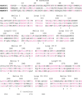
isolate a third opsin-encoding cDNA with the 3′ RACE technique were unsuccessful. While our work was in progress, Chang (1995) isolated fragments of three opsin-encoding cDNAs from the honeybee (Apis mellifera). Two of the honeybee cDNAs were very similar, respectively, to MANOP1 and MANOP2; the third (whose full sequence was recently published by Bellingham et al. 1997) was somewhat different. We isolated the third Manduca sexta cDNA, MANOP3, using a primer (REV-UV, Table 1) based on a sequence in helix VI of the third honeybee cDNA. The three opsin cDNAs isolated from Manduca sexta retinas and their translations are shown in the Appendix. The amino acid sequences of the opsins are compared in Fig. 1.
MANOP1 cDNA contains 1594 base pairs (bp) with a single open reading frame encoding an opsin with 377 amino acids. MANOP2 (1362 bp) also encodes 377 residues, while
N terminus
10 20 30 40 50 MANOP1 MDPGP----GLAALQAWAAKSPAYGAANQTVVDKVPPDMMHMIDPHWYQFPPMNPLWH
MANOP2 MNNQSE--NYYHGAQFEALKSAGAIE---MLGDGLTGDDLAAIPEHWLSYPAPPASAH
MANOP3 MATNFTQELYEIGPMAYPLKMISKDVAEHMLGWNIPEEHQDLVHDHWRNFPAVSKYWH
Helix I Loop I-II Helix II 60 7 0 80 9 0 100 110 ALLGFTIGVLGFVSISGNGMVIYIFM STKSLK TPSNLLVVNLAFSDFLMMCAMSPAMV TALALLYIFFTFAALVGNGMVIFIFS TTKSLR TSSNFLVLNLAILDFIMMAKAPIFIY YVLALIYTMLMVTSLTGNGIVIWIFS TSKSLR SASNMFVINLAVFDLMMMLEMPLLIM * * LoopII-III Helix III Loop III-IV 120 130 140 150 160 170 VNCYYETWVWGPFAC ELYACAGSLFGCASIWTMTMIAFDRY NVIVKGIAAKPMTS NS-AMRGFAVGTVGC QIFALMGAYSGIGAGMTNACIAYDRH STITRPLDGR-LSE NS-FYQRLVGYQLGC DVYAVLGSLSGIGGAITNAVIAFDRY KTISSPLDGR-INT
* * *
Helix IV Loop IV-V 180 190 200 210 220 NGALLRILGIWVFSLAWTLLPFFG-W NRYVPEGNMTACGTDYLSKSWVSR GKVLLMVAFVWIYSTPWALLPLLKIW GRYVPEGYLTSCSFDYLTNTFDTK VQAGLLIAFTWFWALPFTILPAFRIW GRFVPEGFLTTCSFDYFTEDQDTE
*
Helix V LoopV-VI
230 240 250 260 270 280 SYILIYSVFVYFLPLLLIIYSYFFIV QAVAAHEKAMREQAKKMNVASLRSSEAAN-TSAECK LFVACIFTCSYVFPMSLIIYFYSGIV KQVFAHEAALREQAKKMNVESLRANQGGSSESAEIR VFVACIFVWSYCIPMALICYFYSQLF GAVRLHERMLQEQAKKMNVKSLASNKEDNSRSVEIR
*
Helix VI Loop VI-VII Helix VII 290 300 310 320 330 340 LAKVALMTISLWFMAWTPYLVINYTG VFES-APISP LATIWGSLFAKANAVYNPIVYGISHP IAKAALTVCFLFVASWTPYGVMALIG AFGNQQLLTP GVTMIPAVACKAVACISPWVYAIRHP IAKVAFTIFFLFICAWTPYAFVTMTG AFGDRTLLTP IATMIPAVCCKVVSCIDPWVYAINHP
* * a * * C terminus
350 360 370 380 KYQAALYAKFPSLQCQSAPE--DAGSVASGTTAVSEEKPAA MYRQELQRRMPWLQIDEPDDT-VSTATSNTTNS-APPAATA RYRAELQKRLPWMGVREQDPDAVSTTTSVATAGFQPPAAEA
[image:4.609.281.557.397.714.2]MANOP3 (1391 bp) encodes 384 residues. The approximate sizes of the corresponding opsin-encoding mRNAs were measured on northern blots of retinal extracts: MANOP1 mRNA ≈1.8 kb, MANOP2 mRNA ≈1.5 kb and MANOP3 mRNA ≈2.2 kb (Chase et al. 1996; Landry, 1997).
Several considerations indicate that the we have not isolated the full 3′untranslated sequences of MANOP2 and MANOP3. Their 3′UTRs are short in comparison with that of MANOP1 and they lack a polyadenylation signal: AATAAA (Rice et al. 1992). Furthermore, MANOP3 mRNA is almost twice as large as the cDNA we have isolated. It is likely that the RT-1 primer (Table 1) found stretches of adenines in the 3′ UTRs of MANOP2 and MANOP3 rather than annealing to the terminal poly(A) tail. Such internal adenine repeats are present between nucleotides 1424 and 1441 in the 3′ UTR of MANOP1. Secondary structure arising from a GC-rich region prevented sequencing far into the 5′UTR of MANOP1.
The deduced amino acid sequences of the three Manduca
sexta opsins show the various conserved characteristics of
opsins generally and of arthropod opsins in particular (Chang
et al. 1995; Gärtner and Towner, 1995; Yokoyama, 1995;
Popp et al. 1996). Sequence similarities and hydropathy profiles (not shown) confirm the seven membrane-spanning helical domains that characterize opsins and other G-protein-coupled receptors (Baldwin, 1993). A lysine (Lys-330) residue in the middle of helix VI is the site of attachment for the Schiff-base linkage of the retinaldehyde chromophore. Cysteine residues at the extracellular end of helix III (Cys-131) and within extracellular loop IV–V (Cys-209) presumably provide a stabilizing disulfide linkage (Gärtner and Towner, 1995). Asparagine residues susceptible to glycosylation are present in the N-terminal region (MANOP1, Asn-28; MANOP2, Asn-3; MANOP3, Asn-4); as in some other invertebrate opsins (Popp et al. 1996), there is a second potential glycosylation site (Asn-205) in extracellular loop IV–V of MANOP1. There are a number of serine/threonine potential phosphorylation sites in the C termini. Conserved sequences, particularly those that interact with G-protein (transducin), are present in the helix-connecting loops that extend beyond the membrane surfaces (Gärtner and Towner, 1995; Yokoyama, 1995).
Of the amino acids, 25 % are identical in all three opsins of
Manduca sexta. One-third of the residues are found to be the
same when MANOP1 is compared with either MANOP2 or MANOP3. MANOP2 and MANOP3 are more closely related, showing 46 % identity (Fig. 1; Table 2).
Discussion
Opsins and rhodopsins
We isolated opsin-encoding cDNAs from the retina of
Manduca sexta in order to characterize further the three
rhodopsins of the compound eye: P520, P450 and P357. The next step is to decide which opsin forms which rhodopsin. It is likely that MANOP1 is the opsin of P520: MANOP1 mRNA is 10 times more abundant than the other two opsin mRNAs
(Chase et al. 1996; Landry, 1997), and P520, to a similar degree, is the most plentiful photopigment in the retina (Bennett and Brown, 1985; Bennett et al. 1997). We have no equivalent evidence for deciding between P450 and P357 in the assignment of MANOP2 and MANOP3.
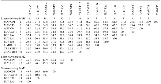
Comparison of the hawkmoth opsins with those of other arthropods offers another avenue to their identification (Fig. 2; Tables 2, 3). The phylogenetic topography of arthropod opsins is growing clearer as more sequences are collected from diverse taxa and the different spectral classes of photoreceptors. Three major divisions are evident in the phylogenetic tree of arthropod opsins (Fig. 2): branch A, opsins from the crab Hemigrapsis sanguineus; branch B, a
crayfish Procambarus clarkii and a number of insects; and branch C, another cluster of insect opsins.
Several of the opsins in branch B have been be identified with rhodopsins absorbing from approximately 480 nm (Rh1 and Rh2 fly opsins: O’Tousa et al. 1985; Cowman et al. 1986; Huber et al. 1990) to 520 nm (Limulus polyphemus opsins: Smith et al. 1993). We therefore classify these opsins as belonging to an ‘LW’ group of functionally similar arthropod photopigments tuned for absorbance at long wavelengths. Similarities at key amino acid positions identified in Table 3 and spectral sensitivity measurements from the crab eye suggest that the opsins of Hemigrapsus
sanguineus should also be included among the LW opsins.
The membership of MANOP1 in the LW group is further support for its identification as P520. It is found in a cluster (designated M1) of opsins from orthopteran and hymenopteran species with amino acid identities of 70 % or greater (Table 2).
Branch C in Fig. 2 includes, at present, only insect opsins: MANOP2 and MANOP3, the Rh3, Rh4 and Rh5 opsins of flies (Montell et al. 1987; Pollock and Benzer, 1988; Chou et al. 1996) and recently characterized opsins from the honeybee (Bellingham et al. 1997) and a locust (Towner et al. 1997). As the only identified rhodopsins of this group are the ultraviolet-absorbing photopigments of Drosophila species, Rh3 and Rh4, we designate this the short-wavelength (SW) cluster of insect opsins (Fig. 2; Table 2). It will be interesting to determine
than insects will join this phyletic group. As both MANOP2 and MANOP3 are situated in the SW group, we infer that the SW division includes both ultraviolet- and blue-absorbing rhodopsins. Certain ultraviolet-, violet- and blue-sensitive photopigments of vertebrate cones similarly constitute a short-wavelength ‘tritope-group’ of related sequences (Hisatomi et
al. 1996; Yokoyama, 1995; Yokoyama and Yokoyama, 1996).
Since MANOP2 is more like the known ultraviolet opsins of flies, we suggest that MANOP2 encodes P357 whereas MANOP3 encodes P450.
Apart from the close similarity of Rh3 and Rh4, the SW opsins are structurally diverse, with amino acid identities ranging from 46 % to 62 % (Table 2). For purposes of comparison (with no necessary functional implication at this point), we have distinguished M2 and M3 subgroups (Fig. 2; Table 2). The M2 group includes only the ultraviolet-sensitive fly rhodopsins and MANOP2. The more disparate M3 group includes, in addition to MANOP3, locust Lo-2, a honeybee opsin and Rh5 of Drosophila melanogaster.
Opsins of the M3 group have been assigned by a variety of criteria. There is indirect evidence that Rh5 encodes a blue-sensitive rhodopsin (Chou et al. 1996). Gärtner and Towner (1995) have suggested that Lo-2, one of two cDNAs isolated from the retina of Schistocerca gregaria, may be the opsin of a blue-sensitive photopigment. The existence of two rhodopsins in the retina of another orthopteran species, Locusta
[image:6.609.38.564.438.715.2]migratoria, one absorbing in the green (λmax500–515 nm) and
Table 2. Percentage residue identities among amino acid sequences of selected arthropod opsins
Long-wavelength M1 16 15 14 13 12 11 10 9 8 7 6 5 4 3 2 1
MANOP1 1 33.2 31.4 33.8 32.7 37.6 33.3 41.2 56.2 60.6 58.5 62.3 71.2 74.5 75.5 76.9 100
MANTIS 2 34.0 32.6 34.9 34.5 38.9 34.2 41.0 56.1 60.3 57.8 62.6 73.7 85.6 77.7 100
ANT Cb 3 35.0 32.8 37.3 36.3 39.2 34.7 41.2 54.8 60.6 57.5 63.9 78.1 77.4 100
LOCUST 1 4 33.9 33.5 34.5 34.8 38.6 34.0 39.7 54.5 59.4 59.8 61.4 70.1 100 BEE LW 5 32.4 31.5 35.7 34.2 37.0 34.4 38.3 55.9 54.6 56.7 63.2 100.0
FLY Rh1 6 35.4 36.8 38.4 37.6 38.9 38.2 44.1 55.2 55.5 53.7 100
FLY Rh6 7 29.8 31.3 32.4 31.6 33.5 30.8 36.8 48.5 50.1 100.0
LIMULUS 8 31.8 35.0 34.8 33.9 41.1 34.6 40.4 56.2 100
CRAYFISH 9 32.0 30.9 30.9 31.7 37.4 32.2 41.7 100 CRAB Rh2 10 36.6 34.0 34.3 34.3 40.3 34.9 100
Short-wavelength M2
MANOP2 11 46.0 45.6 44.9 46.4 62.4 100
FLY Rh3 12 46.0 46.1 47.5 50.9 100
Short-wavelength M3
MANOP3 13 49.7 56.5 59.0 100
LOCUST 2 14 46.2 60.8 100
BEE SW 15 48.9 100
FLY Rh5 16 100
See legend to Fig. 2 for species identification and the basis for grouping insect opsins into long-wavelength and short-wavelength.
another in the blue (λmax 450–430 nm) region, has been
inferred from intracellular spectral sensitivity measurements (Bennett et al. 1967; Lillywhite, 1978). Gärtner and Towner (1995) suggested that Lo-1 and Lo-2 of Schistocerca gregaria are, respectively, the opsins of similar photopigments. However, it would be surprising if locusts lacked ultraviolet receptors, and there is indirect evidence for their existence (Osorio, 1986). Thus, the identification of Lo-2 with a blue-rather than an ultraviolet-sensitive rhodopsin is plausible but not compelling.
Chang (1995) isolated partial sequences of three honeybee opsins that are similar to the three opsins of Manduca sexta. She proposed that the bee homologue of MANOP2 forms its ultraviolet-sensitive pigment and identified the homologue of MANOP3 with the blue-sensitive bee rhodopsin. The latter has since been fully characterized by Bellingham et al. (1997); although they describe it, contrary to Chang’s hypothesis, as the ultraviolet-sensitive rhodopsin, it has not been empirically identified.
Spectral tuning of photopigments
The regulation of rhodopsin absorbance by opsin structure – presumably through the distribution of charged and polar amino acid residues in the binding pocket around the
chromophore – is a challenging problem of current interest. The basis of blue-shifted spectra is particularly uncertain. Recent models and crystallographic data (Baldwin, 1993; Schertler et al. 1993; Schertler and Hargrave 1995; Davies et
al. 1996) provide a foundation for theoretical, comparative and
phylogenetic approaches to the problem (Chang et al. 1995; Gärtner and Towner, 1995; Yokoyama, 1995). Attention has focused on transmembrane helices III, VI and VII (Schertler et
al. 1993; Schertler and Hargrave 1995; Davies et al. 1996).
Amino acid residues of these helices have been shown experimentally to interact with the chromophore in vertebrate rhodopsins (Yokoyama, 1995). However, the details of spectral tuning in vertebrate rhodopsins (Lin et al. 1992; Hunt et al. 1996; Yokoyama, 1995) seem to hold little pertinence for understanding the properties of invertebrate photopigments (Britt et al. 1993; Chang et al. 1995; Gärtner and Towner, 1995). It appears that the assortment of photopigments underlying color vision has evolved independently in insects and vertebrates through distinct opsin/chromophore interactions (Fryxell and Myerowitz, 1991; Chang et al. 1995; Gärtner and Towner, 1995).
[image:7.609.45.566.85.372.2]A number of amino acid positions have been suggested as significant for spectral tuning in invertebrate rhodopsins. For the following discussion, we use the ‘Manduca numbers’ of Table 3. Amino acid positions in transmembrane α-helices that may be significant for spectral tuning in insect rhodopsins
Helix II Helix III Helix IV Helix V Helix VI Helix VII
Manduca no. 103 110 134 145 149 195 198 228 291 298 333 336
Baldwin no. 13 21 3 14 18 24 27 7 8 15 14 17
Long-wavelength M1
MANOP1 Ser (p) Ala Tyr (p) Ser (p) Met Gly Ser (p) Thr (p) Ala Ala Asn (p)
MANTIS * Ser (p) * * * * Ser(p) Thr (p) Ala * *
LOCUST 1 * Thr (p) * * * * Ser(p) Thr (p) Gly * *
BEE LW * Cys * * * * Gly Thr (p) Ala * *
ANT Ca * Cys* * * * * Ser(p) Thr (p) Ala * *
ANT Cb * Ser (p) * * * * Ser(p) Thr (p) Ala * *
FLY Rh1 * Thr (p) * * * * Ser(p) Thr (p) Ala * *
FLY Rh2 * Ser(p) * * * * Ser(p) Thr (p) Ala * *
FLY Rh6 * Thr(p) * * * * Ser(p) Glu(-) Ala * *
Short-wavelength M2
MANOP2 Leu Lys (+) Phe Ala Asn (p) Lys (+) Phe Val Ser (p) Ala Ser (p)
FLY Rh3 Cys * * * * Glu (−) * Ile * * Asp (−)
FLY Rh4 Phe * * * * Gln (p) Asp (−) * Ile * * Asp (−)
Short-wavelength M3
MANOP3 Phe Glu (−) Tyr (p) Gly Asn (p) Arg (+) Phe Ile Ala Ser (p) Asp (−)
FLY Rh5 * Asn (p) * Ala * Gln (p) * * Ala * *
LOCUST 2 * Glu (−) * Ser (p) * Arg (+) * * Ser (p) * *
BEE SW * Glu (−) * Gln (p) * Lys (+) * * Ala * *
Positions are numbered as in Fig. 1 (Manduca numbers), and in each helix according to the model of Baldwin (1933). Most face inward toward the chromophore according to the Baldwin model. Arrowheads indicate positions significant to the external point-charge model of spectral tuning proposed by Towner et al. (1997); position 198 is pertinent only to Rh4. Asterisks indicate that all residues are identical within a group; (p) polar residues, (+) basic residues, (−) acidic residues.
the residues in each helix, according to her model, from 1 to 26 (Table 3). The inward-facing side of helix III is positioned close to the Schiff-base linkage of the chromophore at Lys-330 in helix VI. In vertebrate but not invertebrate photopigments, a highly conserved glutamate residue near the intradiscal (extracellular) end of helix III functions as the negative counterion that stabilizes the protonated Schiff base (Sakmar
et al. 1989, 1991; Lin et al. 1992). There is a polar tyrosine
residue at that position (Tyr-134) in most invertebrate rhodopsins, a residue that may play a role in long-wavelength tuning (Hall et al. 1991; Smith et al. 1993; Chang et al. 1995; Gärtner and Towner, 1995; Popp et al. 1996) since it is replaced by non-polar phenylalanine in the ultraviolet-sensitive rhodopsins of flies (Table 3). Position 134 is also occupied by phenylalanine in MANOP2, whereas MANOP3 and the other members of the M3 group resemble the LW opsins in having tyrosine at that position (Table 3). Chang et al. (1995) focused on another residue in helix III, suggesting that polar Ser-145 cooperates with Tyr-134 in stabilizing the protonated Schiff base in long-wavelength rhodopsins; non-polar residues occupy that position in most members of the M2 and M3 groups.
Bellingham et al. (1997) catalogued five sites in arthropod opsins, all inward-facing according to the model of Baldwin (1993), which consistently differed, with regard to polarity or charge, between long- and short-wavelength-sensitive rhodopsins: positions 103 in helix II, 149 in helix III, 228 in helix V, 291 in helix VI and 336 in helix VII. Table 3 shows that these generalizations hold up well in differentiating LW from SW insect rhodopsins.
Towner et al. (1997) have proposed an external two-point charge model for short-wavelength-shifted insect rhodopsins on the basis of a negatively charged counterion in helix VII (position 336) and a positive/negative pair of charged residues located in helices II (position 110) and IV (position 195 or, in Rh4, position 198) that form a dipole. However, MANOP2 is inconsistent with this model. Position 336 is occupied by a polar residue in MANOP2, as it is in the long-wavelength-absorbing rhodopsins of the M1 group. Furthermore, positions 110 and 195 are both occupied by lysine, rather than a positive/negative pair of residues. Drosophila melanogaster Rh5 lacks charged residues at the proposed dipole positions.
Although the separation of insect SW opsins into M2 and M3 groups is not strongly supported by overall sequence comparison, there are several consistent differences among residues facing inward around the chromophore. Most notable is polar tyrosine residue at position 134 in the M3 opsins, since it may play a crucial role in shifting absorbance to longer wavelengths (Hall et al. 1991). Other sites that consistently differ with regard to residue attributes between the M2 and M3 opsins are position 110 in helix II and position 333 in helix VII. We propose, in accordance with the speculations of others (Gärtner and Towner, 1995; Chang, 1995; Chou et al. 1996; Towner et al. 1997), that the rhodopsins of the M3 group represent a new class of blue-sensitive insect rhodopsins.
with rhodopsin spectra in insect species other than flies. It is a distinct advantage to have the sequences for a complete set of opsins that are expressed in a retina containing the three typical rhodopsins of insect compound eyes. We know enough about the localization of the three characterized Manduca sexta photopigments, both at the cellular level (green- and ultraviolet-sensitive cells occupy different positions in the retinula: Cutler et al. 1995) and across the retina (blue-sensitivity has been localized to the ventral half of the retina: Bennett et al. 1997) to identify our three opsins by in situ hybridization and immunocytochemistry (Xu, 1996).
This work was supported by National Science Foundation Grant IBN-9210933. We thank Kenneth Kleene and Rachel Skvirsky for helpful advice, Richard Kesseli for the use of laboratory facilities, and Belinda Chang for useful discussions and a primer. Lily Landry, Huihong Xu and Amy Tucker provided technical assistance.
References
BALDWIN, J. M. (1993). The probable arrangement of the helices in G protein-coupled receptors. EMBO J. 12, 1693–1703.
BELLINGHAM, J., WILKIE, S. E., MORRIS, A. G., BOWMAKER, J. K. AND HUNT, D. M. (1997). Characterization of the ultraviolet-sensitive opsin gene of the honey bee, Apis mellifera. Eur. J. Biochem. 243, 775–781.
BENNETT, R. R. ANDBROWN, P. K. (1985). Properties of the visual pigments of the moth Manduca sexta and the effects of two detergents, digitonin and CHAPS. Vision Res. 25, 1771–1781. BENNETT, R. R., TUNSTALL, J. ANDHORRIDGE, G. A. (1967). Spectral
sensitivity of single retinula cells of the locust. Z. vergl. Physiol. 55, 195–206.
BENNETT, R. R. AND WHITE, R. H. (1989). Influence of carotenoid deficiency on visual sensitivity, visual pigment and P-face particles of photoreceptor membrane in the moth Manduca sexta. J. comp. Physiol. A 164, 321–331.
BENNETT, R. R., WHITE, R. H. ANDMEADOWS, J. (1997). Regional specialization in the eye of the sphingid moth Manduca sexta: blue-sensitivity of the ventral retina. Vis. Neurosci. 14, 523–526. BERNARD, G. D. AND REMINGTON, C. L. (1991). Color vision in
Lycaena butterflies: spectral tuning of receptor arrays in relation to behavioral ecology. Proc. natn. Acad. Sci. U.S.A. 88, 2783–2787. BRITT, S. G., FEILER, R., KIRSCHFELD, K. ANDZUKER, C. S. (1993). Spectral tuning of rhodopsin and metarhodopsin in vivo. Neuron 11, 29–39.
CARULLI, J. P., CHEN, D.-M., STARK, W. S. ANDHARTL, D. L. (1994). Phylogeny and physiology of Drosophila opsins. J. molec. Evol. 38, 250–262.
CHANG, B. S. W. (1995). Opsin phylogeny and evolution. PhD dissertation, Harvard University, Cambridge, MA, USA.
CHANG, B. S. W., AYERS, D., SMITH, W. C. ANDPIERCE, N. E. (1996). Cloning of the gene encoding honeybee long-wavelength rhodopsin: a new class of insect long-wavelength visual pigments. Gene 173, 215–219.
CHASE, M. R., BENNETT, R. R. ANDWHITE, R. H. (1996). Expression of opsin mRNA in normal and vitamin A deficient retinas of the sphingid moth Manduca sexta. Vis. Neurosci. 13, 353–358. CHOU, W.-H., HALL, K. J., WILSON, D. B., WIDEMAN, C. L., TOWNSON,
S. M., CHADWELL, L. V. ANDBRITT, S. G. (1996). Identification of a novel Drosophila opsin reveals specific patterning of the R7 and R8 photoreceptor cells. Neuron 17, 1101–1115.
COWMAN, A. F., ZUKER, C. S. ANDRUBIN, G. M. (1986). An opsin gene expressed in only one photoreceptor cell type of the Drosophila eye. Cell 44, 705–710.
CUTLER, D. E., BENNETT, R. R., STEVENSON, R. D. ANDWHITE, R. H. (1995). Feeding behavior in the nocturnal moth Manduca sexta is mediated mainly by blue receptors, but where are they located in the retina? J. exp. Biol. 198, 1909–1917.
DAVIES, A., SCHERTLER, G. F. X., GOWEN, B. E. ANDSAIBIL, H. R. (1996). Projection structure of an invertebrate rhodopsin. J. struct. Biol. 117, 36–44.
FROHMAN, M. A., DUSH, M. K. ANDMARTIN, G. R. (1988). Rapid production of full-length cDNAs from rare transcripts: amplification using a single gene-specific oligonucleotide primer. Proc. natn. Acad. Sci. U.S.A. 85, 8998–9002.
FRYXELL, K. J. AND MYEROWITZ, E. M. (1991). The evolution of rhodopsins and neurotransmitter receptors. J. molec. Evol. 33, 367–378.
GÄRTNER, W. ANDTOWNER, P. (1995). Invertebrate visual pigments. Photochem. Photobiol. 62, 1–16.
GOLDSMITH, T. H. (1990). Optimization, constraint and history in the evolution of eyes. Q. Rev. Biol. 65, 281–322.
HALL, M. D., HOON, M. A., RYBA, N. J. P., POTTINGER, J. D. D., KEEN, J. N., SAIBIL, H. R. ANDFINDLAY, J. B. C. (1991). Molecular cloning and primary structure of squid (Loligo forbesi) rhodopsin, a phospholipase C-directed G-protein-linked receptor. Biochem. J. 274, 35–40.
HARIYAMA, T., OZAKI, K., TOKUNAGA, F. ANDTSUKAHARA, Y. (1993). Primary structure of crayfish visual pigment deduced from cDNA. FEBS Lett. 315, 287–292.
HISATOMI, O., SATOH, T., BARTHEL, L. K., STENKAMP, D. L., RAYMOND, P. A. ANDTOKUNAGA, F. (1996). Molecular cloning and characterization of the putative ultraviolet-sensitive visual pigment of goldfish. Vision Res. 36, 933–939.
HUBER, A., SCHULZ, S., BENTROP, J., GROELL, C., WOLFRUM, U. AND PAULSEN, R. (1997). Molecular cloning of Drosophila Rh6 rhodopsin: the visual pigment of a subset of R8 photoreceptor cells. FEBS Lett. 406, 6–10.
HUBER, A., SMITH, D. P., ZUKER, C. S. ANDPAULSEN, R. (1990). Opsin of Calliphora peripheral photoreceptors R1–6. J. biol. Chem. 265, 17906–17910.
HUNT, D. M., FITZGIBBON, J., SLOBODYANYUK, S. J. ANDBOWMAKER, J. K. (1996). Spectral tuning and molecular evolution of rod visual pigments in the species flock of cottid fish in Lake Baikal. Vision Res. 36, 1217–1224.
LANDRY, L. H. (1997). Diurnal and circadian control of opsin mrna in the retina of Manduca sexta. MS thesis, University of Massachusetts Boston, Boston, MA, USA.
LILLYWHITE, P. G. (1978). Coupling between locust photoreceptors revealed by a study of quantum bumps. J. comp. Physiol. A 125, 13–27.
LIN, S. W., SAKMAR, T. P., FRANKE, R. R., KHORANA, H. G. AND MATHIES, R. A. (1992). Resonance Raman microprobe spectroscopy of rhodopsin mutants: effect of substitutions in the third transmembrane helix. Biochemistry 31, 5105–5111.
MACDONALD, R. J., SWIFT, G. H., PRZYBYLA, A. E. ANDCHIRGWIN, J. M. (1987). Isolation of RNA using guanidinium salts. In Methods in Enzymology, vol. 152 (ed. S. L. Berger and A. R. Kimmel), pp. 219–227. San Diego: Academic Press.
MENZEL, R. AND BACKHAUS, W. (1991). Colour vision in insects. InVision and Visual Dysfunction, chapter 14, vol. 6 (ed. P. Gouras), pp. 262–293. Houndsmills: Macmillan Press.
MONTELL, C., JONES, K., ZUKER, C. ANDRUBIN, G. (1987). A second opsin gene expressed in the ultraviolet-sensitive R7 photoreceptor cells of Drosophila melanogaster. J. Neurosci. 7, 1558–1566. OSORIO, D. (1986). Ultraviolet sensitivity and spectral opponency in
the locust. J. exp. Biol. 122, 193–208.
O’TOUSA, J. E., BAEHR, W., MARTIN, R. L., HIRSH, J., PAK, W. L. AND APPLEBURY, M. L. (1985). The Drosophila ninaE gene encodes an opsin. Cell 40, 839–850.
PEARSON, W. R. (1990). Rapid and sensitive sequence comparison with FASTP and FASTA. In Methods in Enzymology, vol. 183 (ed. R. F. Doolittle), pp. 63–98. San Diego: Academic Press.
PEITSCH, D., FIETZ, A., HERTEL, H., DESOUZA, J., VENTURA, D. F. AND MENZEL, R. (1992). The spectral input systems of hymenopteran insects and their receptor-based colour vision. J. comp. Physiol. A 170, 23–40.
POLLOCK, J. A. ANDBENZER, S. (1988). Transcript localization of four opsin genes in the three visual organs of Drosophila; RH2 is ocellus specific. Nature 333, 779–782.
POPP, M. P., GRISSHAMMER, R., HARGRAVE, P. A. ANDSMITH, W. C. (1996). Ant opsins: sequences from the Saharan silver ant and the carpenter ant. Invert. Neurosci. 1, 323–329.
RICE, P. M., ELLISTON, K. AND GRIBSKOV, M. (1992). DNA. In Sequence Analysis Primer, chapter 1 (ed. M. Gribskov and J. Devereux), pp. 1–59. New York: W. H. Freeman.
SAKAMOTO, K., HISATOMI, O., TOKUNAGA, F. ANDEGUCHI, E. (1996). Two opsins from the compound eye of the crab Hemigrapsus sanguineus. J. exp. Biol. 199, 441–450.
SAKMAR, T. P., FRANKE, R. R. ANDKHORANA, H. G. (1989). Glutamic acid-113 serves as the retinylidene Schiff base counterion in bovine rhodopsin. Proc. natn. Acad. Sci. U.S.A. 86, 8309–8313.
SAKMAR, T. P., FRANKE, R. R. ANDKHORANA, H. G. (1991). The role of the retinylidene Schiff base counterion in rhodopsin in determining wavelength absorbance and Schiff base pKa. Proc. natn. Acad. Sci. U.S.A. 88, 3079–3083.
SCHERTLER, G. F. X. AND HARGRAVE, P. A. (1995). Projection structure of frog rhodopsin in two crystal forms. Proc. natn. Acad. Sci. U.S.A. 92, 11578–11582.
SCHERTLER, G. F. X., VILLA, C. ANDHENDERSON, R. (1993). Projection structure of rhodopsin. Nature 362, 770–772.
SMITH, W. C., PRICE, D. A., GREENBERG, R. M. ANDBATTELLE, B.-A. (1993). Opsins from the lateral eyes and ocelli of the horseshoe crab, Limulus polyphemus. Proc. natn. Acad. Sci. U.S.A. 90, 6150–6154.
SWOFFORD, D. L. (1993). PAUP: Phylogenetic Analysis Using Parsimony, Version 3.1.1. Illinois Natural History Survey, Champaign, IL, USA.
THOMPSON, J. D., HIGGINS, D. G. ANDGIBSON, T. J. (1994). CLUSTAL W: improving the sensitivity of progressive multiple sequence alignment through sequence weighting, position-specific gap penalties and weight matrix choice. Nucleic Acids Res. 22, 4673–4680.
TOWNER, P. AND GÄRTNER, W. (1994). The primary structure of mantid opsin. Gene 143, 227–231.
speculative model which may account for ultraviolet wavelength light detection. Vision Res. 37, 495–503.
WHITE, R. H., BANISTER, M. J. ANDBENNETT, R. R. (1983a). Spectral sensitivity of screening pigment migration in the compound eye of Manduca sexta. J. comp. Physiol. A l53, 59–66.
WHITE, R. H., BROWN, P. K., HURLEY, A. K. ANDBENNETT, R. R. (1983b). Rhodopsins, retinula cell ultrastructure and receptor potentials in the developing compound eye of the moth Manduca sexta. J. comp. Physiol. A l50, l53–l63.
WHITE, R. H., STEVENSON, R. D., BENNETT, R. R., CUTLER, D. E. AND
ultraviolet vision in the feeding behavior of hawkmoths. Biotropica 26, 427–435.
XU, H. (1996). Expression of terminal fragments of cDNAs encoding Manduca sexta opsins in Escherichia coli. MS thesis, University of Massachusetts Boston, Boston, MA, USA.
YOKOYAMA, S. (1995). Amino acid replacements and wavelength absorption of visual pigments. Molec. Biol. Evol. 12, 53–61. YOKOYAMA, S. AND YOKOYAMA, R. (1996). Adaptive evolution of