R E S E A R C H
Open Access
Increasing the inspiratory time and I:E ratio
during mechanical ventilation aggravates
ventilator-induced lung injury in mice
Holger C Müller-Redetzky
1*, Matthias Felten
1, Katharina Hellwig
1, Sandra-Maria Wienhold
1, Jan Naujoks
1,
Bastian Opitz
1, Olivia Kershaw
2, Achim D Gruber
2, Norbert Suttorp
1and Martin Witzenrath
1Abstract
Introduction:Lung-protective ventilation reduced acute respiratory distress syndrome (ARDS) mortality. To minimize ventilator-induced lung injury (VILI), tidal volume is limited, high plateau pressures are avoided, and positive end-expiratory pressure (PEEP) is applied. However, the impact of specific ventilatory patterns on VILI is not well defined. Increasing inspiratory time and thereby the inspiratory/expiratory ratio (I:E ratio) may improve oxygenation, but may also be harmful as the absolute stress and strain over time increase. We thus hypothesized that increasing inspiratory time and I:E ratio aggravates VILI.
Methods:VILI was induced in mice by high tidal-volume ventilation (HVT34 ml/kg). Low tidal-volume ventilation (LVT9 ml/kg) was used in control groups. PEEP was set to 2 cm H2O, FiO2was 0.5 in all groups. HVTand LVTmice were ventilated with either I:E of 1:2 (LVT1:2, HVT1:2) or 1:1 (LVT1:1, HVT1:1) for 4 hours or until an alternative end point, defined as mean arterial blood pressure below 40 mm Hg. Dynamic hyperinflation due to the
increased I:E ratio was excluded in a separate group of animals. Survival, lung compliance, oxygenation, pulmonary permeability, markers of pulmonary and systemic inflammation (leukocyte differentiation in lung and blood, analyses of pulmonary interleukin-6, interleukin-1β, keratinocyte-derived chemokine, monocyte chemoattractant protein-1), and histopathologic pulmonary changes were analyzed.
Results:LVT1:2 or LVT1:1 did not result in VILI, and all individuals survived the ventilation period. HVT1:2 decreased lung compliance, increased pulmonary neutrophils and cytokine expression, and evoked marked histologic signs of lung injury. All animals survived. HVT1:1 caused further significant worsening of oxygenation, compliance and increased pulmonary proinflammatory cytokine expression, and pulmonary and blood
neutrophils. In the HVT1:1 group, significant mortality during mechanical ventilation was observed.
Conclusion:According to the“baby lung”concept, mechanical ventilation-associated stress and strain in overinflated regions of ARDS lungs was simulated by using high tidal-volume ventilation. Increase of inspiratory time and I:E ratio significantly aggravated VILI in mice, suggesting an impact of a“stress/strain × time product”for the pathogenesis of VILI. Thus increasing the inspiratory time and I:E ratio should be critically considered.
* Correspondence:holger.mueller-redetzky@charite.de 1
Department of Infectious Diseases and Pulmonary Medicine, Charité -Universitätsmedizin Berlin, Berlin, Germany
Full list of author information is available at the end of the article
Introduction
Mechanical ventilation is a life-saving intervention for patients with acute respiratory failure without alterna-tive. However, mechanical ventilation itself can aggravate or even initiate lung injury, termed ventilator-induced lung injury (VILI) [1]. The impact of VILI on mortality and morbidity in acute respiratory distress syndrome (ARDS) patients is evident. Lung-protective ventilation strategies have been implemented to minimize VILI,
consisting of limitation of tidal volumes (VT) to 6 ml/kg
body weight, use of positive end-expiratory pressure (PEEP), and avoidance of plateau pressures above 30
cmH2O [2,3]. Further, even previously healthy lungs
seem to benefit from lung-protective mechanical venti-lation, and low tidal-volume ventilation causes an inflam-matory response in healthy lungs [4]. Moreover, functional residual capacity is considerably reduced in ARDS (“baby lung concept”), and therefore, ventilated areas of the ARDS lung encounter dramatically increased transpar-enchymal forces, even under low tidal-volume ventilation. Thus, a certain safety threshold for VILI does not seem to exist, and any effort to minimize VILI further might be of relevance, particularly for the most severely ill ARDS patients [5].
Of note, little is known regarding the impact of venti-lator adjustments on VILI. The absolute inspiratory lung strain, which is defined as the end-inspiratory transpul-monary pressure, and the absolute lung strain, defined
as VT/FRC, are central drivers of VILI [5]. We
hypothe-sized that, in addition, the duration of lung stress and strain is relevant, proposing a Time × Stress/strain prod-uct that affects VILI.
Increasing the inspiration-to- expiration ratio (I:E)
and thereby the inspiratory time (ti) of the respiratory
cycle can improve oxygenation. The two main under-lying mechanisms are probably prolonged gas ex-change during inspiration in lung areas that do not take part in gas exchange during expiration, and re-cruitment of lung tissue due to increased intrinsic PEEP generated by dynamic hyperinflation [6,7]. It is tempting to use this intervention to improve oxygen-ation at bedside as, despite all efforts made to stabilize an appropriate residual volume by titrating PEEP, almost always, recruitable lung regions remain. Con-versely, increasing I:E will result in an increased Time × Stress/strain product that might aggravate VILI. A previ-ous experimental study [8] and a recently published re-view of numerous animal models of VILI underscore this hypothesis [9].
In this study, we therefore investigated the impact of I: E on VILI in an experimental VILI mouse model and found that an increased I:E ratio significantly aggravates VILI in mice, suggesting the relevance of a role of a Stress/strain × Time in the pathogenesis of VILI.
Material and methods
Ethics statement
All animal experiments were approved by institutional (Charité-Universitätsmedizin Berlin) and governmental (Landesamt für Gesundheit und Soziales Berlin; G 0130/ 12) authorities.
Mice
Female C57BL/6 mice (8 to 10 weeks; 18 to 20 g; Charles River, Sulzfeld, Germany) were used.
Mechanical ventilation
Mice were anesthetized with intraperitoneal injections of
fentanyl (75μg/kg), midazolam (1.5 mg/kg), and
medeto-medin (0.75 mg/kg). Repetitively, fentanyl (16μg/kg),
mid-azolam (0.33 mg/kg), and medetomedin (0.16 mg/kg) were supplied via an intraperitoneal catheter, when required, to guarantee adequate anesthesia during the experiment. Body temperature was maintained at 37°C by a body temperature-controlled heating pad. Mice were tracheoto-mized, intubated, and ventilated with low tidal-volume
(LVT) respirator settings (tidal volume of 9 ml/kg,
respira-tory rate of 160 per minute, I:E ratio of 1:2, FiO2of 0.5). A
carotid artery catheter was placed for blood pressure moni-toring and infusion of a balanced electrolyte solution (Jonosteril; Fresenius Kabi, Bad Homburg Germany)
con-taining 150Mtrometamolhydrochloride (350μl/h).
A urinary catheter was inserted. Mean arterial blood pressure, heart rate, peripheral oxygen saturation (Mou-seOx; Starr Life Science Corp., Pittsburgh, PA, USA) and urine output were measured. Mice were ventilated by using a special rodent ventilation system, which continu-ously recorded airway pressure, respiratory rate, and tidal volume (flexiVent; Scireq, Montreal, QC, Canada). After preparation, a recruitment maneuver was
per-formed (increasing of the airway pressure to 30 cmH2O),
and mice were ventilated for 4 hours with the following ventilator settings:
Low tidal-volume (LVT) groups
Mice were ventilated with a tidal volume of 9 ml/kg, re-spiratory rate of 160 per minute, and I:E ratio of either
1:2 or 1:1 (LVT 1:2; LVT 1:1). A deep inspiration (30
cmH2O for 1 second), was performed every 10 minutes,
in addition to the applied positive end-expiratory pres-sure (PEEP) to avoid atelectasis. Notably, this protocol does not cause measurable lung injury in mice [10].
High tidal-volume (HVT) groups
Mice were ventilated with a tidal volume of 34 ml/kg, respiratory rate of 70 per minute, and I:E ratio of 1:2 or
1:1, respectively (HVT1:2; HVT1:1).
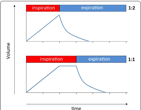
inflation, thereby leaving pressure and flow acceleration during inspiration identical between the corresponding
LVT and HVT 1:2 groups, as schematically illustrated
in Figure 1.
To generate baseline values at the beginning of mechan-ical ventilation, a group of mice referred to as nonventi-lated control (ctr) was anesthetized and operated as outlined earlier. After an identical recruitment maneuver, ctr mice were ventilated for 5 minutes with adjustments
identical to those of the LVT1:2 mice. After measurement
of baseline lung functions and hemodynamics, the experi-ment was terminated.
PEEP of 2 cmH2O and an FiO2 of 0.5 were applied
throughout the experiments in all LVTand HVTgroups.
After 235 minutes of mechanical ventilation (MV), the I: E ratio was switched to 1:2 in all ventilated groups, and
the inspired oxygen fraction was increased to an FiO2of
1.0. After 240 minutes of MV, mice were killed by rapid exsanguination via the carotid artery catheter.
An alternative end point was defined as decrease of mean arterial blood pressure below 40 mm Hg, as this safely predicts death in this model. The I:E ratio was then switched to 1:2, and the inspired oxygen fraction
was increased to an FiO2 of 1.0. After a further 5
mi-nutes of MV, mice were killed by exsanguination via the carotid artery catheter.
To exclude dynamic hyperinflation in the HVT 1:1
group, an additional set of animals was ventilated
ac-cording to the HVT 1:2 ventilation pattern for 30
mi-nutes, and then the I:E ratio was increased to 1:1 for 30 minutes. This procedure was repeated. Mean airway pressure and dynamic compliance were recorded.
Lung function
After the initial recruitment maneuver, dynamic elas-tance, resiselas-tance, and compliance were measured by using a forced oscillation technique. Measurements were repeated every 10 minutes throughout the experiment. In addition, static-compliance values were determined after exsanguination.
Blood gas analyses
Blood samples were analyzed for paO2with a blood-gas
analyzer (ABL-800; Radiometer, Copenhagen, Denmark).
The P/F ratio was calculated as P/F = paO2/FiO2.
Oxy-genation Index was calculated as OI = mean airway
pres-sure × FiO2/paO2.
Lung permeability
After exsanguination, the left-stem bronchus was ligated, and bronchoalveolar lavage (BAL) of the right lung was
performed twice with 400 μl PBS each. From each BAL
fluid (BALF) portion, 250μl was pooled, and BALF
albu-min concentration, as well as plasma albualbu-min concentra-tion, were determined with ELISA (Bethyl Laboratories Inc., Montgomery, AL, USA). Permeability was assessed by calculating the albumin BALF/plasma ratio.
qRT-PCR
Lungs were flushed. RNA was extracted with TRIzol (Ambion; Life Technologies, Darmstadt, Germany) treat-ment of lung homogenates and reverse-transcribed by using a high-capacity reverse transcription kit (Applied Biosystems, Life Technologies, Darmstadt, Germany). Quantitative PCR (qRT-PCR) was performed on ABI 7300 by using TaqMan gene-expression assays (Applied Biosystems). The PCR conditions included initial de-naturation in one cycle of 2 minutes at 50°C and 10 mi-nutes at 95°C, followed by 40 cycles of 15 seconds at 95°C, 20 seconds at 60°C, and 1 minute at 72°C. The in-put was normalized to the average expression of GAPDH. Primer and probe sequences are provided in Table S1 in Additional file 1.
Leukocytes in BAL fluid and blood
Leukocytes in BALF were differentiated with flow cy-tometry, according to their side-scatter/forward-scatter characteristics, and CD45, Gr-1, and F4-80 expression (FACSCalibur; BD, Heidelberg, Germany). Blood leuko-cytes were quantified and differentiated with flow cy-tometry by using TruCount-Tubes according to cellular side-scatter/forward-scatter characteristics and CD45, Gr-1, and CD3 expression.
Quantification of cytokines
[image:3.595.56.291.480.664.2]Cytokines were quantified from total protein of flushed and homogenized left lungs and from plasma samples by Figure 1Schematic graphic of the respiratory cycle during I:E
using the multiplex cytokine assay technique (BioRad, Hercules, CA, USA).
Histopathology
Lung samples were fixed in 4% formaldehyde solution and routinely embedded in paraffin. The 5-μm-thick sec-tions were cut, dewaxed, and stained with hematoxylin and eosin (H&E) or periodic acid-Schiff (PAS). Histo-pathology was performed by two European College of Veterinary Pathologists (ECVP) board-certified patholo-gists, who were blinded to the study groups.
Data analyses
Data are expressed as box-and-whisker plots, or col-umns (mean ± SEM). For comparison between groups, a
Mann–Whitney U test was performed. P values <0.05
were considered statistically significant. For survival ana-lyses, a log rank test was applied.
Results
Increasing inspiratory time and I:E ratio did not result in dynamic hyperinflation
To rule out relevant dynamic hyperinflation, HVTanimals
were ventilated with an alternating I:E ratio (1:2 versus 1:1) in 30-minute intervals for 120 minutes, and mean
air-way pressure and dynamic compliance (Cdyn) were
mea-sured. Mean airway pressure remained stable during the specific interval of MV (see Figure S1A in Additional file 2). The higher but stable mean airway pressure
dur-ing HVT 1:1 is explained by the increased inspiratory
time during HVT 1:1 (see Figure S1A in Additional file
2). Cdyn remained stable during MV, irrespective of the
adjusted I:E ratio (see Figure S1B in Additional file 2).
Thus, HVT 1:1 ventilation did not lead to relevant
dy-namic hyperinflation.
Increasing the inspiratory time and I:E ratio during MV increased mortality in VILI
All mice of the low tidal-volume groups ventilated with
an I:E ratio of either 1:2 or 1:1 (LVT1:2; LVT1:1) and of
the high tidal-volume group ventilated with an I:E ratio of 1:2 survived the procedures. Increasing the I:E ratio in
the HVTgroup to 1:1 resulted in premature termination
of the experiment in 13 of 14 mice because of dropping of mean arterial blood pressure below 40 mm Hg (alter-native end point), corresponding to a 92.1% mortality in
the HVT1:1 group (Figure 2).
Increasing the inspiratory time und I:E ratio increased lung injury
Lungs from ctr, LVT1:2, and LVT1:1 showed no
macro-scopic or histologic signs of lung injury. HVT 1:2
venti-lated mice exhibited significant histopathologic signs of lung injury, whereas only distinct signs of injury were
seen macroscopically. HVT 1:1 led to dramatic
macro-scopic and histopathologic lesions. In both HVTgroups
but not in the controls, the lung architecture was com-promised by severe alveolar collapse and emphysema. Histopathology revealed severe perivascular edema, damage of the alveolar walls with desquamation of al-veolar epithelial cells type I and formation of hyaline membranes, increasing numbers of intraalveolar cells (neutrophils and macrophages) and occasional necrosis of bronchiolar epithelium. Severe lung lesions were ob-served histologically on HE-stained tissues, with no dif-ferences seen between both groups (Figure 3 and Figure S2 in Additional file 3).
Periodic acid-Schiff (PAS) reaction clearly visualized hyaline membranes diffusely distributed throughout the lung parenchyma, indicative of marked damage of the
al-veolar membrane. However, in HVT 1:2 lungs, hyaline
membranes appeared only occasionally as continuous thin layers on the alveolar surface, while lungs of the
HVT 1:1 group had thicker hyaline membranes, which
commonly completely covered the surface of dilated al-veoli (Figure 4). Because pulmonary vascular leakage is a hallmark of ARDS and VILI, we quantified lung perme-ability by measuring the albumin concentration in bron-choalveolar lavage fluid (BALF) and plasma and by calculating the BALF/plasma albumin ratio. Compared
with ctr mice, LVT1:2 and LVT1:1 did not result in
in-creased permeability. In contrast, HVT 1:2 mice showed
a trend toward increased permeability compared with
ctr and LVTgroups, whereas HVT1:1 evoked a dramatic
increase in pulmonary vascular permeability (Figure 5).
Increasing the inspiratory time and I:E ratio reduced oxygenation capacity in VILI
Peripheral oxygen saturation was measured continuously throughout the experiment. Partial pressure of oxygen in
0 60 120 180 240
0 25 50 75 100
***
HVT 1:1 LVT 1:2 LVT 1:1 HVT 1:2
time [min]
[image:4.595.306.540.89.210.2]Survival [%]
arterial blood and mean airway pressure were measured at the end of the experiment, and P/F ratio as well as
oxygenation index (OI) were calculated. Whereas LVT
1:2, LVT1:1, and HVT1:2 groups showed stable
oxygen-ation regarding SpO2 and P/F throughout the
experi-ment (Figure 6A,B), the oxygenation index implied a
reduced oxygenation capacity in HVT 1:2 mice
com-pared to ctr and LVT groups (Figure 6C). HVT 1:1
resulted in severe impairment of oxygenation compared
with ctr, LVT, and HVT1:2 groups (Figure 6A to C).
Increasing the inspiratory time and I:E ratio deteriorated lung function in VILI
Dynamic elastance was quantified every 10 minutes.
While both LVT groups showed stable elastance during
[image:5.595.58.540.89.255.2]the experiment, a slight increase over time in the HVT
Figure 3Increasing the inspiratory time and I:E ratio during MV increased VILI.Mice were mechanically ventilated for 4 hours with either low tidal volume (LVT9 ml/kg) or high tidal volume (HVT34 ml/kg) and an inspiratory/expiratory ratio of 1:2 or 1:1, respectively. An alternative end point was defined as decreasing of mean arterial blood pressure below 40 mm Hg, as this predicts death with certainty in this model. Controls (ctr) were subjected to LVT1:2 ventilation only during operation and were killed before the 4-hour ventilation protocol started. Ctr and LVTgroups showed no signs of lung injury macroscopically. HVT1:2 revealed only subtle macroscopic signs of injury, whereas the HVT1:1 group showed massive edema formation. Representative images from 13 to 14 animals per group are shown.
[image:5.595.58.540.447.626.2]1:2 group, and a strong increase in the HVT 1:1 group
were observed (Figure 7A). Dynamic and static compli-ance at the respective end points of the experiment
showed impaired compliance in the HVT 1:2 compared
with LVT 1:2 mice. HVT 1:1 led to a dramatic decrease
in lung compliance (Figure 7B,C).
Increasing the inspiratory time and I:E ratio: impact on hemodynamics and markers of tissue perfusion
HVTanimals were ventilated with an alternating I:E ratio
(1:2 versus 1:1) in 30-minute intervals for 120 minutes, and mean arterial blood pressure was measured. Changing of the I:E ratio had no impact on mean arterial blood pressure (see Figure S3A in Additional file 4). In animals ventilated
for 4 hours (LVT1:2, LVT1:1, HVT1:2, and HVT1:1),
cu-mulative urine output and blood lactate levels at the re-spective experimental end points were quantified. No difference in urine output between the groups was evident,
whereas HVT1:1 revealed slightly higher lactate levels than
the HVT1:2 group (see Figure S3 B, C in Additional file 4).
Increasing the inspiratory time and I:E ratio exacerbated the inflammatory response in VILI
We measured transcription of the proinflammatory cyto-kines IL-1β, IL-6, KC, and MCP-1 (Figure 8A) and protein ctr 1:2
T
LV
1:1
T
LV
1:2
T
HV
1:1
T
HV
0 25 50 75 100
125
**
Pulmonary Permeability
[albumin
BALF
/albumin
Plasma
[image:6.595.58.291.88.253.2]]
Figure 5Increasing the inspiratory time and I:E ratio during MV increased pulmonary permeability in VILI.Mice were mechanically ventilated for 4 hours with either low tidal volume (LVT9 ml/kg) or high tidal volume (HVT34 ml/kg) and an inspiratory/ expiratory ratio of 1:2 or 1:1, respectively. An alternative end point was defined as decreasing of mean arterial blood pressure below 40 mm Hg, which predicts death with certainty in this model. Controls (ctr) were subjected to LVT1:2 ventilation only during operation and were killed before the 4-hour ventilation protocol started. Albumin concentrations in bronchoalveolar lavage fluid (BALF) and plasma were determined. An increased albumin BALF/plasma ratio indicated enhanced lung permeability.n= 5 to 6 each group; ** < 0.01.
A
C B
0 60 120 180 240
50 60 70 80 90 100
HVT 1:2
HVT 1:1
LVT 1:2 LVT 1:1
time [min]
SpO
2
[%]
ctr 1:2
T
LV
1:1
T
LV
1:2
T
HV
1:1
T
HV
0 100 200 300 400 500
***
paO
2
/FiO
2
[mmHg]
ctr 1:2
T
LV
1:1
T
LV
1:2
T
HV
1:1
T
HV
0 10 20 30 40
50
***
***
***
Oxygenation Index [cmH
2
O/mmHg]
Figure 6Increasing the inspiratory time and I:E ratio reduced oxygenation capacity in VILI.Mice were mechanically ventilated for 4 hours with either low tidal volume (LVT9 ml/kg) or high tidal volume (HVT34 ml/kg) and an inspiratory/expiratory ratio of 1:2 or 1:1, respectively. An alternative end point was defined as decreasing of mean arterial blood pressure below 40 mm Hg, which predicts death with certainty in this model. Controls (ctr) were subjected to LVT1:2 ventilation only during operation and were killed before the 4-hour ventilation protocol started.
[image:6.595.59.537.395.656.2]concentrations of IL-1β, IL-6, KC, and MCP-1 in lung
ho-mogenates (Figure 8B). HVT 1:2 increased IL-1β, IL-6,
KC, and MCP-1 mRNA expression compared with ctr and
LVT mice. Expression of most of these proinflammatory
mediators was further increased in the HVT1:1.
LVT1:2 and LVT1:1 resulted in a certain increment of
BALF neutrophils compared with ctr mice. In line with the elevation of proinflammatory cytokines in the lung,
HVT1:2 led to a further increase of neutrophils, whereas
HVT1:1 was associated with a significant neutrophil
in-filtration of the alveolar space (Figure 9A).
Furthermore, HVT 1:1 exclusively resulted in an
in-creased number of blood neutrophils, indicating a sys-temic inflammatory response (Figure 9B).
Discussion
We provide strong evidence that increasing the I:E ratio during mechanical ventilation can aggravate VILI, indi-cating that not only the absolute lung stress and strain but also the time in which the lung is exposed to stress and strain (the Time × Stress and strain product) may affect the harm of mechanical ventilation.
VILI impairs survival of ARDS patients [2,3]. Besides cyclic opening and closing of lung during tidal ventila-tion, high airway pressures and high tidal volumes have been identified as main drivers of VILI. More precisely,
not absolute airway pressure but the transpulmonary pres-sure termed lung stress, and not the absolute tidal volume, but its relation to the FRC, termed lung strain, are mech-anical determinants of VILI [5]. Besides the amount of lung opening and closing that is correlated with ARDS mortality [11], the concept of intraparenchymal stress raisers during mechanical ventilation may have significant impact on the development of lung injury due to mechan-ical ventilation in the ARDS patient [5,12].
Recent clinical trials revealed that VILI is particularly relevant in patients with severe ARDS, and therefore, optimizing our ventilation strategies especially for those patients is desirable [13,14].
Lung stress and strain are not equally distributed throughout the respiratory cycle under MV, obviously being higher during inspiration than during expiration. The current study now provides evidence that not only absolute lung stress and strain but also increasing lung stress and strain in relation to the cycle time by
prolong-ing the inspiratory time (ti) and increasing the I:E ratio
ag-gravate VILI. This is in line with the theory of weighted lung strain during MV by Carioni and colleagues [9].
Physical forces during mechanical ventilation are sensed by the lung and induce a biochemical response character-ized by inflammation and endoepithelial permeability, referred to as biotrauma [1,15,16]. Therefore we assessed
A
C B
0 60 120 180 240
0 50 100 150 200
HVT 1:2
HVT 1:1
LVT 1:2
LVT 1:1
time [min]
E [cmH
2
O/L]
ctr 1:2
T
LV
1:1
T
LV
1:2
T
HV
1:1
T
HV
0.00 0.01 0.02 0.03 0.04 0.05 0.06 0.07
0.08
***
***
static Compliance
(mL/cmH
2
O)
ctr 1:2
T
LV
1:1
T
LV
1:2
T
HV
1:1
T
HV
0.00 0.01 0.02 0.03 0.04 0.05
***
***
dynamic Compliance
[mL/cmH
2
[image:7.595.60.538.88.342.2]O]
Figure 7Increasing the inspiratory time and I:E ratio deteriorated lung function in VILI.Mice were mechanically ventilated for 4 hours with either low tidal volume (LVT9 ml/kg) or high tidal volume (HVT34 ml/kg) and an inspiratory/expiratory ratio of 1:2 or 1:1, respectively. An alternative end point was defined as decreasing of mean arterial blood pressure below 40 mm Hg, which predicts death with certainty in this model. Controls (ctr) were subjected to LVT1:2 ventilation only during operation and were killed before the 4-hour ventilation protocol started.
lung permeability, detailed lung histology and markers of pulmonary inflammation. Even after the short observa-tional time of 4 hours, we detected a significant impact of
the increased tiand I:E ratio on pulmonary cytokine levels,
pulmonary neutrophil influx, systemic neutrophil counts, lung permeability, and histological signs of lung injury. KC and MCP-1 are chemotactic signals for neutrophils, which
contribute to the development of VILI [17,18]. IL-1βwas
shown to induce endothelial permeability and aggravate
VILI [19]. IL-6 is upregulated under mechanical ventila-tion and, although its exact role in VILI remains contro-versial, IL-6 levels are correlated with organ failure and outcome in ARDS [20-23]. Considering our findings, it is tempting to speculate that not only the absolute physical force or stretch but also its duration is sensed and responded to by the ventilated lung. This implies that mechanotransduction is increased, although the absolute amount of energy added to the system is kept constant, as
mRNA
B
Protein
A
β
β
ctr 1:2
T
LV
1:1
T
LV
1:2
T
HV
1:1
T
HV
0 50 100 150 200
**
***
relative KC expression
0 50 100 150 200 250 300 350
**
***
relative MCP-1 expression
0 100 200 300 400 500 600
*
relative IL-6 expression
0.0 2.5 5.0 7.5 10.0
relative
IL-β expression
0 2.5×10³
5.0×10³
7.5×10³
1.0×10⁴
1.3×10⁴
p=0.0541
IL-1ß [
μ
g/
μ
L]
0 100 200 300
*
***
5.0×10³
1.0×10⁴
1.5×10⁴
IL-6 [
μ
g/µL]
0 1.0×10⁴
2.0×10⁴
3.0×10⁴
4.0×10⁴
*
p=0.0593
MCP-1 [
μ
g/
μ
L]
ctr 1:2
T
LV
1:1
T
LV
1:2
T
HV
1:1
T
HV
0 1.0×10⁴
2.0×10⁴
3.0×10⁴
4.0×10⁴
*
**
KC [
μ
g/
μ
[image:8.595.60.537.87.520.2]l]
pressure and volume remain unchanged throughout the
inspiratory hold in the HVT1:1 group.
To test the hypothesis of this study, we implemented severely injurious ventilation in mice. The tidal volume of 34 ml/kg was extraordinarily high compared with the standard of lung-protective ventilation with 6 ml/kg in humans with ARDS. At first view, this might outrange the stress and strain applied during MV in ARDS pa-tients. However, residual capacity in ARDS lungs is
se-verely reduced, which is referred to as the“baby lung”of
ARDS patients [24]. Notably, the sicker the patient and the lower the oxygenation capacity becomes, the greater is the reduction of the residual capacity and the intention to increase the relative portion of inspiratory time to improve oxygenation. As intensivists do not rou-tinely quantify residual capacity at the bedside, we do not know how much lung strain is generated during MV, despite limiting the tidal volume to 6 ml/kg, espe-cially in very severe ARDS. Further, ARDS is character-ized by a high grade of tissue inhomogeneity, in which open, atelectatic, and collapsed but recruitable lung areas coexist, which locally results in lung stress exceed-ing the measured airway pressure by far [5].
Loss and inactivation of surfactant, a hallmark in ARDS, further aggravate local trauma [1]. Thus, applying high tidal volumes in healthy mouse lungs constitutes a reasonable experimental approach. Further, the currently used model of VILI meets the ATS criteria on lung in-jury in animals, including the evidence of inflammation, microscopic tissue injury, alteration of alveolar barrier function, and impaired oxygenation [25]. Vice versa, the
observation that prolonging tiin the LVTgroups did not
increase detectable lung injury in healthy mice does not argue for the safety of an I:E ratio increase in lung-protective ventilation of ARDS patients.
In this study, it was highly important to control prop-erly factors that might significantly bias the results. (a) Dynamic hyperinflation would increase intrinsic PEEP, which consecutively enhances residual volume and shifts tidal volume upward on the pressure/volume curve, eventually above the upper inflection point, resulting in augmented absolute lung stress and strain. Thus, we
ad-justed respiratory rates in both HVT groups to 70 per
minute to exclude dynamic hyperinflation in the HVT I:
E 1:1 group. (b) To keep dynamics of lung inflation
iden-tical between 1:1 and 1:2 groups, we prolonged ti by
adding an inspiratory hold. This excluded that a differ-ence in pressure acceleration during inspiration or a dif-ference of the total inflation time biased the results of the study. (c) Expiration was most probably similar in
the HVT 1:1 and HVT 1:2, and in the LVT 1:1 and LVT
1:2 groups, respectively. Expiration is a passive process starting after the opening of the expiratory valve with end-inspiratory pressure being the driving force, which was similar in the respective groups. As dynamic hyper-inflation could be excluded, exhalation was complete. (d)
Respiratory rate, PEEP and FiO2 were identical in the
HVT and the LVT groups, respectively and anesthesia
and operation procedures were identical in all groups. Intrathoracic pressure directly affects cardiac function (for an excellent review, see [26,27]), and increased intrathoracic pressure due to increased I:E ratio may decrease cardiac output [28,29]. Reduction of venous re-turn seems to be the central mechanism reducing car-diac output by high intrathoracic pressure, which can be ameliorated by sufficient fluid support. In our study, mice received liberal fluid support to minimize reduc-tion of cardiac output. Blood pressure and urine output were not affected by increased I:E ratio. Nevertheless,
lactate levels were slightly elevated in HVT1:1 compared
A B
ctr 1:2
T
LV
1:1
T
LV
1:2
T
HV
1:1
T
HV 0
5 10 15 20 25 30 35
*
p=0.0823
##
PMN [%]
ctr 1:2
T
LV
1:1
T
LV
1:2
T
HV
1:1
T
HV 0
10 20 30 40 50
60
**
[image:9.595.57.540.89.226.2]PMN [%]
with HVT 1:2 mice. Thus a certain reduction of cardiac
output cannot be excluded. However, circulatory failure and resultant shock as cause of the premature death of
the HVT1:1 animals would have resulted in significantly
higher lactate levels, lower blood pressure, and particu-larly in reduction of urine output.
Thus the data provided here exclude profound hemo-dynamic deterioration as the underlying mechanism for the
devastating outcome of the HVT1:1 group.
Although the data are conclusive and the results are clear, one can only speculate whether the findings can be translated to patients with ARDS. However, the study clearly emphasizes that for further improvement of lung-protective ventilation strategies, a deeper understanding of central factors for VILI is mandatory. In this regard, ex-perimental studies like the present one are essential.
Conclusion
The study design applied aimed to provide a proof of con-cept. The data show that beyond stress and strain, the time in which the lung is exposed to stress and strain (the Time × Stress and strain product) has dramatic impact on VILI. Therefore, it seems reasonable to minimize the Time × Strain product during MV. Particularly, increasing the I:E ratio should be critically revised in patients with ARDS.
Key messages
Increasing inspiratory time und thereby the I:E ratio aggravates VILI.
Beyond stress and strain, the time during which the lung is exposed to stress and strain (the Time × Stress and strain product) has dramatic impact on VILI.
Additional files
Additional file 1: Table S1.Providing primer and probe sequences used for qRT-PCR.
Additional file 2: Figure S1.Giving mean airway pressure and dynamic compliance measurements under alternating I:E ratios (1:2 and 1:1) during HVTventilation.
Additional file 3: Figure S2.Providing HE images of all experimental groups.
Additional file 4: Figure S3.Providing hemodynamic data.
Abbreviations
ARDS:Acute respiratory distress syndrome; BAL: bronchoalveolar lavage; BALF: bronchoalveolar lavage fluid; Cdyn: dynamic compliance; Ctr: control; ELISA: enzyme-linked immunosorbent assay; FiO2: inspiratory fraction of oxygen; FRC: functional residual capacity; HVT: high tidal volume; I:E ratio: inspiratory-to-expiratory ratio; IL-1β: interleukin-1 beta; IL-6: interleukin-6; KC: keratinocyte-derived chemokine; LVT: low tidal volume; MCP-1: monocyte chemoattractant protein-1; MV: mechanical ventilation; OI: oxygenation index; paO2: partial pressure of oxygen; PBS: phosphate-buffered saline; PEEP: positive end-expiratory pressure; qRT-PCR: quantitative
reverse transcription polymerase chain reaction; ti: inspiratory time; VILI: ventilator-induced lung injury; VT: tidal volume.
Competing interests
The authors declare that they have no competing interests.
Authors’contributions
HMR and MW planned and supervised the study, analyzed the data, and drafted the manuscript. MF and KH performed animal experiments, flow cytometry and cytokine assays, and critically revised the manuscript for important intellectual content. OK and ADG performed histology and critically revised the manuscript for important intellectual content. JN, SMW, and BO performed RT-qPCR analyses and critically revised the manuscript for important intellectual content. NS was involved in the study design and participated in drafting the manuscript. All authors read and approved the final version of the manuscript.
Acknowledgements
We thank Marfa Polikarpova for skillful technical assistance. The work is part of the doctoral thesis of Matthias Felten.
Funding
This study was supported by grants from the Deutsche
Forschungsgemeinschaft to HMR (SFB-TR84 C7), MW (SFB-TR84 C3, C6, and DFG OP 86/7-1), NS (SFB-TR84 B1), BO (SFB-TR84 A1, A5), ADG (SFB-TR84 Z1b). The funding sources had no influence on study design or publication.
Author details
1Department of Infectious Diseases and Pulmonary Medicine, Charité
-Universitätsmedizin Berlin, Berlin, Germany.2Department of Veterinary Pathology, Freie Universität Berlin, Berlin, Germany.
Received: 6 October 2014 Accepted: 20 January 2015
References
1. Verbrugge SJC, Lachmann B, Kesecioglu J. Lung protective ventilatory strategies in acute lung injury and acute respiratory distress syndrome: from experimental findings to clinical application. Clin Physiol Funct Imaging. 2007;27:67–90.
2. Amato MB, Barbas CS, Medeiros DM, Magaldi RB, Schettino GP, Lorenzi-Filho G, et al. Effect of a protective-ventilation strategy on mortality in the acute respiratory distress syndrome. N Engl J Med. 1998;338:347–54.
3. The Acute Respiratory Distress Syndrome Network. Ventilation with lower tidal volumes as compared with traditional tidal volumes for acute lung injury and the acute respiratory distress syndrome. N Engl J Med. 2000;342:1301–8.
4. Wolthuis EK, Vlaar APJ, Choi G, Roelofs JJTH, Juffermans NP, Schultz MJ. Mechanical ventilation using non-injurious ventilation settings causes lung injury in the absence of pre-existing lung injury in healthy mice. Crit Care. 2009;13:R1.
5. Gattinoni L, Carlesso E, Caironi P. Stress and strain within the lung. Curr Opin Crit Care. 2012;18:42–7.
6. Kim WH, Hahm TS, Kim JA, Sim WS, Choi DH, Lee EK, et al. Prolonged inspiratory time produces better gas exchange in patients undergoing laparoscopic surgery: a randomised trial: inspiratory time and gas exchange. Acta Anaesthesiol Scand. 2013;57:613–22.
7. Lee SM, Kim WH, Ahn HJ, Kim JA, Yang MK, Lee CH, et al. The effects of prolonged inspiratory time during one-lung ventilation: a randomised controlled trial. Anaesthesia. 2013;68:908–16.
8. Casetti AV, Bartlett RH, Hirschl RB. Increasing inspiratory time exacerbates ventilator-induced lung injury during high-pressure/high-volume mechanical ventilation. Crit Care Med. 2002;30:2295–9.
9. Caironi P, Langer T, Carlesso E, Protti A, Gattinoni L. Time to generate ventilator-induced lung injury among mammals with healthy lungs: a unifying hypothesis. Intensive Care Med. 2011;37:1913–20.
11. Caironi P, Cressoni M, Chiumello D, Ranieri M, Quintel M, Russo SG, et al. Lung opening and closing during ventilation of acute respiratory distress syndrome. Am J Respir Crit Care Med. 2010;181:578–86.
12. Mead J, Takishima T, Leith D. Stress distribution in lungs: a model of pulmonary elasticity. J Appl Physiol. 1970;28:596–608.
13. Papazian L, Forel J-M, Gacouin A, Penot-Ragon C, Perrin G, Loundou A, et al. Neuromuscular blockers in early acute respiratory distress syndrome. N Engl J Med. 2010;363:1107–16.
14. Guérin C, Reignier J, Richard J-C, Beuret P, Gacouin A, Boulain T, et al. Prone positioning in severe acute respiratory distress syndrome. N Engl J Med. 2013;368:2159–68.
15. Han B. Ventilator-induced lung injury: role of protein-protein interaction in mechanosensation. Proc Am Thorac Soc. 2005;2:181–7.
16. Tremblay LN, Slutsky AS. Ventilator-induced lung injury: from the bench to the bedside. Intensive Care Med. 2006;32:24–33.
17. Kawano T, Mori S, Cybulsky M, Burger R, Ballin A, Cutz E, et al. Effect of granulocyte depletion in a ventilated surfactant-depleted lung. J Appl Physiol. 1987;62:27–33.
18. Müller-Redetzky HC, Suttorp N, Witzenrath M. Dynamics of pulmonary endothelial barrier function in acute inflammation: mechanisms and therapeutic perspectives. Cell Tissue Res. 2014;355:657–73.
19. Frank JA, Pittet J-F, Wray C, Matthay MA. Protection from experimental ventilator-induced acute lung injury by IL-1 receptor blockade. Thorax. 2008;63:147–53.
20. Wolters PJ, Wray C, Sutherland RE, Kim SS, Koff J, Mao Y, et al. Neutrophil-derived IL-6 limits alveolar barrier disruption in experimental ventilator-induced lung injury. J Immunol. 2009;182:8056–62.
21. Goldman JL, Sammani S, Kempf C, Saadat L, Letsiou E, Wang T, et al. Pleiotropic effects of interleukin-6 in a“two-hit”murine model of acute respiratory distress syndrome. Pulm Circ. 2014;4:280–8.
22. Gurkan OU, He C, Zielinski R, Rabb H, King LS, Dodd-o JM, et al. Interleukin-6 mediates pulmonary vascular permeability in a two-hit model of
ventilator-associated lung injury. Exp Lung Res. 2011;37:575–84. 23. Meduri GU, Headley S, Kohler G, Stentz F, Tolley E, Umberger R, et al.
Persistent elevation of inflammatory cytokines predicts a poor outcome in ARDS: plasma IL-1 beta and IL-6 levels are consistent and efficient predictors of outcome over time. Chest. 1995;107:1062–73.
24. Gattinoni L, Pesenti A. The concept of“baby lung”. Intensive Care Med. 2005;31:776–84.
25. Matute-Bello G, Downey G, Moore BB, Groshong SD, Matthay MA, Slutsky AS, et al. An official American Thoracic Society workshop report: features and measurements of experimental acute lung injury in animals. Am J Respir Cell Mol Biol. 2011;44:725–38.
26. Feihl F, Broccard AF. Interactions between respiration and systemic hemodynamics, Part I: basic concepts. Intensive Care Med. 2009;35:45–54. 27. Feihl F, Broccard AF. Interactions between respiration and systemic
hemodynamics, Part II: practical implications in critical care. Intensive Care Med. 2009;35:198–205.
28. Chan K, Abraham E. Effects of inverse ratio ventilation on cardiorespiratory parameters in severe respiratory failure. Chest. 1992;102:1556–61. 29. Meinhardt JP, Friess U, Bender HJ, Hirschl RB, Quintel M. Relationship
among cardiac index, inspiration/expiration ratio, and perfluorocarbon dose during partial liquid ventilation in an oleic acid model of acute lung injury in sheep. J Pediatr Surg. 2005;40:1395–403.
Submit your next manuscript to BioMed Central and take full advantage of:
• Convenient online submission
• Thorough peer review
• No space constraints or color figure charges
• Immediate publication on acceptance
• Inclusion in PubMed, CAS, Scopus and Google Scholar
• Research which is freely available for redistribution