Evaluation of non-palpable thyroid nodules by ultra
sound guided fine needle aspiration cytology
*
Khalid Ahmad Al-Sindi1, Mulazim Hussain Bukhari2#, Kanwal Saba3, Wajid Ali3, Madiha Arshad2, Nasir Raza Zaidi3
1
Department of Pathology, King Hamad University Hospital, Al Sayh, Bahrain 2
Department of Pathology King Edward Medical University, Lahore, Pakistan; #
Corresponding Author: drmhbukhari@yahoo.com, mulazim.hussain@gmail.com
3
Department of Radiology, Mayo Hospital/ King Edward Medical University, Lahore, Pakistan
Received 24 November 2012; revised 28 December 2012; accepted 9 January 2013
ABSTRACT
The aim of this study was to see the usefulness of fine needle aspiration by “the Bethesda sys- tem for reporting Thyroid Cytopathology” (TBS- RTC) for non palpable thyroid nodules through ultrasound-guidance for the evaluation and treat- ment planning of nonpalpable thyroid lesions. This study was conducted on 200 patients with non palpable thyroid nodules which are very low lying or felt on swallowing; in Department of Pa- thology and Radiology since January 2011 to June 2012. The patients were scanned and USG- FNAC was performed and reporting was done by “TBSRTC”. Of the 200 specimens 17 samples were nondiagnostic or unsatisfactory (Class I), 145 samples were benign (Class II), 20 samples were showing Atypical of Undetermined Signifi- cance (AUS) or Follicular Lesion of Undetermin- ed Significance (FLUS); (Class III), 6 were show- ing follicular neoplasm or suspicious for a fol- licular neoplasm (Class IV), 7 samples were sus- picious for malignancy (Class V) and 5 samples were positive for malignancy (Class VI). On com- parison of ultrasound guided FNAC with histo- pathology the sensitivity for correct diagnosis was 93%, specificity was 86%, positive predic- tive value was 37%, negative predictive value was 99% and accuracy was 86%. USG-FNAC is a useful modality for the evaluation and treatment planning of nonpalpable thyroid lesions smaller than 5 mm in the maximum diameter. TBSRTC is the best method of reporting but class III and IV are the main pitfall of this system for reporting Thyroid Cytopathology and show high sensitiv- ity, specificity and accuracy.
Keywords: FNAC; USG; The Bethesda Classification; Follicular Adenoma; Follicular Carcinoma; Papillary Carcinoma; Medullary Carcinoma
1. INTRODUCTION
Thyroid nodules are becoming an epidemic problem in the world in the general population, however the thyroid malignancy is rare among these swellings [1-3].
Thyroid tumors are more common in females as com- pared to males with annual worldwide with male to fe- male ration 1:2.6 [2].
The larger thyroid swellings are not a problem to as- pirate and detect through biopsy but smaller or deeper nodules are difficult to diagnose early. These nodules may cause the danger of occult carcinoma. In the litera- ture diverse and contradictory approaches have been sug- gested for the management of non-palpable thyroid nod- ules [4].
Ultrasonography (US) is increasingly able to detect thyroid nodules, and the differentiation between malig- nant and benign nodules has been raising issues among both clinicians and patients, especially patients with non- palpable small nodules [5,6]. It is important to diagnose thyroid cancer at an early stage, because it may reduce the risk of disease recurrence and possible mortality. Fine-needle cytology appears to be a useful diagnostic tool also for nodules of less than 1 cm because the per- centage of unsatisfactory results is not related to the size of the nodule [7,8].
Study was conducted to evaluate a non-Palpable thy- roid nodule through Ultra sound guided thyroid fine ne- edle aspiration cytology (US-FNAC).
2. MATERIAL AND METHODS
*Competing interest: No competing interest to declare by any of the
ales) patients with an age range of 10 - 70 years, in the Department of Radiology, Nuclear Medicine and De- partment of Pathology since January 2011 to June 2012.
A written “informed consent” was taken from patients before performing Ultrasound guided fine needle aspira- tion Cytology (US-FNACs). The study was approved by ethical committee of the University. The FNACs and FNAB samples (Preoperated US-FNAC on admitted pa- tients) were collected from patients presented for a thy- roid nodule after evaluation by thyroid function tests and thyroid scans. Fine Needle biopsy or lobectomy was fol- lowed for histopathology, which was considered as a gold standard to compare the results of these FNAC with the final histopathological reports (Fine needle biopsies and lobotomies), therefore the patients were followed for their biopsies that underwent surgery at our Hospital or Fine needle biopsies.
2.1. Cytopathological Reporting Criteria
Bethesda method was used to report the cytopathology of thyroid aspirates as six classes [9,10].
1) Nondiagnostic or Unsatisfactory: Cyst fluid only virtually acellular specimen (obscuring blood, clotting artifact, etc).
2) Benign: Consistent with a benign follicular nodule (includes adenomatoid nodule, colloid nodule, etc) con- sistent with lymphocytic (Hashimoto) thyroiditis in the proper clinical context consistent with granulomatous (subacute) thyroiditis.
3) Atypica of Undetermined Significance or Follicular Lesion of Undetermined Significance.
4) Follicular Neoplasm or Suspicious for a Follicular Neoplasm Specify if Hürthle cell (oncocytic) type.
5) Suspicious for Malignancy: Suspicious for papillary carcinoma, Suspicious for medullary carcinoma, Suspi- cious for metastatic carcinoma, Suspicious for lymphoma.
6) Malignant: Papillary thyroid carcinoma, Poorly dif- ferentiated carcinoma, Medullary thyroid carcinoma, Un- differentiated (anaplastic) carcinoma, Squamous cell car- cinoma, Carcinoma with mixed features (specify), Me- tastatic carcinoma, Non-Hodgkin lymphoma.
2.2. Inclusion Criteria
1) Age 10 to 70 Years; 2) Both genders;
3) Preoperated US-FNAC on admitted patients; 4) Non palpable nodules size 5 mm - 10 mm in maxi- mum diameter (evaluated after thyroid scan).
2.3. Exclusion Criteria
1) Patients with already diagnosed thyroid lesions; 2) All toxic goiters confirmed by clinical evaluation.
2.4. Evaluation of Patients
Patients presenting with some clinical symptoms re- lated to thyroids were performed scan, thyroid function tests and ultrasonography. Measure serum TSH in the initial evaluation of a patient with a thyroid nodule. If the serum TSH is subnormal, a radionuclide thyroid scan should be performed using either technetium 99 m Tc pertechnetate or 123I. Patients with non palpable thyroid nodules (according to inclusion criteria) in the OPD and fulfilling the inclusion criteria were included in this study. Informed consent from all the patients included in the study was taken. All the patients were recorded for their demographic features, that is, age, sex, and address (for follow up). History of present illness with regard to symptoms and duration was recorded. They were exam- ined for the signs related to the solitary thyroid swelling. All routine investigations and serum T3, T4, and TSH levels were performed by radioimmunoassay (RIA), (nor- mal range of T3, 2.5 - 5.8 pmol/L, T4, 11.5 - 23.0 pmol/L, and TSH, 0.5 - 5.0 mU/L). Patients with thyroid swelling underwent thyroid scan. Thyroid swellings were marked through by nuclear department and then FNAC was per- formed [11].
2.5. USG Reporting Criteria for Thyroid Nodules
High-resolution thyroid ultrasonography (USG) will be used as a diagnostic modality for haunting the non- palpable thyroid nodules. These characteristics of thyroid nodules that would be studied included microcalcifica- tions, an irregular or microlobulated margins, marked hy- poechogenicity, a shape that was taller than it was wide and color flow pattern in Color Doppler ultrasound. The presence and absence of characteristics of nodules will be classified as having positive or negative findings. If even one of these USG features will be presented, the nodule will be classified as positive (malignant). If a nodule has none of the features described, it will be clas- sified as negative (benign). The final diagnosis of a le- sion as benign or malignant will be confirmed by fine needle aspiration cytology and biopsy [12-15] (Figure 1).
2.6. Method of USG FNAC
technique where the needle is moved up and down for a few seconds only by movement of the wrist of the op- erator and without initial aspiration under US guidance. In all cases, only one sampling by one puncture was taken from each nodule. After a sample was obtained, the specimen was mounted immediately onto a glass slide. We obtained four to six slides by performing two or three smears from each nodule. Specimens were fixed with 95% ethanol and were sent for pathological evaluation [11,16] (Figure 1).
3. RESULTS
The mean age of the patients was 33.35 ± 11.77 and male to female ratio was 1:4.5 and most of the patients were from younger group ranging from 21 - 40 (Table 1). Of the 200 specimens on FNACs, 17 samples (8.5%) were non-diagnostic or unsatisfactory for diagnosis (Class I), 145 (72.5%) were benign (Class II), 20 (10%) cases were of follicular neoplasm or suspicious for a fol- licular neoplasm (Class III), 6 (3%) smears were show- ing atypia of undetermined significance or follicular le- sion of undetermined significance (Class IV), 7 (3.5%) smears were of suspicious for malignancy and 5 (2.5%) cases were malignant lesions of thyroid (Tables 2, 4 and 5). There were 24 false positive cases from correct diag- nosis and only 1 smear was false negative when com- pared with histopathology biopsy (Table 3).
On histopathological examination of these smears, there were 15/200 (7.5%), cases malignant thyroid le- sions; 10/15 (66.6%) cases were papillary carcinomas, 4/15 (26.6%) cases were follicular carcinomas and 1/14 (6.6%) case was of medullary carcinomas (Table 4).
Of 168/200 (84%) benign specimens; there were 25/ 168 (15%) follicular adenomas, 119/158 (71%) speci- mens were multinodular goiters, 20/168 (12%) cases were of colloid goiter and 4/168 (2%) specimens were of Hashimoto thyroiditis. Of the Class III smears only 1 case (follicular carcinoma) was found malignant and 18 cases were of follicular adenomas on histopathology (Table 5).
The comparison of other classes is given in Table 2. On FNAC, the clear cut malignant smears were 5/200 (2.5%), 7/200 smears were showing suspicious smears, 6/200 and 20/200 smears created impact of undetermined morphology, while on histopathology, there were 15/200 (7.5%) specimens proved to be malignant (Tables 1 and 3). All the false positive smears were found follicular adenoma (Table 1, Figures 2-5).
On cytohistologic correlation, the sensitivity was 93%, specificity was 86%, positive predictive value was 37%, negative predictive value was 99% and accuracy was 86%. The false negative specimen was papillary thyroid carcinoma which was improperly aspirated from targeted nodules and was diagnosed as benign lesions (Tables 2 and 3).
Table 1. Distribution of subjects by age and sex.
Number1:4 Age
Male 36 Females 164
Percentage
10 - 20 2 8 10
21 - 30 12 69 81
31 - 40 10 56 66
41 - 50 6 17 23
51 - 60 4 9 13
61 - 70 2 5 7
Total 36 164 200
Mean ± SD 33.35 ± 11.77
Male to female ration 1:4.5
Table 2. Distribution of different lesions diagnosed by histopa-
thologists.
Group B FNA by USG Histopathology
Classes Numbers Benign Malignant
Non-diagnostic
or Unsatisfactory 17 (8.5%)
Not operated
Not operated
II Benign 145 (72.5%) 144 1 (PTC)
III
Follicular Neoplasm or Suspicious for a Follicular Neoplasm
20 (10%) 18
(FA) 2 (FTC)
IV
Atypia of Undetermined Significance or Follicular
Lesion of Undetermined Significance
6 (3%) 4 (FA) 2 (FTC)
V Suspicious for Malignancy 7 (3.5%) (FA) 2 5 (PTC)
VI Malignant 5 (2.5%) 0 4 (PTC) and 1 (MTC)
Total 200 168
(84%) 15 (7.5%)
Key PTC: papillary thyroid carcinoma, FA: follicular adenomas, MTC: medullary thyroid carcinoma, FTC: follicular thyroid carcinoma.
Table 3. Comparison of FNAC of with histopathology.
Cytopathology Histopathology
FNAC + ve 14 024 038
FNAC – ve 01 144 145
Total 15 168 183
Table 4. Distribution of malignant lesions on histopathology.
Malignant Lesions (n = 15: 7.5%)
Numbers Percentage
Papillary carcinoma 10 66.6%
Follicular carcinoma 4 26.6%
Medullary carcinoma 1 6.6%
Total 15 100
[image:4.595.57.286.101.202.2]Note: Non-palpable nodules have lower prevalence of malignancy as com- pared to palpable one in our country.
Table 5. Distribution of benign lesions on histopathology.
Benign Lesions
(n = 168: 84%) Numbers Percentage
Follicular Adenoma (20 = Follicular adenoma and 5 Hurthle cell lesions)
25 15%
Multinodular goiters 20 12%
Colloid goiters 119 71%
Hoshiomot’s thyroiditis 04 02%
Total 168 100
Note: Most of the non-palpable nodules were found benign (84%).
4. DISCUSSION
Ultrasound guided fine needle aspiration technique is anon-invasive, simple and an accurate modality to evalu- ate a non-palpable thyroid nodular nodule. The American Thyroid Association (ATA) guidelines suggest that, in general, only nodules larger than 10 mm in diameter should be evaluated as the nodules having the potential to represent a clinically significant cancer [17]. Some- times occult carcinoma may arise from the nodules less than 10 mm therefore a careful evaluation is necessary from the patients with these smaller lesions [18,19].
In this study, the sensitivity and negative predictive value of performing an US-FNAC for the diagnosis of non-palpable thyroid nodules were high as compared
Figure 1. (A)Photograph of the patients, USG and needle po-
[image:4.595.56.288.251.372.2]sitions for FNAC of thyroid lesions; (B) USG picture showing needle entry in the thyroid nodule.
Figure 2. Photomicrograph of (A) (H&E 20×), The Bethesda
System for reporting Thyroid Cytopathology (TBSRTC) Class II smear with colloid back ground, macrophages and follicular cells (TB); (B) On histopathology (H&E 20×) of colloid goiter; (C) TBSRTC Class II smear, with hurthle cells and lympho- cytes (H&E 20×); (D) Hashimoto’s thyroiditis.
Figure 3. Photomicrograph of (A) (H&E 20×), The Bethesda
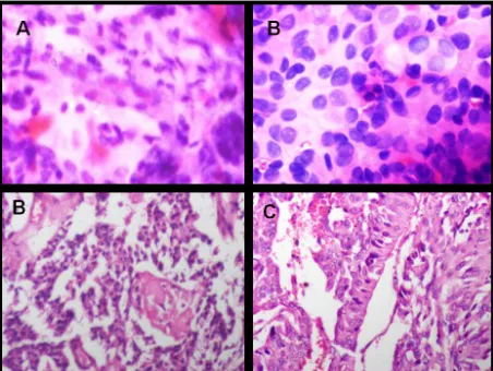
system for reporting Thyroid Cytopathology (TBSRTC) Class III smear atypical arrangement of follicular cells (long arrow) and few hurtle cells (small arrow); (B) On histopathology (H&E 20×) Follicular Carcinoma with vascular invasion (arrow); (C) TBSRTC Class V (Suspicious for malignancy) smear showing two of the features of papillary carcinoma (H&E 20×); (D) Papil- lary Carcinoma with ground glass nuclei and psamaoma bodies.
with values obtained for other studies; however, positive predictive value was low [16].
[image:4.595.309.538.330.500.2] [image:4.595.58.288.596.699.2]Figure 4. Photomicrograph of (A) (H&E 20×), The Bethesda system for reporting Thyroid Cytopathology (TBSRTC) Class IV smear follicular cells against scanty colloid; (B) On histo- pathology (H&E 20×) follicular carcinoma with capsular in- vasion (Arrow); (C) TBSRTC Class IV (follicular cells against scanty colloid) smear showing (H&E 20×); (D) Follicular ade- noma (False positive lesions).
Figure 5. Photomicrograph of (A) (H&E 20×), The Bethesda
system for reporting Thyroid Cytopathology (TBSRTC) Class VI (malignant) smear, showing round and spindle cells with giant cells; (B) On histopathology (H&E 20×) medullary car- cinoma; (C) TBSRTC Class VI (malignancy) smear (H&E 20×) showing all features of papillary carcinoma (overlaping, groves, inclusions,ground and smuged chromatic); (D) Tall columnar cell variant papillary carcinoma.
sensitivity was 93%, specificity was 86%, positive pre- dictive value was 37%, negative predictive value was 99% and accuracy was 86%. The false negative speci- men was papillary thyroid carcinoma which was im- properly aspirated from targeted nodules and was diag- nosed as benign lesions.
We categorized cytological results into five classes according to Bethesda description for thyroid cytology. Such categorization of FNAC smears results is necessary to allow clinicians to use cytology results to guide patient
management with specific reference to the need for thy- roidectomy [20].
With USG FNAC, the sensitivity was 93%, specificity was 86%, positive predictive value was 37% and nega- tive predictive value was 99%. According to our reports patients of Classes III-VI underwent thyroid surgery.
None of them were showing any distance metastasis or lymph node involvement and only 15/200 (7.5%) cases showed true positivity. There were only 10 cases of pap- illary thyroid carcinoma, while only 4 cases were suffer- ing from follicular carcinoma and one case was found to be medullary carcinoma. None of these cases were show- ing multifocality or bilateralism. These findings are also different from Kim et al. [16]. The justification of our findings is that we performed our study on 5 - 10 mm nodules. Our findings show that ignorance of these small lesions may lead to spread of carcinoma to distance or- gans while early management proves to better prognosis and long survival of these patients.
Our study showed that use of better reporting system (Bethesda methods) there was relatively low rate of non diagnostic or inadequate smears, allowing it to be useful method for determining the treatment plan for non pal- pable smaller thyroid nodules. In our study the incidence of adequate specimens was 183/200 (91.5%) with US- FNAC as compared to the Kim et al who found it 81% in his series of 201 patients [16].
5. CONCLUSION
USG-FNAC is a useful modality for the evaluation and treatment planning of nonpalpable thyroid lesions smaller than 5 mm in the maximum diameter. TBSRTC is the best method of reporting but class III and IV are the main pitfall of this system for reporting Thyroid Cy- topathology and shows high sensitivity, specificity and accuracy
6. JUSTIFICATION OF STUDY
Thyroid nodules are very common in the general population, but malignancy is relatively rare. The goal of the ultrasound guided evaluation of nonpalpable thyroid nodules by FNAC is the early detection of lesions and to save the overt spread of malignancies, while identifying and avoiding unnecessary surgery in those with benign, asymptomatic thyroid nodules. UGS is very helpful in locating the non-palpable thyroid nodules for FNAC and increases the quality of diagnosis which is helpful for the clinical management of such patients. Our study showed good sensitivity, specificity for US-FNAC with relatively low rate of non diagnostic or inadequate smears.
7. LIMITATIONS OF THE STUDY
[image:5.595.59.285.344.514.2]as cohort one on a large scale study. The high false rate was our limitation for reporting the Classes III and IV le- sions with Bethesda methods that needs more training of cytopathologists. We could not control some of our con- founding factors like underlying diffuse thyroid disease which influenced the outcome of the US-FNACs like thyroiditis and diffuse colloid disease but we excluded the hot nodules to overcome negative influences on our study. In our study we did not follow the patients with inadequate swellings and we could not convince the sur- geons for delaying the surgery for Class II smears (Be- nign). Classes III and IV are the main pitfall of The Be- thesda System for reporting Thyroid Cytopathology (TBSRTC).
8. IMPORTANCE OF THIS STUDY
The evaluations of these smaller nodules for the early diagnosis of thyroid cancers with ultrasound guided FNAC were helpful for detection of 7% malignant thy- roid lesions. These techniques will be fruitful to improve patient outcome and selection of treatment modalities (e.g., a lobectomy or total thyroidectomy or delaying the surgery option) and will save for distance metastasis and progressing to high grade anaplastic or poorly differenti- ated thyroid carcinomas.
9. ACKNOWLEDGEMENTS
We are thankful to the support of Radiological Department and Pa- thology Department of King Edward Medical University.
REFERENCES
[1] Anil, G., Hegde, A. and Chong, F.H. (2011) Thyroid nod-
ules: Risk stratification for malignancy with ultrasound and guided biopsy. Cancer Imaging, 11, 209-223.
[2] Ghassi, D. and Donato, A. (2009) Evaluation of the thy-
roid nodule. Postgraduate Medical Journal, 85, 190-195. doi:10.1136/pgmj.2008.072140
[3] Sclabas, G.M., Staerkel, G.A., Shapiro, S.E., Fornage, B.
D., Sherman, S.I., Vassillopoulou-Sellin, R., et al. (2003) Fine-needle aspiration of the thyroid and correlation with histopathology in a contemporary series of 240 patients.
The American Journal of Surgery, 186, 702-709. doi:10.1016/j.amjsurg.2003.08.015
[4] Accurso, A., Rocco, N., Palumbo, A. and Leone, F. (2005)
Usefulness of ultrasound-guided fine-needle aspiration cytology in the diagnosis of non-palpable small thyroid nodules. Tumori, 91, 355-357.
[5] Khurana, K.K., Richards, V.I., Chopra, P.S., Izquierdo, R.,
Rubens D. and Mesonero, C. (1998) The role of ultra- sonography-guided fine-needle aspiration biopsy in the management of nonpalpable and palpable thyroid nodules.
Thyroid, 8, 511-515. doi:10.1089/thy.1998.8.511
[6] Rago, T. and Vitti, P. (2008) Role of thyroid ultrasound in
the diagnostic evaluation of thyroid nodules. Best Prac-
tice & Research: Clinical Endocrinology & Metabolism,
22, 913-928. doi:10.1016/j.beem.2008.09.016
[7] Kwak, J.Y., Kim, E.K., Kim, M.J. and Son, E.J. (2009)
Significance of sonographic characterization for manag- ing subcentimeter thyroid nodules. Acta Radiologica, 50,
917-923. doi:10.1080/02841850903062724
[8] Frates, M.C., Benson, C.B., Charboneau, J.W., Cibas,
E.S., Clark, O.H., Coleman, B.G., et al. (2005) Manage-
ment of thyroid nodules detected at US: Society of radi- ologists in ultrasound consensus conference statement.
Radiology, 237, 794-800. doi:10.1148/radiol.2373050220
[9] Cibas, E.S. and Ali, S.Z. (2009) The Bethesda system for
reporting Thyroid Cytopathology. American Journal of
Clinical Pathology, 132, 658-665. doi:10.1309/AJCPPHLWMI3JV4LA
[10] Wong, L.Q. and Baloch, Z.W. (2012) Analysis of the
Bethesda system for reporting Thyroid Cytopathology and similar precursor Thyroid Cytopathology reporting
schemes. Advances in Anatomic Pathology, 19, 313-319.
doi:10.1097/PAP.0b013e3182666398
[11] Basharat, R., Bukhari, M.H., Saeed, S. and Hamid, T.
(2011) Comparison of fine needle aspiration cytology and
thyroid scan in solitary thyroid nodule. Pathology Re-
search International, 2011, Article ID: 754041.
[12] Cai, X.J., Valiyaparambath, N., Nixon, P., Waghorn, A.,
Giles, T. and Helliwell, T. (2006) Ultrasound-guided fine needle aspiration cytology in the diagnosis and manage- ment of thyroid nodules. Cytopathology, 17, 251-256. doi:10.1111/j.1365-2303.2006.00397.x
[13] Robinson, I.A. and Cozens, N.J. (1999) Does a joint ul-
trasound guided cytology clinic optimize the cytological
evaluation of head and neck masses? Clinical Radiology,
54, 312-316. doi:10.1016/S0009-9260(99)90561-5
[14] Kovacevic, D.O. and Skurla, M.S. (2007) Sonographic
diagnosis of thyroid nodules: Correlation with the results of sonographically guided fine-needle aspiration biopsy.
Journal of Clinical Ultrasound, 35, 63-67. doi:10.1002/jcu.20287
[15] Papini, E., Guglielmi, R., Bianchini, A., Crescenzi, A.,
Taccogna, S., Nardi, F., et al. (2002) Risk of malignancy
in nonpalpable thyroid nodules: Predictive value of ultra-
sound and color-Doppler features. The Journal of Clinical
Endocrinology & Metabolism, 87, 1941-1946. doi:10.1210/jc.87.5.1941
[16] Kim, D.W., Park, A.W., Lee, E.J., Choo, H.J., Kim, S.H.,
Lee, S.H., et al. (2009) Ultrasound-guided fine-needle
aspiration biopsy of thyroid nodules smaller than 5 mm in the maximum diameter: Assessment of efficacy and
pathological findings. Korean Journal of Radiology, 10,
435-440. doi:10.3348/kjr.2009.10.5.435
[17] Gharib, H., Papini, E., Valcavi, R., Baskin, H.J., Cres-
cenzi, A., Dottorini, M.E., et al. (2006) American Asso-
ciation of Clinical Endocrinologists and Associazione Medici Endocrinologi medical guidelines for clinical practice for the diagnosis and management of thyroid nodules. Endocrine Practice, 12, 63-102.
[18] Cooper, D.S., Doherty, G.M., Haugen, B.R., Kloos, R.T.,
Thyroid Association management guidelines for patients with thyroid nodules and differentiated thyroid cancer.
Thyroid, 19, 1167-1214. doi:10.1089/thy.2009.0110
[19] Cooper, D.S., Doherty, G.M., Haugen, B.R., Kloos, R.T.,
Lee, S.L., Mandel, S.J., et al. (2006) Management guide-
lines for patients with thyroid nodules and differentiated thyroid cancer. Thyroid, 16, 109-142.
doi:10.1089/thy.2006.16.109
[20] Bongiovanni, M., Spitale, A., Faquin, W.C., Mazzucchelli,
L. and Baloch, Z.W. (2012) The Bethesda system for re-
porting Thyroid Cytopathology: A meta-analysis. Acta