ORIGINAL RESEARCH
FUNCTIONAL
Anatomic Location of Tumor Predicts the Accuracy of Motor
Function Localization in Diffuse Lower-Grade Gliomas
Involving the Hand Knob Area
X S. Fang,XJ. Liang,XT. Qian,XY. Wang,X X. Liu,XX. Fan,XS. Li,XY. Wang, andXT. Jiang
ABSTRACT
BACKGROUND AND PURPOSE: The accuracy of preoperative blood oxygen level– dependent fMRI remains controversial. This study assessed the association between the anatomic location of a tumor and the accuracy of fMRI-based motor function mapping in diffuse lower-grade gliomas.
MATERIALS AND METHODS: Thirty-five patients with lower-grade gliomas involving motor areas underwent preoperative blood oxygen level– dependent fMRI scans with grasping tasks and received intraoperative direct cortical stimulation. Patients were classified into an overlapping group and a nonoverlapping group, depending on the extent to which blood oxygen level– dependent fMRI and direct cortical stimulation results concurred. Tumor location was quantitatively measured, including the shortest distance from the tumor to the hand knob and the deviation distance of the midpoint of the hand knob in the lesion hemisphere relative to the midline compared with the normal contralateral hemisphere.
RESULTS:A 4-mm shortest distance from the tumor to the hand knob value was identified as optimal for differentiating the overlapping and nonoverlapping group with the receiver operating characteristic curve (sensitivity, 84.6%; specificity, 77.8%). The shortest distances from the tumor to the hand knob ofⱕ4 mm were associated with inaccurate fMRI-based localizations of the hand motor cortex. The shortest distances from the tumor to the hand knob were larger (P⫽.002), and the deviation distances for the midpoint of the hand knob in the lesion hemisphere were smaller (P⫽.003) in the overlapping group than in the nonover-lapping group.
CONCLUSIONS: This study suggests that the shortest distance from the tumor to the hand knob and the deviation distance for the midpoint of the hand knob on the lesion hemisphere are predictive of the accuracy of blood oxygen level– dependent fMRI results. Smaller shortest distances from the tumor to the hand knob and larger deviation distances for the midpoint of hand knob on the lesion hemisphere are associated with less accuracy of motor cortex localization with blood oxygen level– dependent fMRI. Preoperative fMRI data for surgical planning should be used cautiously when the shortest distance from the tumor to the hand knob isⱕ4 mm, especially for lower-grade gliomas anterior to the central sulcus.
ABBREVIATIONS:BOLD⫽blood oxygen level– dependent; DCS⫽direct cortical stimulation; DD⫽deviation distance for the midpoint of the hand knob on the lesion hemisphere; D-min⫽the shortest distance from the tumor to the hand knob
T
he sensorimotor area is divided by a central sulcus. Because the anterior structure is responsible for motor function, braintumors in this area cause transient motor function deficits and even permanent paralysis.1It is crucial to identify the motor cortex accurately, especially areas relevant to hand movement. Previous studies have investigated various ways of localizing the motor cortex. The hypothesis that the hand knob (hand
Received March 13, 2017; accepted after revision June 4.
From the Department of Neurosurgery (S.F., Y.W., T.J.), Beijing Tiantan Hospital, Capital Medical University, Beijing, China; Beijing Neurosurgical Institute (S.F., J.L., Y.W., X.L., X.F., Y.W., T.J.), Capital Medical University, Beijing, China; MR Collabora-tions NE Asia (T.Q.), Siemens Healthcare, Beijing, China; Functional Neuroradiology Center (S.L.), Beijing Neurosurgical Institute, Beijing, China; and Beijing Institute for Brain Disorders Brain Tumor Center (T.J.), Beijing, China.
S. Fang, J. Liang, and T. Qian contributed equally to this article.
This study was supported by funds from the National High Technology Research and Development Program of China (863 program) (2015AA020504), the National Basic Research Program of China (No. 2015CB755500), and the National Natural Science Foundation of China (No. 81601452).
Please address correspondence to Tao Jiang, MD, PhD, Beijing Neurosurgical Institute, Capital Medical University, 6, Tiantanxili, Beijing, 100050, China; e-mail: [email protected]; Yinyan Wang, MD, Department of Neurosurgery, Beijing Tiantan Hospital, Capital Medical University, 6, Tiantanxili, Beijing, 100050, China; e-mail: [email protected]
Indicates open access to non-subscribers at www.ajnr.org Indicates article with supplemental on-line appendix. Indicates article with supplemental on-line photos.
motor cortex), whose shape resembles the Greek letter⍀the in axial plane, represents the hand motor cortex is well-recog-nized.2However, due to morphologic deviations, tumor infil-tration, and edema, hand knobs cannot be localized through typical structural landmarks.3-5 Hence, a novel method is needed to localize the motor cortex to achieve higher accuracy and reliability.
Blood oxygen level– dependent (BOLD) fMRI is a reliable pre-operative method for mapping functional regions of the brain that allows assessment of the function of cortical and subcortical regions.6BOLD fMRI helps neurosurgeons determine surgical strategy before an operation.7,8Recently, fMRI was applied to help guide intraoperative direct cortical stimulation (DCS), the criterion standard for mapping the functional cortex.9,10 Motor cortical mapping was considered more accurate com-pared with language mapping via BOLD fMRI.11-15However, inaccurate localizations may sometimes misguide the process of motor functional preservation.11,16,17In individual cases, BOLD fMRI identified the motor cortex on the posterior cen-tral gyrus18or failed to localize the motor cortex.19Here, we investigated the factors that affect the accuracy of motor area localization with BOLD fMRI.
Previous studies have suggested that inaccurate BOLD fMRI results are associated with certain tumor locations, neurovascular uncoupling, and functional reorganization.3,20-23That tumor lo-cations affect BOLD fMRI signals has been verified.21,24 Unfortu-nately, how tumor locations impact BOLD fMRI accuracy has not been quantified, to our knowledge. Tumors localized posterior to the central sulcus may not invade the precentral gyrus because of the barrier formed by the central sulcus and the ascending branch of the cingulate sulcus.25It is unclear whether tumor location (anterior or posterior to the central sulcus) influences the accu-racy of BOLD fMRI mapping.
This study investigated the association of tumor locations with the accuracy of BOLD fMRI–based localization using quantita-tively assessed radiographic characteristics. Two parameters were assessed, namely the shortest distance from tumor to the hand knob (D-min) and the deviation distance for the mid-point of the hand knob on the lesion hemisphere (DD). We hypothesized the following: 1) Smaller D-min and larger DD would be associated with relatively poor BOLD fMRI accuracy, and 2) tumors anterior to the hand knob would notably de-crease BOLD fMRI accuracy.
MATERIALS AND METHODS
PatientsThirty-five patients with lower-grade gliomas whose tumor in-volved motor areas were enrolled in this retrospective study. All patients underwent preoperative fMRI evaluations and awake craniotomy with intraoperative brain mapping between January 2014 and February 2016 at the Glioma Therapy Center at Beijing Tiantan Hospital. The criteria for inclusion were the following: 1) age older than 18 years, 2) no history of surgical treatment or radiation therapy, 3) no preoperative paralysis, 4) no contra-indications to MR imaging, 5) distance from the tumor to the hand knob area of⬍20 mm, and 6) pathologically confirmed diffuse lower-grade gliomas. Subjects were subdivided into
group A (the glioma was located anterior to the central sulcus) or group P (the glioma was located posterior to the central sulcus). We recruited 16 healthy subjects as the control group. This study was approved by the ethics committee of the Beijing Tiantan Hospital. Informed consent was obtained from all par-ticipants included in this study.
Anatomic Images: fMRI
MR imaging was performed with a Magnetom Prisma 3T scanner (Siemens, Erlangen, Germany). Anatomic images of each lesion were collected with T1 magnetization prepared rapid acquisition of gradient echo (TR, 2300 ms; TE, 2.3 ms; flip angle, 8°; FOV, 240⫻240 mm2; voxel size, 1.0⫻1.0⫻1.0 mm3; sections, 192; section thickness, 1 mm) and T2-weighted imaging (TR, 5000 ms; TE, 105 ms; flip angle, 150°; FOV, 240⫻240 mm2; voxel size, 0.5⫻0.5⫻3 mm3; sections, 33; section thickness, 3 mm).
Each subject received training for grasping before BOLD fMRI scans within 3 days of the scan. During the fMRI scan, subjects performed a motor task 4 times (twice with the ipsilateral hand and twice with the contralateral hand). The subjects grasped their hands at 1 Hz for 30 seconds, then rested for 30 seconds, and repeated this sequence 3 times during each scan. Before each scan, the magnetic field required 8 seconds to stabilize (On-line Fig 1). The subject relaxed for 20 seconds before each scan. The echo-planar imaging sequence collected the fMRI data (TR, 2000 ms; TE, 30 ms; flip angle, 90°; FOV, 240⫻240 mm2; voxel size, 3.0⫻3.0⫻3.0 mm3; sections, 30; section thickness, 3 mm).
DCS during Awake Craniotomy
Within 3 days after BOLD fMRI scans, all patients underwent awake craniotomy and intraoperative brain mapping with DCS to identify the hand motor cortex by one of the authors, who has considerable experience in awake craniotomy. The procedure was similar to that in previously published reports.16,26We used an Ojemann Cortical Stimulator (Integra Life, Plainsboro, New Jersey) to stimulate the cortical and subcortical structures 3 times (intensity, 2– 6 mA; frequency, 60 Hz; square wave and duration, 1 ms). We recorded unconscious hand movements during stimulation as positive controls. If at least 2 of the 3 stimulations induced hand movement, we labeled that posi-tion as a positive hand motor area, which should be protected during tumor removal. Each positive site was recorded by in-traoperative photographs. Patients were anesthetized again af-ter brain mapping and tumor resection. During DCS, no sei-zures occurred.
BOLD fMRI Data Processing
Anatomic Structure Data Analysis
Two parameters were established for this study. The first, D-min, represented the shortest distance from the tumor to the midpoint between both ends of the hand knob. The other, DD, represented the distance deviation between the hand knob midpoint in the lesion hemisphere and the equivalent hand knob midpoint in the healthy, contralateral hemisphere.
MRIcron (http://www.mccauslandcenter.sc.edu/mricro/mricron) was used to define the reference plane. In the axial plane, the reference plane was defined as the point where both sides of the hand knob were completely visible. Both D-min and DD were calculated at the reference plane.
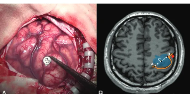
D-min (Fig 1A), the midpoint of the hand knob (point C) in the lesioned hemisphere, was calculated on the basis of both of the
hand knob end point coordinates (ie, points A and B). Subsequently, the point nearest on the tumor boundary (point D) to the midpoint of the hand knob (point C) was identified. Finally, D-min was calculated according to the coor-dinates of points C and D (calculative details are in the On-line Appendix, Part 1).
A mathematic model called WAN was generated to calculate the DD (Fig 1B). In the model, the coordinates of the midpoint in the healthy hemisphere (point H) and the midpoint in the le-sioned hemisphere (point I) were calcu-lated on the basis of both of their hand knob end point coordinates. The mid-line (blue dotted mid-line) was calculated on the basis of points E and F, which were located on either side of the falx cerebri intersection. In addition, the midpoint (point H⬘) that mirrored point H was calculated on the basis of the line EF. Fi-nally, the DD was calculated according to the coordinates of point H⬘and point I (calculative details are in the On-line Appendix, Part 2).
DCS Results and Overlap Index Calculation
To characterize the region of positive stimulation by DCS, a global region of 5-mm diameter was defined on the basis of each positive site. This region was manually drawn by 2 neuroradiologists independently according to intraopera-tive photographs (Fig 2A). If their selec-tions varied from each other by ⬎5%, another neuroradiologist with 20 years of experience made the final decision re-garding the region location.
The number of overlapping voxels between BOLD fMRI results and DCS regions was calculated via Matlab 2014a (MathWorks, Natick, Massachusetts). The overlap index was cal-culated via the formula (in this study, the total voxels of BOLD⫽ 300):
Overlap Index⫽Number of Overlapping Voxels
Total Voxels of BOLD ⫻100%.
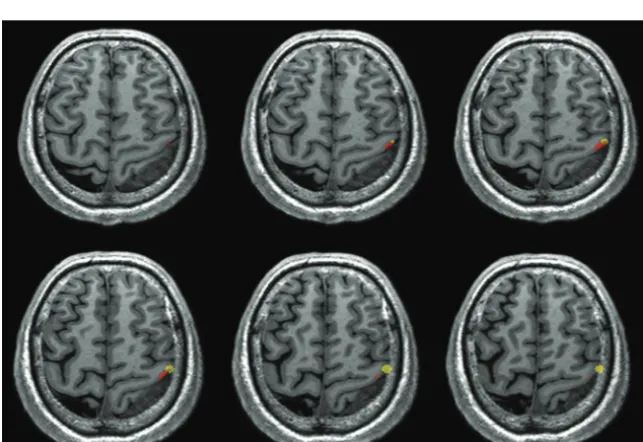
The nonoverlap phenomenon was defined as the overlap in-dex equal to zero (Fig 3). In contrast, the overlap phenomenon was defined as the overlap index greater than zero (Fig 4).
Statistical Analysis
A2test was performed for the distribution of several attributes (including sex, age, lesion hemisphere, World Health Organiza-FIG 1. The mathematic model used to quantify the anatomic characteristics (the original
magni-fication of the smaller pictures was⫻200%).A, Representation of the calculation of D-min. Points A and B represent the ends of the hand knob. Point C represents the midpoint of the hand knob on the lesion hemisphere. Point D is a point on the tumor boundary that is closest to the Point C. D-min is shown as thegold line.B, The WAN model was used to calculate the DD. Thedotted blue lineindicates the midline of the brain. Points H and I are the midpoints of the hand knobs on the healthy and lesion hemispheres, respectively. Point H⬘is the point that mirrors H on the basis of the midline of the brain (dotted blue line).Thepink linerepresents the DD.
[image:3.594.58.375.47.228.2] [image:3.594.56.378.319.479.2]tion grade, and preoperative epilepsy history). A Studentttest was used to an-alyze the differences in D-min and DD between the 2 patient groups. All results are presented as mean⫾SD. The re-ceiver operating characteristic curve was generated with GraphPad Prism 6.0c software (GraphPad Software, San Di-ego, California). The level of signifi-cance was .05 (2-tailed) for each statisti-cal test.
RESULTS
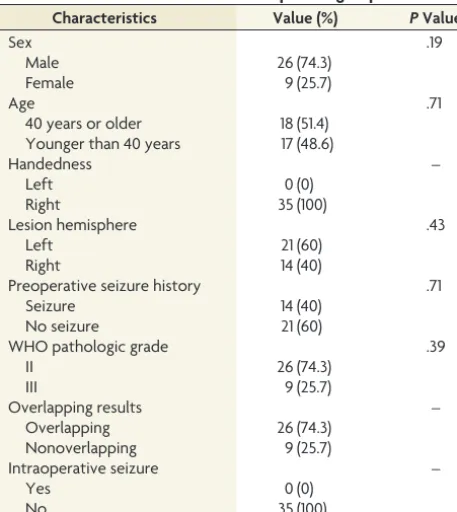
PatientsThirty-five patients met our inclusion criteria (Table 1). Eighteen of them were older than 40 years of age. All were right-handed. Fourteen patients (40%) had a preoperative seizure history. Twenty-six (74.3%) had grade II gliomas, and the others had grade III gliomas. There were no statistical differences in general char-acteristics with respect to overlapping or nonoverlapping phenomena.
In terms of anatomic characteristics (Table 2), 20 gliomas were anterior to the hand knob and 15 gliomas were pos-terior to it (Fig 5). The mean tumor vol-ume was 50.76⫾25.12 mm3, the mean D-min was 7.50 ⫾ 5.28 mm, and the mean DD was 9.38⫾1.36 mm.
There were 16 subjects (1 women, 15 men) in the control group. Their mean age was 25.6⫾1.0 years, and only 2 pa-tients were right-handed.
Association between D-min and Motor Area Localization
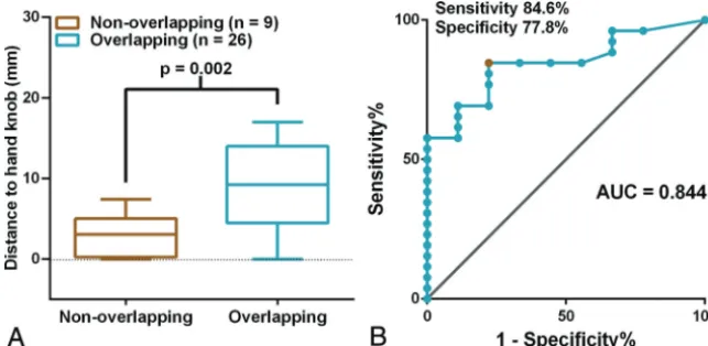
The mean D-min for the nonoverlap-ping group was smaller than that for the overlapping group (3.02 ⫾ 0.88 mm versus 9.07 ⫾ 1.01 mm, respectively; P⫽.002,ttest;Fig 6A). The receiver operating characteristic curve analysis indicated that 4 mm was the optimal cutoff value (84.6% sensitivity; 77.8% specificity; area under the curve, 0.844) to predict nonoverlapping phenomena (Fig 6B).
Overlap Index between BOLD fMRI and DCS Results
In group A, a significant difference in the overlap index was identified be-tween the D-minⱕ4-mm group and the D-min⬎4-mm group (17.2%⫾ 7.5% versus 59.1%⫾ 10.5%, respec-tively;P⫽.005, On-line Fig 2B). Con-FIG 3. An example of the nonoverlapping phenomenon (patient No. 9). There was no region
overlapping between the BOLD fMRI and DCS results. The red region is the BOLD fMRI results. The yellow region is the DCS results.
[image:4.594.55.376.47.398.2] [image:4.594.55.377.475.696.2]versely, no difference was found between the D-minⱕ4-mm group and the D-min⬎4-mm group in group P (33.5%⫾ 22.5% versus 56.6%⫾9.4%, respectively;P⫽.275, On-line Fig 2C).
Hand Knob Deviation and Nonoverlapping Phenomena
In the WAN model, we evaluated the symmetry of the bilateral hand knob in patient and control groups by measuring the DD. The DD of the patient group was significantly larger than that of the control group (9.38⫾1.36 mm versus 4.16⫾0.74 mm;P⫽ .015,ttest; On-line Fig 3).
The DD for the patient group was larger in the nonoverlapping group than in the overlapping group (17.59⫾4.00 mm versus 7.06⫾0.71 mm, respectively;P⫽.003,ttest; On-line Fig 4A). In group A, a statistical difference in DD was found between the overlapping group and the nonoverlapping group (nonover-lapping group, 20.88⫾5.45 mm; overlapping group, 6.30⫾ 0.90 mm;P⫽.001,ttest; On-line Fig 4B), whereas no signif-icant difference existed in group P (overlapping group, 7.95⫾ 1.12 mm; nonoverlapping group, 10.99⫾3.21 mm, respec-tively;P⫽.280,ttest; On-line Fig 4).
DISCUSSION
Identifying the hand motor cortex accurately is crucial when plan-ning surgery for gliomas involving the functional motor cortex. At present, BOLD fMRI is the prevalent noninvasive preoperative method used to localize the motor cortex. However, the accuracy of BOLD fMRI remains contested. Previous studies have con-cluded that unreliable BOLD fMRI results are associated with neurovascular uncoupling, particular tumor locations, tumor grades, cooperation of patients, task selection, and functional re-organization.9,27-30Although the BOLD fMRI signals are affected by tumor locations,21,24the impact of tumor location on BOLD fMRI accuracy has not been quantified. Thus, in our study, we investigated how tumor location affected BOLD fMRI accuracy via some locational characteristics such as D-min and DD.
On the basis of the regression model, the calculated voxel val-ues were positively related to the degree of activation. A previous study31considered the top 2% of the highly activated voxels as the functional area. Similarly, we allowed a maximum of 300 voxels from the BOLD fMRI results, to exclude a large number of irrel-evant voxels.
BOLD fMRI takes advantage of variations in blood hemoglo-bin levels to identify the relevant func-tional cortex.32The mechanism relies on neurovascular coupling. When neurons are activated, the oxygen-hemoglobin level increases rapidly and remains high until activation ceases in relevant areas. Coincidentally, the oxygen-hemoglobin exhibits a type of diamagnetism that acts as an endogenous contrast medium, thus revealing relevant activated areas.33 Due to limitations of BOLD fMRI,34 false-negative results may appear in the presence of neurovascular uncoupling. Thus, the actual functional area may not be displayed completely, and the BOLD fMRI results may be unreliable.28,35,36 Alterations in hemodynamics and the microenvironment are the primary rea-sons.21,33,37First, gliomas impair cere-bral vascular reactions and reduce CBV FIG 5. An example of tumor and hand knob locations.A, The tumor was anterior to the hand
[image:5.594.54.283.134.390.2]knob.B, The tumor was posterior to the hand knob. Theorange lineis the central sulcus, and the blue regionis the hand knob. Points A and B represent both ends of the hand knob.
Table 1: General characteristics for the patient group
Characteristics Value (%) PValue
Sex .19
Male 26 (74.3)
Female 9 (25.7)
Age .71
40 years or older 18 (51.4) Younger than 40 years 17 (48.6)
Handedness –
Left 0 (0)
Right 35 (100)
Lesion hemisphere .43
Left 21 (60)
Right 14 (40)
Preoperative seizure history .71
Seizure 14 (40)
No seizure 21 (60)
WHO pathologic grade .39
II 26 (74.3)
III 9 (25.7)
Overlapping results –
Overlapping 26 (74.3)
Nonoverlapping 9 (25.7)
Intraoperative seizure –
Yes 0 (0)
No 35 (100)
[image:5.594.54.375.436.694.2]Note:—WHO indicates World Health Organization.
Table 2: Anatomic characteristics of the patient group
Characteristics Value
Tumor volume (mean) (mm3) 50.76⫾25.12
D-min (mean) (mm) 7.50⫾5.28
DD (mean) (mm) 9.38⫾1.36
Tumor location
and CBF.21,34This process raises the level of deoxygenated hemo-globin that is paramagnetic and reduces BOLD signals.22Second, gliomas change the microenvironment of surrounding tis-sues.33,38Abnormal astrocyte proliferation decreases potassium reabsorption, which reduces the oxygen supply and elevates de-oxygenated hemoglobin levels.39Previous studies illustrated that gliomas induce neurovascular uncoupling.35,36Furthermore, the higher-grade gliomas are associated with a greater probability of neurovascular uncoupling.15,17,21,22 Our results demonstrated that if D-min were ⱕ4 mm, nonoverlapping phenomena oc-curred more frequently than when D-min was⬎4 mm. We spec-ulate that this outcome is due to neurovascular uncoupling. A previous study verified that gliomas caused a decline in BOLD fMRI activation by neurovascular uncoupling when the glioma was near the primary motor cortex.40Our results were consistent with this theory to some extent. However, another study sug-gested that the distance between the tumor and primary motor cortex was no more likely than tumor type in influencing BOLD fMRI activation.41This finding may be because all patients in that study had glioblastomas rather than lower-grade gliomas. On the basis of previous studies,15,17,21,22we believe that the degree of neurovascular uncoupling in our patients was not greater than that in patients with glioblastomas. Hence, we believe that D-min significantly affects BOLD fMRI accuracy in lower-grade gliomas. Distortion and movement of hand knobs may decrease the accuracy of BOLD fMRI results. Hand knobs will be moved and become distorted when gliomas grow and invade surrounding structures.3-5 By comparing DD, we found that hand knobs moved more obviously in the patient group than in the control group. Hand knob distortion decreased the number of true-pos-itive voxels in relevant regions. However, the number of postrue-pos-itive voxels stimulated by DCS was unchanged.26Hence, the overlap indexes decreased. This result could explain the significant differ-ence in overlap between the group with a D-min of⬍4 mm and the group with a D-min of⬎4 mm. This finding may also explain
the larger DD in the nonoverlapping group than in the overlapping group. Hence, we believe that distortion and movement of hand knobs may decrease the accuracy of BOLD fMRI results.
We found that the accuracy of BOLD fMRI results was associated with glioma location. The structure of the primary motor cortex is unique. The cingulate sulcus and the central sulcus constitute an obstruction that divides the precen-tral and posterior cenprecen-tral gyri com-pletely.25A prior study showed that the pre- and postcentral sulci were only linked by the paracentral lobule, which was responsible for foot movement.42 Because of its special structure, it is eas-ier for gliomas to invade the precentral gyrus from anterior areas. In contrast, it is difficult for gliomas that grow on the posterior central gyrus to invade the pre-central gyrus directly. Hence, we hy-pothesized that gliomas that grow anterior to the central sulcus compared with those growing posterior to it would influence the BOLD fMRI accuracy more. To test our theory, we divided pa-tients into group A or P. We found significant differences in both D-min and DD in group A, but not in group P. This finding corresponded with our hypothesis. Thus, we concluded that an-terior gliomas influenced the BOLD fMRI accuracy more obvi-ously than posterior gliomas. The limited number of samples in our study made it difficult to verify this finding. In the future, we will recruit more patients to verify our hypothesis.
Besides neurovascular uncoupling and tumor locations, there are other factors that may cause the inaccuracy of BOLD fMRI mapping, including task selection, the cooperation of patients, and cognitive status.12,33 Grasping is one of the traditional tasks used to identify the motor cortex and has been used in many previous studies.16,43,44Poor cooperation and impaired cognitive status of the patient could reduce the quality of BOLD fMRI.33,45To reduce the above influences, we enrolled patients without preoperative paralysis, and each patient was trained to achieve good performance in hand grasping before the BOLD fMRI scans.
CONCLUSIONS
This study suggests that D-min and DD are predictive of the accuracy of BOLD fMRI results. A smaller D-min and larger DD are associated with less accuracy of motor area localization with BOLD fMRI. One should be cautious in the use of preoperative fMRI data for surgical planning when D-min is smaller than 4 mm, especially for a lower-grade glioma located anterior to the central sulcus.
ACKNOWLEDGMENTS
We thank Stefan Huwer from Siemens for support with image collection.
Disclosures: Tianyi Qian—UNRELATED:Employment: Siemens.
[image:6.594.54.376.48.205.2]REFERENCES
1. Chang EF, Clark A, Smith JS, et al.Functional mapping-guided re-section of low-grade gliomas in eloquent areas of the brain: im-provement of long-term survival— clinical article. J Neurosurg
2011;114:566 –73CrossRef Medline
2. Yousry TA, Schmid UD, Alkadhi H, et al.Localization of the motor hand area to a knob on the precentral gyrus: a new landmark.Brain
1997;120(pt 1):141–57CrossRef Medline
3. Hou BL, Bhatia S, Carpenter JS.Quantitative comparisons on hand motor functional areas determined by resting state and task BOLD fMRI and anatomical MRI for pre-surgical planning of patients with brain tumors. Neuroimage Clin 2016;11:378 – 87 CrossRef Medline
4. Pujol J, Deus J, Acebes JJ, et al.Identification of the sensorimotor cortex with functional MRI: frequency and actual contribution in a neurosurgical context. J Neuroimaging 2008;18:28 –33 CrossRef Medline
5. Gabriel M, Brennan NP, Peck KK, et al.Blood oxygen level depen-dent functional magnetic resonance imaging for presurgical plan-ning.Neuroimaging Clin N Am2014;24:557–71CrossRef Medline
6. Majos A, Tybor K, Stefan´czyk L, et al.Cortical mapping by func-tional magnetic resonance imaging in patients with brain tumors.
Eur Radiol2005;15:1148 –58CrossRef Medline
7. Talacchi A, Turazzi S, Locatelli F, et al.Surgical treatment of high-grade gliomas in motor areas: the impact of different supportive technologies—a 171-patient series.J Neurooncol2010;100:417–26
CrossRef Medline
8. Spena G, D’Agata F, Panciani PP, et al.Supratentorial gliomas in eloquent areas: which parameters can predict functional outcome and extent of resection?PLoS One2013;8:e80916CrossRef Medline
9. Pirotte B, Voordecker P, Neugroschl C, et al.Combination of func-tional magnetic resonance imaging-guided neuronavigation and intraoperative cortical brain mapping improves targeting of motor cortex stimulation in neuropathic pain.Neurosurgery2008;62:941–56
Medline
10. De Witt Hamer PC, Robles SG, Zwinderman AH, et al.Impact of intraoperative stimulation brain mapping on glioma surgery outcome: a meta-analysis.J Clin Oncol2012;30:2559 – 65CrossRef Medline
11. Bartos R, Jech R, Vymazal J, et al.Validity of primary motor area localization with fMRI versus electric cortical stimulation: a com-parative study.Acta Neurochir (Wien)2009;151:1071– 80CrossRef Medline
12. Trinh VT, Fahim DK, Maldaun MV, et al.Impact of preoperative functional magnetic resonance imaging during awake craniotomy procedures for intraoperative guidance and complication avoid-ance.Stereotact Funct Neurosurg2014;92:315–22CrossRef Medline
13. Ille S, Sollmann N, Hauck T, et al.Impairment of preoperative lan-guage mapping by lesion location: a functional magnetic resonance imaging, navigated transcranial magnetic stimulation, and direct cortical stimulation study.J Neurosurg2015;123:314 –24CrossRef Medline
14. Ille S, Sollmann N, Hauck T, et al.Combined noninvasive language mapping by navigated transcranial magnetic stimulation and func-tional MRI and its comparison with direct cortical stimulation.
J Neurosurg2015;123:212–25CrossRef Medline
15. Holodny AI, Schulder M, Liu WC, et al.The effect of brain tumors on BOLD functional MR imaging activation in the adjacent motor cortex: implications for image-guided neurosurgery.AJNR Am J Neuroradiol2000;21:1415–22Medline
16. Lehe´ricy S, Duffau H, Cornu P, et al.Correspondence between func-tional magnetic resonance imaging somatotopy and individual brain anatomy of the central region: comparison with intraopera-tive stimulation in patients with brain tumors.J Neurosurg2000;92: 589 –98CrossRef Medline
17. Holodny AI, Schulder M, Liu WC, et al.Decreased BOLD functional MR activation of the motor and sensory cortices adjacent to a
glio-blastoma multiforme: implications for image-guided neurosur-gery.AJNR Am J Neuroradiol1999;20:609 –12Medline
18. Due-Tonnessen P, Rasmussen I, Berntsen EM, et al.Identifying the central sulcus in patients with intra-axial lesions: a multicenter study comparing conventional presurgical MRI to topographical analysis and BOLD-fMRI. J Comput Assist Tomogr 2014;38:1– 8
CrossRef Medline
19. Zhang D, Fox MD, Leuthardt EC, et al.Preoperative sensorimotor mapping in brain tumor patients using spontaneous fluctuations in neuronal activity imaged with functional magnetic resonance imaging: initial experience.Neurosurgery2009;65:226 –36Medline
20. Vassal M, Charroud C, Deverdun J, et al.Recovery of functional connectivity of the sensorimotor network after surgery for diffuse low-grade gliomas involving the supplementary motor area.J Neu-rosurg2017;126:1181–90CrossRef Medline
21. Hou BL, Bradbury M, Peck KK, et al.Effect of brain tumor neovascula-ture defined by rCBV on BOLD fMRI activation volume in the primary motor cortex.Neuroimage2006;32:489 –97CrossRef Medline
22. Fujiwara N, Sakatani K, Katayama Y, et al.Evoked-cerebral blood oxygenation changes in false-negative activations in BOLD con-trast functional MRI of patients with brain tumors.Neuroimage
2004;21:1464 –71CrossRef Medline
23. Agarwal S, Sair HI, Yahyavi-Firouz-Abadi N, et al.Neurovascular uncoupling in resting state fMRI demonstrated in patients with primary brain gliomas. J Magn Reson Imaging 2016;43:620 –26
CrossRef Medline
24. Wang L, Chen D, Olson J, et al.Re-examine tumor-induced alterations in hemodynamic responses of BOLD fMRI: implications in presurgi-cal brain mapping.Acta Radiol2012;53:802–11CrossRef Medline
25. Shah KB, Hayman LA, Chavali LS, et al.Glial tumors in Brodmann area 6: spread pattern and relationships to motor areas. Radiograph-ics2015;35:793– 803CrossRef Medline
26. Duffau H, Capelle L, Denvil D, et al.Usefulness of intraoperative electrical subcortical mapping during surgery for low-grade glio-mas located within eloquent brain regions: functional results in a consecutive series of 103 patients. J Neurosurg 2003;98:764 –78
CrossRef Medline
27. Picht T, Wachter D, Mularski S, et al.Functional magnetic resonance imaging and cortical mapping in motor cortex tumor surgery: comple-mentary methods.Zentralbl Neurochir2008;69:1– 6CrossRef Medline
28. Majos A, Bryszewski B, Kos´la KN, et al.Process of the functional reor-ganization of the cortical centers for movement in GBM patients: fMRI study.Clin Neuroradiol2017;27:71–79CrossRef Medline
29. Ulmer JL, Hacein-Bey L, Mathews VP, et al.Lesion-induced pseudo-dominance at functional magnetic resonance imaging: implica-tions for preoperative assessments.Neurosurgery2004;55:569 –79; discussion 580 – 81CrossRef Medline
30. Xie J, Chen XZ, Jiang T, et al.Preoperative blood oxygen level-de-pendent functional magnetic resonance imaging in patients with gliomas involving the motor cortical areas.Chin Med J (Engl)2008; 121:631–35Medline
31. Qiu TM, Yan CG, Tang WJ, et al.Localizing hand motor area using resting-state fMRI: validated with direct cortical stimulation.Acta Neurochir (Wien)2014;156:2295–302CrossRef Medline
32. Ogawa S, Lee TM, Kay AR, et al.Brain magnetic resonance imaging with contrast dependent on blood oxygenation.Proc Natl Acad Sci U S A1990;87:9868 –72CrossRef Medline
33. D’Esposito M, Deouell LY, Gazzaley A.Alterations in the BOLD fMRI signal with ageing and disease: a challenge for neuroimaging.
Nat Rev Neurosci2003;4:863–72CrossRef Medline
34. Ulmer JL, Krouwer HG, Mueller WM, et al.Pseudo-reorganization of language cortical function at fMR imaging: a consequence of tu-mor-induced neurovascular uncoupling.AJNR Am J Neuroradiol
2003;24:213–17Medline
35. Zaca` D, Jovicich J, Nadar SR, et al.Cerebrovascular reactivity map-ping in patients with low grade gliomas undergoing presurgical sensorimotor mapping with BOLD fMRI.J Magn Reson Imaging
36. Chen CM, Hou BL, Holodny AI.Effect of age and tumor grade on BOLD functional MR imaging in preoperative assessment of pa-tients with glioma.Radiology2008;248:971–78CrossRef Medline
37. Sakatani K, Murata Y, Fujiwara N, et al.Comparison of blood-oxy-gen-level-dependent functional magnetic resonance imaging and near-infrared spectroscopy recording during functional brain acti-vation in patients with stroke and brain tumors.J Biomed Opt2007; 12:062110CrossRef Medline
38. Schwarzbauer C, Heinke W.Investigating the dependence of BOLD contrast on oxidative metabolism.Magn Reson Med1999;41:537– 43
Medline
39. Paulson OB, Newman EA.Does the release of potassium from astro-cyte endfeet regulate cerebral blood flow?Science1987;237:896 –98
CrossRef Medline
40. Liu WC, Feldman SC, Schulder M, et al.The effect of tumour type and distance on activation in the motor cortex.Neuroradiology2005; 47:813–19CrossRef Medline
41. Fraga de Abreu VH, Peck KK, Petrovich-Brennan NM, et al.Brain tumors: the influence of tumor type and routine MR imaging char-acteristics at BOLD functional MR imaging in the primary motor gyrus.Radiology2016;28:876 – 83CrossRef Medline
42. Wang Y, Hao Y, Zhou J, et al.Direct current stimulation over the human sensorimotor cortex modulates the brain’s hemodynamic response to tactile stimulation. Eur J Neurosci 2015;42:1933– 40
CrossRef Medline
43. Krings T, Reinges MH, Erberich S, et al.Functional MRI for presur-gical planning: problems, artefacts, and solution strategies.J Neurol Neurosurg Psychiatry2001;70:749 – 60CrossRef Medline
44. Mo¨ller M, Freund M, Greiner C, et al.Real time fMRI: a tool for the routine presurgical localisation of the motor cortex.Eur Radiol
2005;15:292–95CrossRef Medline