Studies on Simuliidae(Diptera), with particular
reference to
Austrosimulium tillyardianumVolume 1
A
thesis presented for the
degree of Doctor of Philosophy in Zoology
in the University of Canterbury,
Christchurch, New Zealand
by
'In a general way the habits of the Simuliidae are pretty well known and the group is so homogeneous that we can expect little that is novel even from a good observer in another part of the globe.'
PREFACE i
v.
PrefaceThe aim of my thesis is to present information on population changes in the common New Zealand simuliid Austrosimulium (Austrosimulium)
tillyardianum Dumbleton. Larvae of this species live in riffle areas of open-country streams. Before this aim could be achieved, it was necessary to define the identity of the study animal and its relationships with other New Zealand Simuliidae, and gain an understanding of its life history.
Wainui Valley Stream (430 49'S, 1720 54IE), Banks Peninsula was
chosen as the study area to undertake these investigations on population changes (p.165). A. tillyardianum was the only simuliid present in the stream, larvae were readily available throughout the year, and also there were few other species present thus reducing the amount of possible
interspecific interactions. The stream was sufficiently small in size to simplify sampling procedures. Counteracting these favourable aspects w'ere the unfavourable factors of the Wainui Valley being a popular holiday area, and the presence of a Y.M.C.A. camp site by the stream. Because of these two factors, it was considered inadvisable to leave equipment at the stream to moni<tor continuously stream tempera.ture and water level which were thought to influence population size.
The results of my investigations are contained in the series of papers which follow. The three main papers of the study are presented first, followed by four supporting papers on aspects not restricted to A. tillyardianum. Finally, three general interest papers and data of the ins tar determination and population sampling studies are included, as appendices. Each paper is complete in itself, and has been written in the style and format of the journal in which it has been (or will be) published, or to which it will be submitted.
r-1cdn papers
PREFACE i i
which could be expected within species and between species. After examin-ation of this material I was certain that.A. tilJyardianum was the only simuliid present in the Wainui Valley Stream.
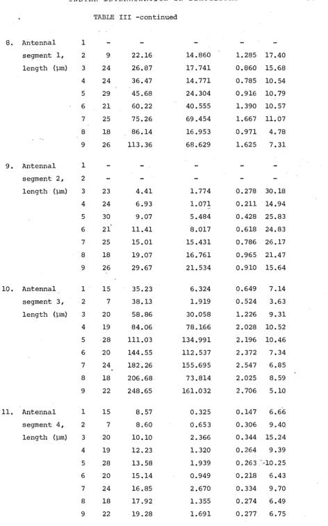
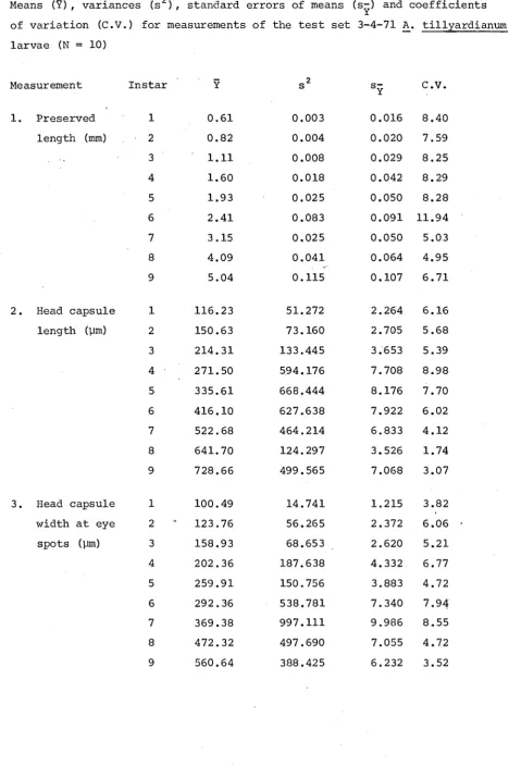
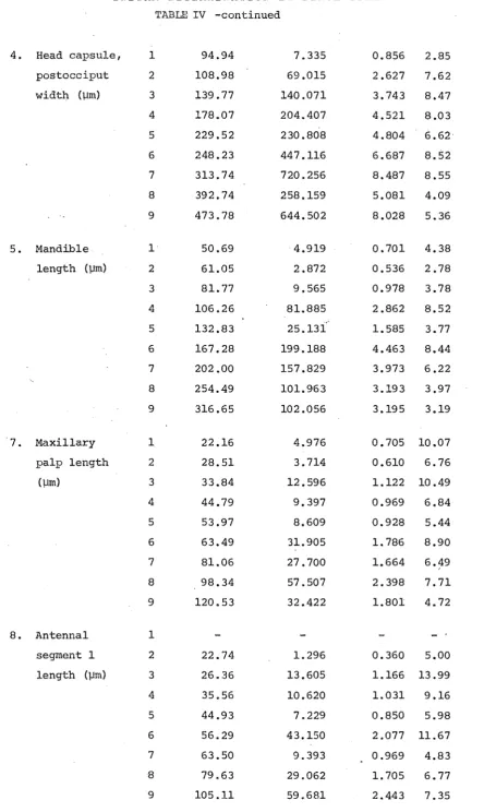
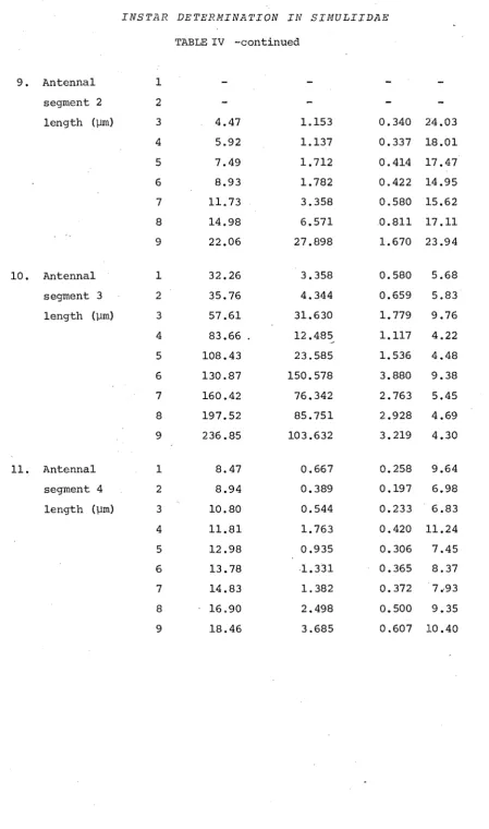
2. Instar determination. The nine larval instars of A. tillyardianum are shown to be biometrically distinguishable, and that larval instars separated on the basis of measurements correspond to the larval ins tar groupings
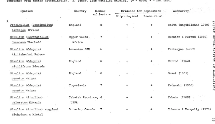
separated on the basis of morphoiogical characters. Nine larval instars is the highest recorded for any simuliid, and hence a detailed study on ins tar determination was required. Four methods of analyzing measurements are critically evaluated: frequency distributions, Brooks' rule (Dyar's rule), Student's t-test, and multiple discriminant function analysis. Previous instar determination studies are evaluated, and most are shown to be
capable of being re-interpreted to give a higher number of larval ins tars.
3 •. Population changes. A 10 x 1 m experimental channel was constructed in a bend of the Wainui Valley Stream, and the bed was covered with small stones to be used as sampling units. Weekly sampling over a seven month period when stream temperatures were relatively uniform was undertaken. In this time nine cohorts were followed through their life histories. Although every larval instar was collected on each sampling occasion, the cohorts could be separated by changes in ins tar composition of samples between weeks as .shm'ffi by the positions of "peaks" and "troughs". It was
concluded that intraspecific competition for attachment sites and floods were the main factors influencing the size of A. tillyardianum popUlations.
Supporting papers
4. Food of two New Zealand Trichoptera. Hydrobiosis parumbripennis and Hydropsyche colonica appeared to be the only potential invertebrate predators present in thewainui Valley Stream that could affect A. tillyardianum
numbers. However, gut analyses revealed that their predation on A. till·-yardianum was unimportant.
PREFACE i i i
6. Wing and haltere venation of larvae. The main features of the larval and adult wing venation of the Austrosimulium australense group are shown to correspond, but two differences are of interest in terms of the phylogeny of the genus Austrosimuliuln and the proposed homology for a vein. The
presence of a venation pattern in haltere buds of larvae is interpreted as further evidence for the view that halteres of Diptera are highly modified hind wings.
7. Dyar's rule predated by Brooks' rule. The rule credited to Dyar -that there is a geometric increase in the size of a sclerotized structure with each ins tar - is shown to have been formulated before Dyar by Brooks.
Appendices
Appendix 1. The historical use of the common names "sandfly" and "black fly" for species of Simuliidae is outlined.
Appendix 2. Gynandromorph of Austrosimulium australense. This is the first report of a primary somatic hermaphroditic simuliid; that is, an individual possessing parts of the internal and external secondary sexual .apparatus of both sexes •
Appendix 3. Obtaining literature through interlibrary loan. This summarizes my use of the University of Canterbury inter loan system. Information on time for interloans to arrive is the first published from the point of view of a user. Compilation of the data for this paper was initially for use in a survey of New Zealand university library resources.
ACKNOWLEDGMENTS
iv
ACKNOWLEDGMENTS
I wish to thank my supervisors Drs M.J. Winterbourn and V.M. stout for their assistance and discussions during the course of this study, and also Professor E.C. Young (now at the University of Auckland) for his initial supervision. Other assistance received from individuals has been
acknowledged in the different papers.
Specialized advice received from the Technical Staff and the provision of equipment for the study by them is gratefully acknowledged. I also thank colleagues who helped in various ways, especially G. Habib, G.S. Knight, F.H. Wood and Abdul Moeed.
Financial support for the study was provided by a New Zealand Postgraduate Scholarship.
Finally, I am grateful to my family and friends for their understanding and interest in this study over the.years, and who helped to make it a
CONTENTS
v
Contents Volume 1
Page Paper 1. Life history stages and taxonomy of Austrosimulium
(Austrosimulium) tillyardianum (Diptera : Simuliidae) Introduction
Previous taxonomic studies on Austrosimulium Tonnoirrs use of "tillyardi"
Materials and methods Descriptions
Male Female Pupa
Generalized description of larva Instar 1
Ins tar 2
Instar 3 Instar 4 Instar 5
Instar 6 Instar 7
Instar 8
Instar 9
1 2 3 3 4 5 6 11 15 20 29 29 30 30 31 31 32 32 33
Egg 33
Material examined 33
Discussion 34
Distribution of A. (A.) tillyardianum 34
Relationship of A. (A.) tillyardianum with other New Zealand
species 35
Acknowledgments 36
Literature Cited 36
Paper 2. Critique of ins tar determination in Simuliidae (Diptera)~ 41 with particular reference to methods applied to a study
of Austrosimulium tillyardianum Contents
Introduction
43
CONTENTS
vi
A. tillyardianum material 50
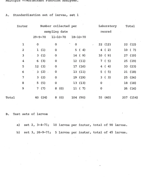
Sourc~ of larvae 50
Collection dates 50
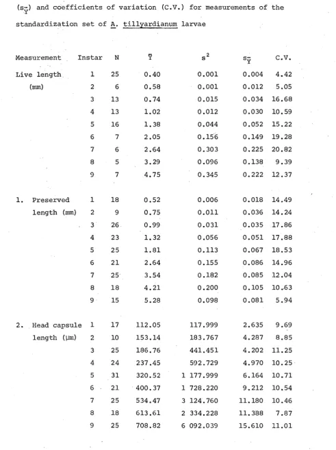
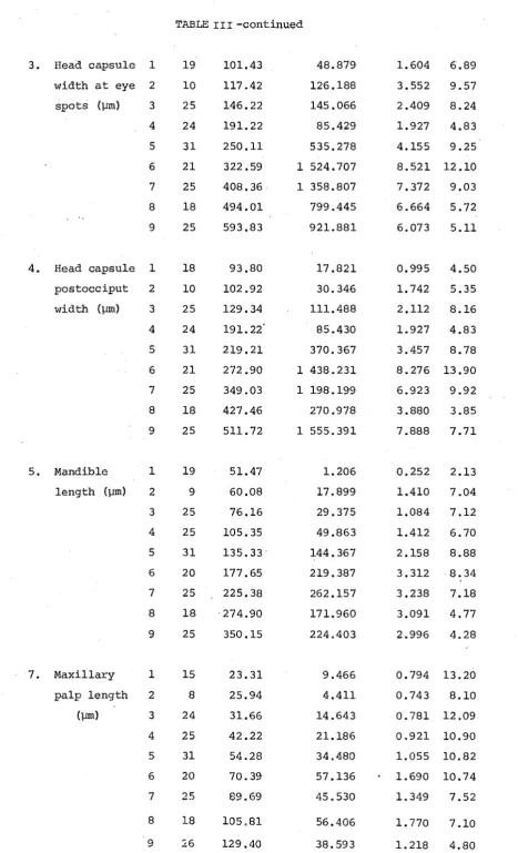
Measurements and morphological features used 52 Measuring methods and their effects on statistical tests 64
Measuring methods 64
Sampled randomization tests 65
Effect of measuring scale on t-test statistic 66 Effect of measuring scale on F-ratio statistic 69 Student's t-test or adjusted t-test to test means 70
Division into instars 71
Methods of analyzing ins tars 72
Separating ins tars by using frequency distributions of
measurements 73
Brooks' rule (Dyar's rule) 76
Application of Brooks' rule to A. tillyardianum measurements 76
Patterns of growth 78
Detection of missing ins tars 80
Growth ratios 80
Graphing logarithmic progressions 84
Application of Brooks' rule to other simuliid studies 87
student's t-test 89
Sensitivity of the t~test 89
Effect of lumping instars on t values 93 Multiple discriminant function analysis 94
Quadratic discriminant functions 97
Discriminant :t::uns undertaken 98
Results of the discriminant function analyses 100 A. Discriminating between 9 ins tars
B. Discriminating between 2 instars
Assignment according to mean discriminant value
102 102
confidence limits 104
Cross-validation of standardization discriminant
equations 110
Test set 3-4-71 data as standardization set 116 Effect of lumping ins tars on generalized Mahalanobis
CONTENTS
Fisher's linear discriminant function
Efficiency of classification
Seasonal effects on ins tars
Discussion
Evaluation of previous simuliid ins tar determination studies
Application .of A. tillyardianum results to field situation Significance of nine instars in A. tillyardianum
summary References
vii
128 l32 133 l35 135 150 151 153 156Paper 3. Population changes of Austrosimulium tillyardianum in an 165 experimental channel of a New Zealand stream
(Diptera : Simuliidae)
Introduction 166
Study area 169
Methods 173
Description of the experimental channel 173
Sampling 178
Analysis of data 180
Separation of cohorts 180
Effectiveness of placing larvae into size categories to follow
population changes 182
Life history of A. tillyardianum 184
Results and discussion 187
Developmental times of cohorts 187
Factors affecting larval numbers 199
1. Water level
2. Behaviour of larvae
Factors affecting pupae and adults
Survival rate
Summary
Acknowledgments
References
Paper 4. Food of the New Zealand trichopterans Hydrobiosis parumbripennis McFarlane and Hydropsyche colonica McLachlan
Summary
Introduction
Study area
Metli.ods sampling Gut analyses
CONTENTS
Life histories of the predators during the sampling period Life histories of the two main prey species during the sampling
period
Results and discussion Food of larvae
Food of predaceous larvae Number and size of prey taken
Forage ratios and prey availability
Importance of H. parumbripennis as a predator of A. tillyardianum
Acknowledgments References Key words
viii
218 218 219 220 220 222 222 223 223 226 228 230 230 232Paper 5. Trichomycetes (Harpellales) of New Zealand Austrosimulium 233 larvae (Diptera : Simuliidae)
Introduction
Midgut trichomycete, Harpella melusinae Observations on reproduction
Abundance and occurrence in different species Instar at which infection occurs
Hindgut trichomycetes Sununary
Acknowledgments References
Paper 6. Wing and haltere venation in larvae of the
Austrosimulium (Austrosimulium) australense group .from New Zealand (Diptera : Simuliidae)
234 234 236 237 238 239 240 240 240
. 242
Introduction 243
Occurrence of larval venation in Simuliidae 243 comparison of larval and adult wing venation 246
Submedian fork 246
Radial sector vein, Rs 247
Specific and individual differences in the presence of
the Rs fork 247
Phylogenetic implications for the origin of Austrosimulium 248
SUmmary References
CONTENTS
Paper 7. Dyar's rule predated by Brooks' rule Acknowledgments
References
Volume 2 -- Appendices
Appendix 1. Use of the common names "sandfly" and "black. fly" for species of Simuliidae (Diptera)
Historical use Acknowledgments Literature cited
Appendix 2. A gynandromorph of Austrosimulium (Austrosimulium) australense from New Zealand (Diptera : Simuliidae) Introduction
Description of the A. (A.) australense gynandromorph Discussion
Summary
Acknowledgments References
Appendix 3. Obtaining literature through interlibrary loan Efficiency of the University of Canterbury interloan system Evaluation of Garfield's (14) proposal
Acknowledgments References
Appendix 4. Instar det.ermination data and programs
Appendix table 1 -- Measurements of the standardization set of larvae
Appendix table 2 Appendix table 3
Measurements of the test sets of larvae Standardization set of discriminant function equations
Appendix table 4 -- Discriminant values of the standardization set of larvae in the standardization set of discriminant function equations
AP AP AP AP AP AP AP AP AP AP AP AP AP AP AP AP
ix
249 250 252 253 253 1 2 3 4 5 6 6 7 8 8 8 10 13 14 16 17AP 18 AP 19
AP 23 AP 27
CONTENTS X
Appendix table 5 -- Means and variances of the discriminant values AP 52 of the standardization set of larvae in the
standardization set of discriminant function equations
Calculation of quadratic discriminant function equations Multiple discriminant function analysis program -- MDISC
Program for evaluating discriminant function equations -- DSEV Program for counting outlier discriminant values -- LIMIT
Appendix 5. Wainui Valley stream sampling programme da~a
Set out of data
The data for each sample
AP 56
AP 61
AP 70
AP 72
AP 74
AP 75
'How often an inquirer asks "What is this organism?" and, on being told "Melania alba", considers himself somehow wiser than before. Anthropologists know well the supposed power conferred by knowledge of a name. I
LIFE HISTORY STAGES AND TAXONOMY
Paper 1
Life history stages and taxonomy of Austrosimtzli um (Austrosimulium) tillyardianum (Diptera : Simuliidae)
Accepted for publication in: Hew Zealand Joul.'nal of Zoology 1 (l), 1974
1
LIFE HISTORY STAGES AND TAXONOMY 2 INTRODUCTION
Austrosimulium (Austrosimulium) tillyardianum Durnbleton, 1973 was first described by Tonnoir (1925) under the name of tillyardi, and brief descriptions were given of the male, female, pupa, final instar larva and egg. Durnbleton (1973) amplified the original descriptions and noted that the name tillyardi was already preoccupied in Austrosimulium by an earlier use of the name by Tonnoir (1923a) (as Simulium tillyardi) , and hence the new· name tillyardianum was required (see p. 3) •
The redescription of tillyardianum by Durnbleton was in two parts; the first part was given in a general description of the New Zealand species of Austrosimulium, whereas the second part described the diagnostic features of tillyardianum itself. Although many new features of tillyardianum were described, many characters of use for comparing tillyardianum with other world species were not mentioned. Further, the larval instars of tillyard-ianum, except the final, remained unrecognized and undescribed.
My work has shown that tillyardianum has nine larval instars in its life history. This was determined after detailed morphological examination of 342 larvae, and was verified by applying univariate and mUltivariate statistical techniques to ten measurements made .on 289 of these larvae (in preparation). The discovery of nine instars for tillyardianum was surprising since it is the highest number recorded for any simuliid species, and is probably the highest ins tar number recorded for any nematoceran (Hennig, 1948). Most simuliids that have been s·tudied are believed to have six
(Terteryan, 1957; Harrod, 1964) or seven (Grenier & Feraud, 1960; Kacanski, 1968; Jedlicka, 1972) instars in their life histories, although two species have been reported to have only four instars (Yakuba, 1960), and one species has been shown to have eight (Smith, unpublished 1969).
LIFE HISTORY STAGES AND TAXONOMY 3 illustrating each of the life stages for the first time, many characteristic features of Austrosimulium (Austrosimulium) are given that may be used for a basis of comparison with other genera. The incomplete synonymy of
Dumbleton· (1973) is expanded.
Previous taxonomic studies on Austrosimulium
The genus Austrosimulium was erected in 1925 by Tonnoir for the simuliids with lO~segmented antennae found in New Zealand and Australia. A South
American species, Paraustrosimulium anthracinum (Bigot), has been assigned to Austrosimulium by several workers (Edwards, 1931; Wygodzinsky &
Coscaron, 1962; Dumbleton, 1973), but this relationship has now been rejected (Crosskey, 1969; Wygodzinsky & Coscaron, 1973),
Since Tonnoir, taxonomic s·tudies on Austrosimulium have been carried out in Australia by Mackerras &; Mack'erras (1948-55) and in Nm·, Zealand by
Dumbleton (1963a, 1973). Additionally, diagnoses of Austrosimulium sensu lato have been given by Edwards (1931), Smart (1945), Mackerras & Mackerras
(1949), Wygodzinsky & Coscaron (1962), stone (1963), and Dumbleton (1963a, 1973), whereas Crosskey (1969) has enumerated the differences between Austrosimulium and the genera of Simuliini occur%ing in Africa.
Austrosimulium and Gigantoaax were combined in the tribe Austrosimuliini by Smart (1945), but subsequently Crosskey (1969) has considered that
Austrosimulium belongs in the tribe Simuliini and that Gigantodax should be assigned to the tribe Prosimuliini; this arrangement is accepted,
Tonnoir's use of "tillyardi"
LIFE HISTORY STAGES AND TAXONOMY
In 1925, Tonnoir formally described a species under the nruoe of Aus'trosimulium ti11 yardi , but this was not the tillyardi of the 1923 paper. Tonnoir (1925) stated that he had pUblished a note on n • • • how
one of the New Zealand species builds its cocoon" but the species was not . mentioned by name. Presumably Tonnoir realized that he had misidentified
his tillyardi of 1923, but probably did not realize that his use of
tillyardi constituted a valid description, and therefore his tillyardi of 1925 should have been described under another name. Dumbleton (1973) briefly outlined the nomenclatural problems of tillyardi, and renamed
tillyard.i Tonnoir, 1925 as tillyardianum.
4
The misuse of tillyardi by Tonnoir has resulted in two different species being called tillyardi by subsequent workers (see synonymy for authors). Those authors describing cocoon construction and the hatching of the adult are using tillyardi to denote the species australense (Schiner), whereas the remaining authors are using tillyardi. to mean the species
tillyardianum Durnbleton. It is likely that this confusion will persist in the literature for some time to come.
MATERIALS AND ME'I'HODS
All descriptions are based on specimens collected from the Wainui Valley Stream, Banks Peninsula, Canterbury (430 49'S, 1720 54'E), although
specimens of tillyard.ianum ",ere examined from throughout its geographical range. The restriction to one locality for the description was for two reasons: firstly, tillyardianum was the only species present in the Wainui and thus no cunfusion with larval instars of other species was possible, and secondly, since many well-studied "species" have now been found to be complexes of sibling species, a description based on material from the Wainui Valley stream study area only is desirable (Dunbar (1972), for example, has recognized 16 chromosomally distinct entities, exhibiting biological differences in many cases, in the widely distributed African "species" Simulium (Edwardsellmn) damnosl.1m Theobald) •
Terminology in the descriptions is that outlined by Crosskey (1969) and, for the larval head capsule, that of Craig· (1969) i consequently I many
LIFE HISTORY STAGES AND TAXONOMY 5 Specimens used for the descriptions were those that had been preserved in 90°/0 ethanol. All drawings ~7ere made with the aid of a camera lucidaj whole mounts of specimens were used for low ~agnification drawings, but specimens used for dra'l>lings made at higher magnifications were first cleared with lOolo KOH before the required structures were mounted in glycerine.
DESCRIPTIONS
AUSTROSIMULIUM (AUSTROSIMULIUM) TILLYARDIANUM DUMBI.ETON, 1973
Austrosimulium (Austrosimulium) tillyardianum Dumbleton,1973, New Zealand Journal of Science 15(4) :513. Replacement name for tillyardi Tonnoir, 1925, Bulletin of Entomological Resea.rch 15 (3) : 253, junior secondary homonym of tillyardi Tonnoir,1923a, Annales de Biologie Lacustre 11
..
(3-4) :165 australense Schiner,1868, "Reise der Osterreichischen Fregatte 'Novara' urn die Erde" ZoologischerTheil. 2:15].
Austrosimulium tillyardi Tonnoir,1925, Bulletin of Entomological Research 15(3) :253, (original description);---pulikowsky 1929, zeitschrift fur
..
Morphologie und Okologie der Tiere 13:656-9i---Rubzov 1940, Fauna SSSR 6(6) :117;---Smart 1945, Transactions of the Royal Entomological Society of London 95(8) :499;-~-Mackerras & Mackerras 1949, Proceedings of the Linnean Society of New South Wales 73(5-6) :404;---Miller 1950, New Zealand Department of Scientific and Industrial Research Bulletin
100:61;---Hennig 1950, "Die Larvenfol:lIlen der Dipteren", Akademie-Verlag, Berlin, 2:384;---Wisely 1952, (Unpublished M.Sc. thesis, lodged in
library of Universi·ty of Canterbury, New Zealand) p.5l; ---Wisely 1962, Transactions of the Royal Society of New Zealand, Zoology 2(25):213;---Dumbleton 1963a, New Zealand Journal of Science 6(3) : 334;---DumbLeton 1963b, New Zealand Entomologist 3 (2) : 35 i----Wise 1965, Pacific Insects 7(2) :208;---Craig 1966, (Unpublished Ph.D. thesis, lodged in library of University of Canterbury, New Zealand) chapter 3:23;---Dumbleton J.969 , In Knox, G.A. (Ed.) "The Natural History of Canterbury", .A.H. & A.W. Reed, v\Tellington, p.485j---Stout 1969, In Knmc, G.A. (Ed.) "The Natural History of Canterbury", A.H. & A.W. Reed, Wellington, p.491; ---Dumbleton 1970, New Zealand Entomologist 4(3) :20.
LIFE IHSTORY STAGES AND TAXONOMY
Not tillyardianum, species referred to is Austrosimulium (Austrosimulium) australense (Schiner).
6
Simulium tillyardi Tonnoir,1923a, Annales de Biologie Lacustre 11(3-4) :165; ---Tonnoir 1923b, Bulletin de la Societe Entomolog.ique de Belgique 5:85;-:"-Puri 1925, parasitology 17(3) :301;---Wu 1931, Papers from the Michigan Academy of Science, Arts and Letters 13:552;--~Bequaert 1934, In strong, R.P.; Sandground, J.H.; Bequaert, J.C.; Ochoa, M.M. "Oncho-cerciasis with particular reference to the Cen-tral American form of the disease", Harvard University Press, Cambridge, p.189;---Smart 1934, Proceedings of the Royal Physical Society of Edinburgh 22(4) :236;---Ussova 1955, Dokladg Akademii nauk SSSR 105(4) :846;---Burton 1966, Annals of Tropical Medicine and Parasitology 60(1) :48.
Austrosimulium tillyardi; ---Mackerras & Mackerras 1948, Austra_Iian Journal of Scientific Research (B) 1(2) :246;---Peterson 1956, Canadian Entom-o.Iogist 88(8) :500.
Medium sized species, approximate measurements: Male: body length 2.5-3.0 mm, wing length 2.2-2.5 mm. Female: body length 2.6-3.1 mm, wing length 2.8-3.2 nun. Pupa: body length 3.0-3.5 mm.
9th instar larva: body length 4.6-6.0 nun.
Egg: length 0.20-0.24 mm, greatest breadth 0.12-0.15 mm.
MALE: General body colour brownish-black; pinned specimens with overlying silvery pruinose appearance.
Head: Normal (Figs 2, 4, 6), wider than thorax. Head holoptic (Figs 2, 6), enlarged upper eye facets occupying 2/3 area of head, areas of lower eye facets and clypeus correspondingly reduced. About 20 vertical columns and 28 horizontal rows of upper eye facets; diameter of upper eye facets 2.0-2.5 times that of lower facets (Fig. 7). Clypeus dark brown, sparsely haired. Antenna 10-segmented (Fig. 8), pale yellowish-brown, covered with fine hair; scape and pedicel slightly enlarged, bearing hairs, these about 1/2 length of pedicel; segment 3 sligh-Uy smaller -than pedicel; segments 4-9 small; segment 10 elongated cone. Maxillary palp (Fig. 9) 5-segmented, pale yellowish-brown, but segment 3 darker brown with longer hairs; segment 5 abou1: 1.5 times as long as segment 3; Lauterborn I s organ (Fig. 10) small
LIFE HISTORY STAGES AND TAXONOMY
.emale ;;!SOllm I male
4
FIGS l-6--Austrosimulium tillyardianum, adult head form. (1) Female, facial aspect, antennae omitted. (2) Male, facial aspect, antennae omitted. (3) Female, posterior aspect. (4) Male, posterior aspect. (5) Female, profile. (6) Male, profile.
LIFE HISTORY STAGES AND TAXONOMY 8
9
8
11 12
16
FIGS 7-16--Austrosimulium tillyardianum, male. (7) Franta-ocular triangle
and eye facets. (8) Antenna. (9) Maxillary palp. (10)
Lauterborn's organ (sensory vesicle) of 3rd segment of maxillary palp. (11) Tip of mandible. (12) of maxilla. (13) Fore leg. Stippling indicates regions of darker coloration. (14)
Mid leg. (15) Hind leg. Calcipala and pedisulcus present.
LIFE HISTORY STAGES AND TAXONOMY 9
---~M1
I
500..,m ~~--. submedian fork
17
[image:22.550.47.498.53.623.2]LIFE HISTORY STAGES AND TAXONOMY 10
'."7/
coxile-I 50 pm
posterior
I 50P~ 21 anterior
FIGS 18-21--Austrosimulium tillyardianum, male. Genitalia in ventral view. (18) Ventral plate, with median keel. (19) Aedeagal membrane with combs of fine spinules. (20) Coxite, style and paramere.
LIFE HISTORY STAGES AND TAXONOMY
11
poorly developed; mandible (l"ig. 11) with 2 spine-like projections apicallYi maxilla tt,ig. 12) with fine projections apically and subapically. Posterior surface of head (Fig. 4) with postgenal lobes separated by postgenal mem-braneioccipital foramen ovoid.
Thorax: Scutum wi thOl..1t pattern, unicolorous dark reddish-brown;
covered with evenly distributed brassy yellow (in pinned specimens) decumbent hair. Prescutellar depression with longer dark hairs. Scutellum with long black hair, more numerous laterally. Postnotum bare. Pleural membrane bare. Katepisternum bare. Mesepisternal sulcus deep, well defined. Legs
(Figs 13.,..15): light yellowish-brown, with darker brown femoral, tibial and tarsal bases; hairs yellowish, short. Fore, mid and hind tarsus slender, parallel-sided; hind tarsus widest. Fore basitarsus about 8 times as long as its greatest breadth. Hind leg with pedisulcus and well developed calci-palai hind basitarsus about 5.0-5.5 times as long as its greatest breadth. Claw (Fig. 16) partially covered by.a finely striate sclerotized pad extend-ing distally from tarsus 5. Wextend-ings (Fig. 17): fore veins well developed, dark brown. Costal vein with well developed spinules as well as hairs (Fig. 35) i spinules present from a point halfway between hm and Sa apex, to C apexi Rl haired but without spinules. Rs not forked; Rl and Rs meeting C about 2/3 from distal end of wing. M with short stem arising from r-m.
,MI, Mz, CUI, CUZI 2A, well defined; submedian fork and lA ill defined. CU2
with double curva'ture; basal section of radius haired; basal cell present; membrane evenly covered with normal microtrichia. Halteres brownish-black.
Abdomen: Tergi tes and sterni tes blackish-brown, lateral areas dull black. Tergites occupying whole width of dorsum; sternites 1, 3-9 present,
2 absent; sternite 1 small, semicircular; sternites 3-8 subquadrate, increas-ing in size from 3 to 8; sternite 9 small, semicircular, at base of coxites. Abdominal hairs present in pleural region, longest in mid-abdominal region. Genitalia (Fig. 21) of normal Austrosimulium type. Styles (Fig. 20) taper-ing and slightly truncate at tips, shorter than coxites, with 2 or 3 apical teeth; coxite not produced beyond base of style; ventral plate (Fig. 18) broad with median keel, all but posterior side ha.ired, anterior arms short and stout; median sclerite not visible; parameres (Fig. 20) short, without hooks or spines i aedeagal membrane (Fig. 19) 'Vlith forward-directed combs of
fine spinules.
LIFE HISTORY STAGES AND TAXONOMY
12
Head: Normal (Figs 1, 3, 5) slightly narrower than thorax. Head dichoptic (Figs 1,3), eye facets uniformly small (Fig. 22). Frons with short hairs; narrowest just above fronto-ocular triangles, being about 1/4 of head width (Fig. 1) f with median longit:udinal depression. Clypeus dark brown (Fig. 1) with sparse hairing. Antenna 10-segmented, pale yellowish-brown, covered with fine hair; proportions of segments similar to male. Maxil palp (Fig. 24) 5-segmented, similar to male. Lauterborn's organ
(Fig. 25) small and dark; wider than male. Proboscis light brown, with dark brown sclerotized supports; short and stout, twice as long as wide. Mouthparts well developed, capable of biting man; mandible (Fig. 26) with about 35 inner teeth, increasing in size towards apex, no outer teeth; maxilla (Fig. 27) with about 20 outer and 15 inner inwardly directed teeth; posterior border of cibarium (Fig. 28) without spines or sculpturing in median area; well sclerotized lateral arms. Posterior surface of head (Fig. 3) with postgenal lobes separated by postgenal menfurane; occipital foramen ovoid.
Thorax (Fig. 34): Scutum unicolorous semi-shiny dark reddish-brown; sparsely covered with decunfuent hairs; in pinned specimens, a median and 2 lateral longitudinal dark lines visible between overlying silvery pruinosity. other features as in male. Legs (Figs 29-31): yellowish-brown, with darker brown bases to femoral, tibial and tarsal segments; hairs short, yellowish. Fore, mid and hind tarsus slender, sub-parallel sided; hind tarsus widest. Fore basi tarsus about 8 times as long as its greatest breadth. Hind leg
with pedisulcus and well developed semicircular calcipala (Fig. 32), calcipala with a comb of small teeth as in male; hind basitarsus about 6.5~7.0 times as long as its greatest breadth. Claw (Fig. 33) without basal tooth. vHngs (Figs 17, 35): as in male. Halteres light yellowish-brown.
Abdomen: Tergi tes and sterni tes blackish-brown, lateral areas dull greyish-black. Tergites (Fig. 36) of variru)le shape and size; tergites 2 and 7 , tergite 10 smallest and only visible caudally. Sternite 1 present, small; sternites 2-6 absent; sternites 7 and 8 (Fig. 37);
sternite 8 crescentic, convex posteriorly, lateral lobes acute and located between 8 and 9. Tufts of abdominal hair in pleural areas, longest distally. Genitalia (Figs 38-41) of normal Austrosimulium type. Spermatheca ovoid, without surface pattern, and with internal hairs 5~7 ].lm long usually arranged in sho:t:t rows i spermathecal duct not sclerotized. Anterior
gonapo-physes (l"ig. 39) triangular, not produced, unpigmented, imperfectly
separated from sternite 8. Genital fork (Fig. 38) broad, lightly sclerotized; lateral arms short; stem directed anteriorly in median ventral line,
LIFE HISTORY STAGES AND TAXONOMY
hind basHarous
'y
Gale'pal.
ped,sulcus ~r
2nd hind tarsal _
segment \
tibtB:
tarsus 1 _
tbasHarsus)
FIGS 22-33--Austrosimulium tillyardianum, female. (22) Franta-ocular triangle and eye facets. (23) Antenna. (24) Maxillary palp.
(25) Lauterborn's organ (sensory vesicle) of 3rd segment of maxillary palp. (26) Tip of mandible. (27) Tip of maxilla.
(28) Posterior border of cibarium. (29) Fore leg. Stippling indicates regions of darker coloration. (30) Mid leg. (31) Hind leg. (32) Second hind tarsal segment and apex of hind basitarsus showing calcipa1a and pedisu1cus. (33) Claw of hind leg.
LIFE HISTORY STAGES AND TAXONOMY
T7
FIGS 34-41--Austrosimulium tillyardianum, female.
38
ar'lcul .. t~on
poInl toTO
9
~
core.oD
40 41
(34) Thorax, lateral
14
view. (35) Costal vein of wing, showing spines, macrotrichia and microtrichia. (36) Abdomen, dorsal view, showing shape of tergites. Tl-T9, tergites 1-9. (37) Terminalia, lateral view. T7-T9, tergites 7-9, S7 and SS, sternites 7 and S. (3S)
Genital fork, dorsal view. (39) Anterior gonapophyses, ventral view. (40) Paraproct and cercus, ventrolateral view. (41)
LIFE HISTORY STAGES AND TAXONOMY
15
triangular, not joined. Cerci (:Pigs 40, 41) \vider than long, thin; semi-circular in lateral view.
PUPA (Fig. 42): General colour of head and upper thorax dark reddish-brown to bro\<mish-black, lower thorax and legs pale yellmvish-reddish-brown, abdomen greyish-black to dull black.
Head: Cephalic apotome of male (Fig. 47) strongly tuberculate on upper halfi that of female (Fig. 48) tuberculate laterally only, concave; paler spots present, more in male (Fig. 47). Antennal sheath of male
extending 1/2 or slightly more of distance to hind margin of head; antennal sheath of female reaching hind margin of head. Frontal setae absent;
facial setae present, 1 on each side between antennal bases; 2 short epi-cranial setae present on margin of cephalic apotome and antennal sheath at mid length; postorbital spine absent.
Thorax: Dorsum coarsely granulated; tubercles raised, circular,
distinct, contiguous (Fig. 45); tubercles fonu trident-shaped pattern, median line (along ecdysal suture), posterior transverse line and 2 lateral long-itudinal lines arising on each end of posterior line (Dumbleton, 1973:Fig. 56), between lines and in pleural areas, tubercles are aggregated in groups of 12 or more. Setae simple, small and difficult to see, sometimes apparently absent, distributed as in Dumbleton (1973). Gill (Fig. 46): Horn short, about 2 1/2 times as long as its greatest breadth; basal 1/3 light ye 11 mv , distal 2/3 black, <covered with trabeculae, tips anteriorly directed, trab-eculae longest distally. Filaments 15-20, light yellow, about 3-4 times the length of horn. Filaments arising from apex and apical 1/3 of external
margin, and from basal 1/3 to apex of internal margin; thick, stiff, tapering, inward curving, of reticulated type.
LIFE HISTORY .STAGES AND TAXONOMY
o
<:)
47
48
45 ecdyssl suture
FIGS 42-48--Austrosimulium tillyardianum, pupa. (42) Pupa in cocoon, dorsal view. Note inward curving of the respiratory filaments of the pupal gill. (43) Cocoon in profile (shoe-shaped).
(44) Cocoon, dorsal view. Note ill-defined paramedian ridges. (45) Left side of thoracic notum showing arrangement of
tubercles. (46) Left pupal gill, dorsal view. (47) Cephalic apotome, male. (48) Cephalic apotome, female.
LIFE HISTORY STAGES AND TAXONOMY
2 3
6
6
II
500~m
-I
terminal abdominal
hook 52
17
FIGS 49-52--Austrosimulium tillyardianum, pupa. (49) Abdomen, dorsal surface, showing arrangement of anteriorly directed hooks and weakly developed erect terminal abdominal hooks. (50)
LIFE HISTORY STAGES AND TAXONOMY
18
nerve cora....;::,.:.:...:. ..
ventral dorsal
LIFE HISTORY STAGES AND TAXONOMY
cephalic fan
maxillary
submental brildaE~-~.
55
I-250 ~mFIG. 55--Austrosimulium tillyardianum, 9th ins tar larva. Lateral view
of head.
[image:32.549.86.501.139.592.2]LIFE HISTORY STAGES AND TAXONOMY 20
Cocoon
(Figs 42-44): Shoe-shaped, light yellowish-brown, oval,close-fitting to pupa, closely woven, thin, with high collar; posterior margin of aperture irregular; collar margin passing immediately below gills and cover-ing head; posterior 1/4 with floor, loosely woven; 2 ill-defined paramedian ridges dorsolaterally, arising from distal edge of aperture and converge near posterior of cocoon.
GENERALIZED DESCRIPTION OF LARVA; (Detailed description of each instar follows this generalized description which is based on the 9th instar larva).
General colour:
head capsule shi!1y pale-yellow to light reddish-brown,light greyish-brown dorsal, lateral and anterior ventral surfaces, whitish-transparent posterior ventral surface, nerve cord black; pigmentation patterns as in Figs 53, 54, 69. Body shape (Figs 53, 54, 69); abdomen slightly clubbed with posterior segments clearly set off from anterior seg-ments, thorax somewhat swollen.
Head: Sides convex (Fig. 60) i cephalic apotome widest posteriorly
with rounded posterolateral sides (Fig. 60); head spots positive, slightly darker brown than cephalic apotome but poorly demarcated (Fig. 60); 2 well marked black lateral eye spots- (Fig. 55), anterior smaller; no eyebrow stripe dorsad of eye spots; postoccipital ring black; cervical sclerites
(Fig. 60) black, transverse triangular, separated from postocciput. Sub-mental cleft (Fig. 74) rounded apically, small, 1/2 length of subSub-mental bridge; large oval posterior tentorial pit either side. Suboesophageal ganglion (Fig. 53) pigmented, grey-black. Mentum (Figs 74, 109) with 13
teeth, all but median and corner teeth concealed by ventral anterior exten-sion of mentum, teeth black; single row of mental setae about parallel with each lateral side, simple. Antenna 4-segmented (Fig. 90); about 1. 5-2.0
times as long as stem of cephalic fan; segments 1 and 2 darker than cephalic apotome, segments 3 and 4 pale; segment 3 sclerotized, especially on apical 1/4 and around mandibular articulation; apical teeth (Fig. 100) stout, third largest; third tooth not conspicuously enlarged; other inner teeth well developed, regularly decreasing in sizel tips pointed; 2
raand-ibular serrations, no supernumerary serrations. Maxillary (Fig. 73) about 2 1/4 times as long as its basal width; outer dorsal hair tuf-!: of cardo small and inconspicuous; teeth of lacinia as in Pig. 73. Hypopharynx
LIFE HISTORY STAGES AND TAXONOMY
21
burster
56
57
250 ~m
I I
59
60
FIGS 56-60--Austrosimulium tillyardianum larvae, head region in dorsal
view. Only the pigmentation pattern for ins tar 9 has been indicated, earlier instars are similar. (56) Instar 1. Note that the ends of the postoccipital collar are contiguous.
LIFE HISTO~~ STAGES AND TAXONOMY 22
61
62
63
64
65
66
67
1 mm
FIGS 61-69--Comparative size of ins tars of Austrosimulium tillyardianum larvae, lateral view., Only the pigmentation pattern of ins tar 9 has been indicated, earlier instars are similar. (61)
labial
74
LIFE HISTORY STAGES AND TAXONOMY 23
~ 71
mental teeth
- - - submental bndge
I
'OltmWll m.lgnum
brush
anterior margin of
hvpopharyngeal
lobe
hypopharyngeal sclerita
sonsilia basiconica
73
as~oclated sensory
...
~
'\
75
sensor y 's pme
) teeth 01 lac,n,a
. . . /~lilClOm
/ 1_50pm I .
72
76 77
FIGS 70-77--Austrosimulium tillyardianum, 9th ins tar larva. (70) Labiohypo-pharyngeal complex, dorsal view. Opening of silk canal is medially located between hypopharyngeal lobe and labial lobe.
(71) Sensory lobes, showing grouping of sensilla basiconica. (72) Maxillary lobe, dorsal view. (73) Maxillary palp, dorsal view. (74) Head capsule, ventral view. (75) Primary ray of cephalic fan. (76) Middle section of primary ray showing pattern of microtrichia. (77) Basal section of primary ray showing
25,um
LIFE HISTORY .STAGES AND TAXONOMY
inner side
)
18
posterior circlet of hooks
- - mea lOan plale
posterior arm
terminal expansion
FIGS 78-81--Austrosimulium tillyardianum, 9th instar larva. (78) Lateral sclerite of proleg. Bristles in groups of 2, rarely 3.
24
LIFE HISTORY STAGES AND TAXONOMY
82 83 84
85
I 50.um I
86
87
88
89
90
3
sensilla basiconica
FIGS 82-90--Austrosimulium tillyardianum. Larval antennae. (82) Instar 1, 2-segmented. (83) Instar 2, 3-segmented. (84) Instar 3, 4-segmented. (85) Instar 4. (86) Instar 5. (87) Instar 6.
(88) Instar 7. (89) Instar 8. (90) Instar 9.
LIFE HISTORY STAGES AND TAXONOMY
A
_ _ _ _ _ _ _ artiCUlation pOint
10 postantenl)ai
50,um I
D
ml)
D
93
92
buttress
apical teeth - - - ' "
mandibular
serralions--B
FIGS 9l-100--Austrosimulium tillyardianum. Comparative size of larval mandibles. (91) Instar 1. (92) Instar 2. (93) Instar 3.
(94) Instar 4. (95) Instar 5. (96) Instar 6. (97) Instar 7. (98) Instar 8. (99) Instar 9. AB, length measured.
(100) Tip of mandible, in.tar 9, showing arrangement of teeth and mandibular serrations.
LIFE HISTORY STAGES AND TAXONOMY
~
~ 101102
~
menial row 01OD'A,,_-103
!OO)lm
mid Ime
I
~
106 108
FIGS 101-109--Mental of Austrosimulium til1yardianum larvae. (101) Instar 1, 9 mental teeth. (102) Instar 2, 13 mental teeth. (103) Instar 3. (104) Instar 4. (105) Instar 5.
(106) Instar 6. (107) Instar 7. (108) Instar 8. (l09) Instar 9. Note that one of the intermediate teeth on the left side is missing.
LIFE HISTORY STAGES AND TAXONOMY
110
112
I-- 50J.,lm
,
)
"
111
FIGS 110-113--Austrosimulium tillgardianum. Development of anal and semicircular sclerites of larvae. (110) Instar 2. Anal sclerite only present. (Ill) Instar 3. Anal sclerite and beginning of semicircular sclerite. (112) Instar 4.
Complete semicircular sclerite, interarm struts of anal sclerite present. (113) Instar 5. Terminal expansions on semicircular sclerite present.
LIFE HISTORY STAGES AND TAXONOMY
29
Thorax: cuticle bare. Spiracular scars faint. Lateral sclerite (Fig. 78) of pro leg lightly sclerotized, anterior margin with lobed appear-ancei about 25 bristles, arranged in groups of 2, sometimes· 3, longest on outer (ventral-most.) side.
Abdomen: cuticle bare ,- Spiracular scars very inconspicuous. 2 sub-conical ventral papillae present posteriorly (Fig. 69). Rectal gills
simple (Fig. 69), milky white without pattern, 3 lobes, each bluntly rounded. Accessory sclerites and rectal scales absent. Anal sclerite (Figs 79, 80) X-'shaped; anterior arms broader than posterior arms i apices of anterior
arms bluntly pointed, apices of posterior arms sharply pointed; anterior margin of anterior arms lightly sclerotized; interarm struts arise from apical 1/3 of anterior arms, run ventral to but lateral of median plate to near base of posterior arm, do not fuse to posterior armSi posterior margin of median plate with about 9 tubercles, Semicircular sclerite (Fig. 80) stout, with terminal expansions. Hooks-of posterior circlet of hooks (Fig. 81) normal.
INSTAR 1 (Fig. 61)
Mean preserved length: 0.52 rom.
Egg burster (Fig. 56): present on cephalic apotome, in a median line, _level with eye spots. Consis-ts of a raised point surrounded by 7 small
points, black. This is absent in instar 2.
Antenna (Fig. 82): 2-segmented. Mean total length: 43.8 ].1m.
Cephalic rays: about 10 primary and 8 secondary rays, weakly sclerotized.
~landible (Fig. 91): mean length AB: 51.5 ].1m. 3 apical -teeth, 2-3 comb teeth. All subsequent instars have 3 apical. teeth.
Maxillary palp: weakly sclerotized, nearly transparent.
Men tum (Fig. 101): 9 mental teeth i in 3 groups of 3 , central tooth of
median group mo:r:e anterior than others. 1 seta in each mental row. Cervical scleri (Fig. 56): not developed, ends of postocc:i.pi tal collar contiguous.
Proleg circlet of hooks: 12 rows with 1 hook per rmV".
Posterior circlet of hooks: about 45 rows with 2-·3 hooks per row. Anal scleri te: not developed.
Semicircular scleri te: not developed.
INSTAR 2 (Fig. 62)
Mean preserved -length: 0.75 rom.
~IFE HISTORY STAGES AND TAXONOMY
Cephalic rays: about 32 primary and 9 secondary rays, moderately
sclerotiz~d.
30
Mandible (Fig. 92): mean length AB: 60.1 l.lIll. 3 comb teeth. All subse-quent instars have 3 comb teeth.
Maxillary palp: moderately sclerotized.
Mentum (Fig ~ 102): 13 mental teeth as in final instar, median one most anterior. 1 seta in each mental row.
Cervical sc S: not developed, slight separation of ends of post-occipital collar, as in Fig. 57.
Proleg Girclet of hooks: 12 rows with 1-2 hooks per row.
Posterior ci.rclet of hooks: about 50 rows with 3-4 hooks per row. Anal scleri te (Fig. 110): present, backwardly directed interarm strut from each anterior arm absent.
Semicircular sclerite: not developed.
INSTAR 3 (Fig. 63)
Mean preserved length: 0.99 nun.
Antenna (Fig. 84): 4-segmented, as are all subsequent instars. Mean total length: 110.2 ~m.
Cephalic about 21-25 primary and 11 secondary rays; strongly sc1erotized, similar to all subsequent instars.
Mandible (Fig. 93): mean length AB: 76.2 ~m. 1-2 inner teeth.
Maxillary lp: strongly sclerotiz,ed, as are all subsequent instars. Men tum (Fig. 103): 2 setae in each mental row I posterior seta shorter
than anterior seta.
Cervical scleri tes (Fig. 57): not developed, increasing separation of ends of postoccipital collar.
Proleg circlet of hooks: about 14 rows with 2 hooks per row.
Posterior circlet of hooks: about 50-55 rows with 5-6 hooks per row. 1\_nal s te (Fig. Ill): present, interarm strut from each anterior arm absent.
Semic ircular scleri te (Fig. 111): ei·ther not developed, or partly developed.
INST.AR 4 (Fig. 64)
Mean length: 1. 32 rom.
Antenna (Fig. 85): mean total length: 139.5 ~m.
LIFE HISTORY STAGES AND TAXONOMY
Mentum (Fig. 104): 2, or sometimes 3, setae in each mental row; the posterior seta shorter than anterior seta(-e).
31
Cervical s: not developed, ends of the postoccipital collar drawing apart, as in Fig. 57.
Imaginal buds (Fig. 64): all present (pupal respiratory histoblast, adult wing bud, haltere bud and 3 leg buds) , just visible as clear patches
against pigmented thorax. No differentiation of tissue within buds.
Prothoracic leg bud sometimes absent.
Proleg circlet.' hooks: about 14 rows with 4 hooks per row.
Posterior circlet of hooks: about 52-60 rows with 6-7 hooks per row. Anal scleri te (:E'ig. 112): present, interarm strut from each anterior arm present but weakly sclerotized.
Semicircular sc rite (Fig. 112): developed, but without terminal expansions.
INSTAR 5 (Fig. 65)
Mean pre length: 1. 81 rom.
Antenna (Fig. 86): mean total length: 179.4 11m.
Cephalic rays: about 30-34 primary and 13 secondary rays. Mandib Ie (Fig. 95): mean length AB: 135.3 llID. 3-7 inner teeth. Mentum (Fig. 105): 3,. or sometimes 4, setae in each mental row; post-erior 2 setae shorter than anterior seta(-e).
Cervical scleri tes : not developed, postoccipital collar as in Fig. 58. Imaginal buds (Fig. 65): all present, no differentiation of tissue within buds.
Proleg of hooks: about 17-18 rows with 4-6 hooks per row. Posterior circlet of hooks: about 63-67 rows with 6-8 hooks per rO';,l. Anal sc
te
(Fig. 113): present, interarm strut strongly sclerotized, similar ,to all subsequent instars.Semicircular Bcleri te (Fig. 113): developed, \"ith terminal expansions, width of an expansion no more than twice wid,th of rest of scleri te, shallow
notch to expansion.
INSTAR 6 (Fig. 66)
Mean served lengt,h: 2,64 mm,
An
tenna
(Fig. 87): mean total length: 231. 3 11m.LIFE HISTORY STAGES AND TAXONOMY
Cervical scleri tes (Fig. 58): slightly differentiated from ends of post~ccipital collar.
Imaginal buds (Fig. 66): pupal respiratory histoblast and adult wing bud slightly
buds.
than other buds, no differentiation of tissue wi thin 32
Proleg circlet of hooks: about 22-25 rows with 5-7 hooks per row. Posterior circlet of hooks: about 78-88 rows with 9-12 hooks per row. Semicircular scleri te: width of terminal expansions about 3 times width of rest of sclerite. Sides of expansions more heavily sclerotized
than centre, notch about 1/3 length of expansion.
INSTAR 7 (Fig. 67)
Mean preserved length: 3.54 mm.
Antenna (Fig. 88): mean total length: 289.4 11m.
Cephalic rays: about 35-44 primary and 14-16 secondary rays. Mandible (Fig. 97): mean length AB: 225.4 11m,· 6-9 inner teeth. Mentum (Fig. 107): 4-5 setae in each mental row, posterior 2 setae shorter than anterior setae.
Cervical scler slightly differentiated from ends of postoccipital collar, as in Fig. 58.
Imaginal buds (Fig. 67): diameter of pupal respiratory histoblast and adult wing bud twice that of other buds, no differentiation of
within buds.
Proleg circlet of hooks: about 24-25 rows with 7-9 hooks per row. Posterior circlet hooks: about 85-90 rows with 11-12 hooks per row. Semicircular scleri te: notch of terminal expansion about 1/2 its
length, as in all subsequent instars.
INSTAR 8 (Fig. 68)
Mean preserved 4.21 mm.
Antenna (Fig. 89): mean total length: 329.8 pm.
Cephalic rays: about 40-45 primary and 15~16 secondary rays. Mandible (E'ig. 98): mean length AB: 279.4 llm. 7-11 inner teeth. Mentum (Fig. 108): 4-5 sometimes 6 setae in each mental row, 2 setae shorter than anterior setae.
Cervical sclerites (Fig. 59): dHferentiat.ed, but still attached to ends of postoccipital c011a.ri moderately sclerotized.
Imag inal buds (Fig. 68): diamet.er of pupal respiratory histoblast and adult wing bud -twice that of other , no differentiation of tissue
LIFE HISTORY STAGES AND TAXONOMY 33 Proleg circlet of hooks: about 25-27 rows with 8-10 hooks per row. Posterior circlet of hooks: about 90-105 rows with 12-15 hooks per row.
INSTAR 9 -the pharate pupa (Hinton, 1958) (Figs 53, 54, 69) Hean preserved length: 5.28 mm.
Antenna (Fig. 90): mean total length: 411. 0 Vm.
Handible (Fig. 99): mean length AB: 350.1 }im. 9-12 inner teeth (Fig. 100). Hentum (Fig. 109): 5-7 setae in each mental row, posterior 2 setae
shorter than anterior setae. Sometimes 1 less seta in 1 row than in other.
Cervical scleri tes (Fig. 60): separated from ends of postoccipital
collar; strongly sclerotized.
Imag inal buds (F ig. 69): pupal respiratory histoblast with folded filaments white in early stage, but black just before pupation. Wing and
haltere buds with a venation pattern taking form of pigmented lines,
appearing as instar matures. Leg buds with inflexions present.
Pro leg circlet of hooks: about 27-32 rows with 9-12 hooks per row. Posterior circlet of hooks: about 100~120 rows with 14-18 hooks per row (Fig. 81).
EGG: Normal simuliid type; triangular-ovoid, asymmetrical because of
bulge on one side. Surface smooth. Whitish-yellow when laid, dark
yellowish-brown prior to hatching. Laid on downstream surface of rocks
15-100 rom below water level, 250-330 eggs in egg mass 6-8 mm by 3-5 rnm,
single layer, fixed to stone by transparent gelatinous materia:J...
MATERIAL EXAHINED
(L = larvae, P := pupae, A = adults; T.K.C. == T.K. Crosby)
Type material pi.nned; specimens collected during study preserved in
90
0/0
ethanol, some adults pinned.Holotype male, Maitai River, Nelson, -vi.23; bred, A.L. Tonnoir;
allotype female, same data; paratypes, 2 males, 3 females, same data; 2 males, Nelson, -i.22, bred, A.L. Tonnoir: all in Entomology Division,
D.S.I.R., Auckland.
53 vials containing specimens collected by Dumbleton, preserved in alcohol:
all in Entomology Division, D.S.I.R., Auckland. (Distribution gi.ven in
LIFE HISTORY STAGES AND TAXONOMY 34
Maitai River, Nelson (type locality), 41°18'S, 173°19'E, 6.viii.72, T.K.C. & Z.J. Mazur, LPA. Cold Stream, 42°40'S, 173°19IE, 4.xi.69, T.K.C., LP.
Cave Stream, Broken River Ski Field Entrance, 43°09'S, 1710
441E, 2.ii.70,
T.K.C., L. Cave Stream, cave exit, 43°12'S, 171°44'E, S.iii.70, T.K.C., LPA. Broken River, , 43°12'S, 1710
44'E, 5.iii.70, T.K.C., P. Kmvai River, 43°19'S, 171°47'E, 2.ii. 70, S.iii. 70, T.K.C., LP. Stream near The Spectacles, 430
20'S, 171°3S ' E, -vii.70, W.J. Crumpton, LP. South Branch, Shipley'sfarm, 43°28'S, 172°34'E, l.ix.69, T.K.C., L. Kaituna Valley Stream, 43°44'S, 172°43'E, 14.vi.69, T.K.C., LP. Prices Valley stream, 43°46'S, 172°43'E, 14.vi.69, T.K.C., LPA. Stream crossing Puaha Road, 43°4S'S, 172°S1'E, 3.vii.69, 7.vii.69, 26.viii.69, T.K.C., L. Opuahau Stream, Little River, 43°46'S, 172°49'E, 3.vii.69, 26.viii.69, 19.vii.70, T.K.C., LP. Wainui Valley Stream, 43°49'S, 172°S4iE, 31.vii.69, 26.viii.69,
9.x.69, 1.xi.69, 2S.xi.69, 27.i.70, 19.vii.70, 30.viii.70, 29.ix.70, 18.x.70, 23.i.71, 13.ii.71, 14.iii.71, 3.iv.71, 25.iv.71, 2.v.71, 9.v.71, 16.v.71, 30.v.71, 6.vi. 71, 13.vi. 71,' 20.vi. 71, 27.vi. 71, 4.vii. 71, 11. vii. 71, 18.vii.71, 2S.vii.71, 1.viii.71, 8.viii.71, lS.viii.71, 22.viii.71,
29.viii.71, 5.ix.71, 12.ix.71, 19.ix.71, 26.ix.71, 3.x.71, 10.x.71, 17.x.71, 24.x.71, 31.x.71, 7.xi.71, 14.xi.71, 21.xi.71, 19.xii.71, 16.i.72, T.K.C., LPA. Opihi River, 44°05'S, 170040'E, 23.i.71, A.M. Fallis, LP.
DISCUSSION
Distribution of
A:
(A.) ti 11 yardianumAustrosimulium ti11ya2'dianum is a common found in both the
North and South Islands (Dumbleton, 1973). 'The larvae are characteristically found in open-country lowland streams, and the stones to which the' larvae attach are usually rounded arid free of algae. Often other Austrosimulium species are associated wi th tillyal:dianumj these include austra1ense I
laticorne Tonnoir, longicorne Tonnoir, and mu1ticorne Tonnoir~
LIFE HISTORY STAGES AND TAXONOMY
Relationship of
A. (A.) tillyardianumwith other
New Zealand species
35
A. til1yardianum is a member of the endemic australense species group (Dumble'con, 1973). The seven species of the group are divided into two subgroups: the australense stmgroup containing two New Zealand-wide species australense and longicorne; and the tillyardianum subgroup containing five species (tillyardianum, m. multicorne and m. fiordense Dumbleton, stewart-ense Dumbleton, '1. 1aticorne and 1. a1veo1atum Dumbleton, and albovelatum Dumbleton) with restricted distributions in either the North or South
Islands. A. ti11yardianum is considered to be closest related to multicorne (Dumbleton, 1973).
Since the similarities and differences between ti11yardianum and the other New Zealand species have been described and illustrated recently by Dumbleton (1973), they will not be repeated here. Some observations and interpretations given by DUmbleton (1973) differ'from those made on
till-yardianum in the present study, however, and these will now be discussed.
1. The mandible of the first instar larva of ti11yardianum possesses three apical and two or three comb teeth. In his general description of a first instar AL1strosimtzlium larva Dumbleton (1973) stated that there are ". • • two apical and tvlO subapical paler teeth" present. Since this statement appears to be based on an examination of one species only (pro-bably stewartense) (Dumbletol1, 1964), this number of teeth can not be regarded as a generic characteristic.
2. Under the heading "subsequent instars", meaning from the third instar, Dumbleton (1973) mentions that the interarm struts of the anal
"
scleri te and the semicircular seleri te around the posterior circlet o'f hooks are present. In t.il1yardiarmm, however, the interarm struts do not appear un'til the fourth instar. Also, whereas the semicircular sclerite may be partly developed in the thiJ::d instar, it is never fully developed until the fourth instar.
3. The interpretation of the simuliid wing venation presented by Dumbleton (1973) differs from the generally accepted interpretation
LIFE HISTORY STAGES AND TAXONOMY 36 defined 2A vein is labelled as the only anal vein.
4. In many pupae of tillyardiamlm from the Wainui Valley stream, the setae located on the head and thorax \vere extremely difficult to see, and, at times, appeared to be absent. This suggests that the comparatively isolated Wainui population has some slight differences from the populations of tillyardianum examined by Dumbleton (1973).
ACKNOWLEDGMENTS
I wish to thank Dr M.J. Winterbourn for his helpful discussions and criticisms of this paper and the Entomology Division, D.S.I.R., Auckland, for the loan of -type material and Dumbleton' s collection. The above work was supported by a New Zealand Postgraduate Scholarship.
LITERA'rURE CITED
BEQUAERT, J. C. 1934: Part III. Notes on the black-flies or Simuliidae, with special reference to those of the Onchocerca region of Guatemala.
In Strong, R. P.i Sandground, J.B.; Bequaert, J.C.; Ochoa, M. M. "Onchocerciasis with special reference to the Central American form of the disease", pp. 175-224. Contributions from the Department of Tropical Medicine and the Institute for Tropical Biology and Medicine, No.6. Harvard University Press, Cambridge, Massachusetts.
BURTON, G. J. 1966: Observations on cocoon formation, the pupal emergence of the adult of Simulium damnosum 'J:heobald in Ghan~.
of Tropica.l Medicine "and Parasi tology 60 (1): 48-56, 1 plate.
, and Annals
CRAIG, D. A. 1966: The biology of some New Zealand Blepharoceridae (Diptera: Nematocera) • (Unpublished Ph.D. -thesis, lodged in library of University of Canterbury, New Zealand).
--- 1969: The embryogenesis of the larval head of Simulium venustum Say (Diptera: Nematocera.). Canadian Journal of Zoology 47(4): 495-503, 1 plate.
CROSBY, T. K. in press: Wing and hal tere venation in larvae of the Austro~'
LIFE HISTORY STAGES AND TAXONOMY 37 CROSSKEY, R. W. 1969: A re-classification of the Simuliidae (Dip·tera) of
Africa and i'ts islands. Bulletin of the British Museum (Natural Hist-ory), Entomology, Supplement 14: 1-196,1 plate.
DUMBLETON, L. J. 1963a: The classification and distribution of the Simuliidae (Diptera) with particular reference to the genus Austrosimulium. New Zealand Journal of Science 6(3): 320-57.
--- 1963b: Evolution in some aquatic Nematocera (Diptera). New Zealand Entomologist 3(2): 34-41.
--- 1964: The first ins tar larva in the genus Austrosimulium (Diptera: Simuliidae).· New Zealand Journal of Science 7(1); 32-7.
--- 1969: Canterbury Diptera. In Knox, G. A. (Ed.) "The Natural History of Canterbury", pp. 484-6. A. H. & A. W. Reed, Wellington.
--- 1970: Pleistocene climates and insect distributions. New Zealand Entomologist 4(3): 3-23.
--- 1973: The genus Austrosimulium Tonnoir (Diptera: Simuliidae) with particular reference to the Ne,'l Zealand fauna. New Zealand Journal of Science 15(4): 480-584.
DUNBAR, R. W. 1972: Speciation in the Simulium (Edwa:i:dsellum) damnosum complex (Diptera: Simuliidae). [Abstract only]. 14 International Congress of Entomology, Canberra. p. 286.
EDWARDS, F. W. 1931: Simuliidae. In "Diptera of Patagonia and south Chile". Part II, fascicle 4.-Simuliidae, ceratopogonidae, pp. 121-54. British r.1useum (Natural History) I London.
GRENIER, P. 1949: Contribution
a
1'etude biologique des simuliides de France. Physiologia Comparata et Oecologia 1: 165-300.GRENIER, P.; FERAUD, L. 1960: Etude biometrique et morpho1ogique de la croissance 1arvaire chez Sillluli.um damnosum 'l'heobald. Bullet_i.n de la SociEd::e de Pathologie Exotique 53 (3): 563-81.
HARl,{OD, J. J. 1964: 'l'he instars of Simu.Iium ornatum var. nitidifrons
Edwards (Dipt., Simuliidae). Entomologist's Monthly Magazine 100: 34-5. HENNIG, W. 1948: "Die Larvenformen der Dipterenlt Volume 1. Aka.demi-Verlag,
Berlin. 185 p.
--- 1950: "Die Larvenformen der Dipteren" Volume 2. Akademi-Ver1ag, Berlin. 456 p.