A Thesis Submitted for the Degree of PhD at the University of Warwick
Permanent WRAP URL:
http://wrap.warwick.ac.uk/
131751
Copyright and reuse:
This thesis is made available online and is protected by original copyright.
Please scroll down to view the document itself.
Please refer to the repository record for this item for information to help you to cite it.
Our policy information is available from the repository home page.
For more information, please contact the WRAP Team at:
wrap@warwick.ac.uk
RNA synthesis in Candida albicans
bjr
David W i H i a m Peters B.Sc. (Honours)
A thesis submitted for the degree of Doctor of Philosophy at the
University of Warwick, England. The research presented in this
thesis was conducted at the Department of Chemistry and Molecular
Sciences, University of Warwick and at Imperial Chemical Industries
Limited, Pharmaceuticals Division, Mereside, Alderley Park,
Macclesfield, Cheshire.
i
-CONTENTS OF THIS THESIS
Table of contents i
Acknowledgements vili
Summary ix
Abbreviations X
Pre face xii
Chapter 1 RNA synthesis in C. albicans 1.
1.1 Introduc tion 1.
1.2.1 C. albicans - Characteristic morphology 1.
1.2.2 C. albicans - Biochemistry and Physiology 4.
1.2.3 C. albicans - Epidemiology and Ecology 6.
1.2.A C. albicans - Candidosis and pre-disoosing of the host
factors
7. 1.2.5 C. albicans - Antifungal agents currently available
for the treatment of candidosis 10.
1.3 RNA synthesis 15.
1.3.1 Transcription of DNA coded RNA species 16.
1.3.2 Organisation of the eukaryotic DNA into chromasomes 19.
1.3.3 DNA directed RNA polymerases 20.
1.3.4 Some aspects of the transcription mechanism 29.
1.4 RNA synthesis in C. albicans - Aims of this thesis 39.
Chapter 2 Materials and Methods 42.
2.1 Introduction 42.
2.2 Chemicals, enzymes, radiochemicals, scintillation
fluids, buffers and media 42.
2.3.1 C. albicans - Use of a pathogen in biochemical
research 44.
2.3.2 C. albicans - Strains used 44.
ii
-2.3.4 C. albicans - Identification 46.
2.3.5 C. albicans - Recovery from oral flora 47.
2.3.6 C. albicans - Preparation of protoplasts 48.
2.3.7 C, albicans - Preparation of nuclei 49.
2.3.8 C. albicans - Purification of RNA polymerases 51.
2.4 S. cerevisiae - Strain and growth conditions used 51.
2.5 Enzyme assays 51.
2.5.1 Measurement of RNA synthesis by protoplasts and yeast
cells 51.
2.5.2 Measurement of RNA synthesis in vitro 52.
2.5.3 Assay for RNase activity 53.
2.5.4 DNase treatment of nucleic acids from C. albicans 53.
2.5.5 Lactate dehydrogenase assay 54.
2.6 Estimation of protein and nucleic acid content 54.
2.6.1 Determination of DNA content 54.
2.6.2 Determination of RNA content 55.
2.6.3 Estimation of protein content 55.
2.7.1 Extraction of RNA synthesised in vivo 56.
2.7.2 Extraction of RNA synthesised in vitro 57.
2.8 Analytical techniques 58.
2.8.1 Electrophoretic analysis of RNA 58.
2.8.2 Polyethyleneimine thin layer chromatography 60.
2.8.3 Gel filtration 61.
2.8.4 Ion exchange chromatography 61.
2.8.5 Affinity chromatography 62.
2.8.6 Glycerol gradient centrifugation 62.
2.8.7 Non-denaturing polyacrylamide gel electrophoresis 63.
2.8.8 Protein staining 63.
2.8.9 Preparation of autoradiographs 64.
iii
-2.9 Data analysis 64.
Chapter 3 A comparison of systems used to study In vivo and
in vitro RNA synthesis by C. albicans 65.
3.1 Introduction 65.
3.2 Materials and methods 68.
3.3 Results 68.
3.3.1 RNA synthesis by protoplasts 68.
3.3.2 RNA synthesis by C. albicans blastospores 68.
3.3.3 Extraction and analysis of RNA synthesised by
protoplasts in vitro 70.
3.3.4 Extraction and analysis of RNA synthesised in vivo
by C. albicans yeast cells 70.
3.3.5 Isolation of nuclei 73.
3.3.6 RNA synthesis in vitro by nuclei 74.
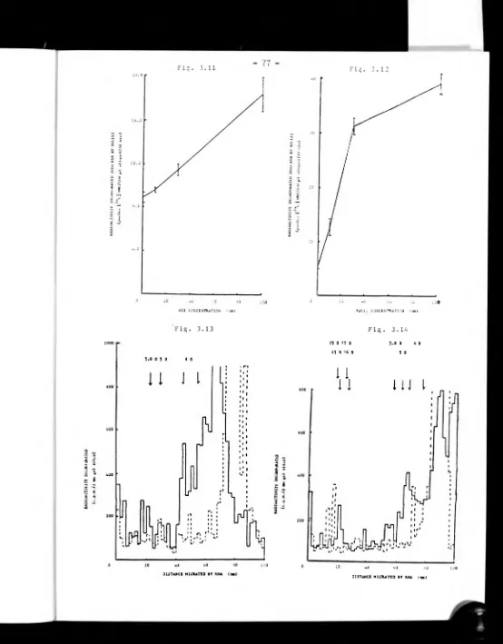
3.3.7 Effect of magnesium and salt concentration on nuclear
RNA synthesis 74.
3.3.8 Analysis of RNA synthesised in vitro by nuclei 76.
3.4 Discussion 76.
3.4.1 RNA synthesis in C. albicans blastospores and
protoplasts 76.
3.4.2 Preparation of C. albicans nuclei 81.
3.4.3 Nuclear RNA synthesis in vitro 84.
3.4.4 RNA species synthesised in vitro by C. albicans
nuclei 91.
Chapter 4 Purification of RNA polymerases from yeast and
partial purification from mycelial form C_j_albican^ 93.
4.1 Introduction 93.
4.2 Materials and methods 96.
4.2.1 Preparation of cell free extracts 96.
4.2.2 Protamine sulphate precipitation of nucleic acids 97.
iv
-4.2.4 Ion exchange chromatography 97.
4.2.5 Affinity chromatography 98.
4.2.6 Glycerol gradient centrifugation 98.
4.2.7 Polyacrylamide gel electrophoresis 98.
4.3 Results 99.
4.3.1 Purification of RNA polymerases from C. albicans 99.
4.3.2 Initial stages in the purification of RNA polymerases
from yeast C. albicans; preparation of homogenate 99.
4.3.3 Initial stages of RNA polymerase purification:
preparation of sonicate 103.
4.3.4 RNA polymerase activity at various times after
preparation of homogenate 105.
4.3.5 Gel filtration of yeast form RNA polymerases 105.
4.3.6 Ion exchange chromatography of yeast form RNA
polymerases 105.
4.3.7 Affinity chromatography of RNA polymerases from yeast
form C. albicans 107.
4.3.8 Glycerol gradient centrifugation of yeast RNA
polymerases 107.
4.3.9 Non-denaturing PAGE of purified yeast RNA polymerases 107.
4.3.10 Divalent cation optima for partially purified RNA
polymerases 110.
4.3.11 Salt optima for partially purified RNA polymerases 110.
4.3.12 <x -Amanitin sensitivities of partially purified RNA
polymerases 110.
4.3.13 Apparent Km for CTP for yeast form partially purified
RNA polymerases 114.
4.3.14 Partial purification of mycelial form RNA polymerase 116.
4.3.15 Gel filtration of mycelial form RNA polymerase 116.
4.3.16 Ion exchange chromatography of mycelial form RNA
polymerase 116.
4.4 Discussion 116.
4.4.1 Purification of RNA polymerases from yeast form
C. albicans 116.
V
-P a g e
4.4.2 Characterisation of RNA polymerases from yeast form
C. albicans 125.
4.4.3 Preparation of RNA polymerase from mycelial form
C. albicans 131.
Chapter 5 Some studies on inhibitors of RNA svnthesis in
C. albicans 134.
5.1 Introduction 134.
5.2 Materials and methods 135.
5.3 Results 135.
5.3.1 Effect of lomofungin on C. albicans exponential
phase growth 135.
5.3.2 Effect of lomofungin on growth of several strains
of C. albicans 136.
5.3.3 Effect of various concentrations of lomofungin on
RNA synthesis by protoplasts of C. albicans 136.
5.3.4 Effect of lomofungin at a concentration of 20 /ig/ml
on growth of S. cerevisiae 138.
5.3.5 Effect of some known inhibitors of transcription and nucleoside analogues on in vitro RNA svnthesis bv
C. albicans 138.
5.3.6 Effect of some known inhibitors of transcription and nucleoside analogues on in vivo RNA svnthesis bv
C. albicans 142.
5.4. Discussion 142.
5.4.1 Studies using the inhibitor lomofungin 142.
5.4.2 Inhibitors of in vivo and in vitro RNA svnthesis 144.
Chapter 6 The involvement of RNA svnthesis in the veast-
mycelial transformation of C. albicans 147.
6.1 Introduction 147.
6.2 Materials and methods 149.
6.2.1 Organism and growth conditions used 149.
6.2.2 Germ tube formation bv C. albicans 150.
6.2.3 Measurement of RNA synthesis during germ tube
vi
-6.2.4 Extraction and analysis of RNA synthesised during
germ tube formation 150.
6.3 Results 151.
6.3.1 Pretreatment of C. albicans for eenti tube formation 151.
6.3.2 Germ tube formation by C. albicans in various buffered systems and the effect of varying the
concentration of the stimulant 153.
6.3.3 RNA and protein content of C. albicans durine eerm
tube formation 156.
6.3.4 RNA synthesis bv C. albicans durine eerm tube
formation 156.
6.3.5 Inhibitors of transcription and translation on germ
tube formation by C. albicans 163.
6.3.6 Analysis of RNA synthesised by C. albicans durine
germ tube formation 168.
6.4 Discussion 169.
6.4.1 Pretreatment of C. albicans for eerm tube formation 169.
6.4.2 Media used to induce germ tube formation by
C. albicans 171.
6.4.3 Protein and RNA content during the yeast-mycelial
transformation of C. albicans 177.
6.4.4 RNA synthesis by C. albicans durine the veast-
mycelial transformation 181.
6.4.5 The effect of inhibitors of transcription and
translation on eerm tube formation by C. albicans 186.
6.4.6 RNA species synthesised by C. albicans durine eerm
tube formation 189.
Chapter 7 General discussion 194.
7.1 Introduction 194.
7.2 RNA synthesis in C. albicans 194.
7.3 Other potential sites for chemotherapy 195.
7.4 Further research on RNA synthesis in C. albicans 196.
7.4.1 Further research on RNA polymerases of C. albicans 196.
«Si*.
VÜ
-7.4.3 Studies on the transport of drugs into C. albicans
7.4.4 Combination chemotherapy and alternative approaches to candidosis
7.5 Final conclusion
Page
2 0 0
.
2 0 1
.
2 0 2.
- Till -
ACKNOWLEDGEMENTS
I would like to thank the Chairman of the Department of Chemistry
and Molecular Sciences, Professor G.H. Wallbridge for the use of a
laboratory and facilities to pursue this research. In addition, I
must also thank the Science and Engineering Research Council for
financial support throughout this research. I would also like to thank
my supervisor Dr. B.E.P. Swoboda for help and encouragement. In
addition, I would like to express my appreciation to Imperial Chemical
Industries p.l.c. for allowing me the use of a laboratory and facilities
at Mereside, Alderley Park and Dr. J.F. Ryley, Dr. K. Barrett-Bee and
Mr. R.G. Wilson for showing me how to use them. I would also like to
thank Dr. A.M. Coles for his help during the initial stages of this
research. Finally, I must thank my sister, Miss C. Peters for typing
SUMMARY
The thesis describes the results from investigations into RNA synthesis in the dimorphic fungus Candida albicans. Methods were described, and evaluated, for the preparation of protoplasts and nuclei - two in vitro systems that were used to study RNA synthesis. It was found that most radiolabelled precursor was incorporated into low molecular weight RNA by nuclei. In contrast, the precursor was associated with higher molecular weight RNA species from protoplasts, from which the nuclei were prepared. Contamination by RNases was a serious problem in the preparation of this
in vitro system from protoplasts.
The RNA polymerases were purified from yeast and part-purified from mycelial C . albicans. Three classes of RNA polymerase were resolved by ion exchange chromatography of cell-free extract from yeast, whilst only one was found in mycelia. Some characteristics of RNA polymerases from the yeast were described. All three isozymes had optimal activity in vitro when the media contained mono and divalent ion concentrations that were similar to those reported for Saccharomyces cerevlsiae - a yeast often compared with C. albicans. In addition, the three isozymes from C. albicans had similar Km values for CTP to that of these enzymes from other eukaryotes. RNA polymerases I and III from yeast C. albicans showed similar sensitivi ties to <*.— amanitin as the corresponding isozymes from S. cerevisiae. RNA polymerase II was far more insensitive to the amatoxin than the corres ponding enzyme from higher eukaryotes.
A variety of nucleoside analogues were suggested as potential anti fungal agents warranting further investigations. These were capable of inhibiting RNA synthesis by C. albicans in vitro and/or in vivo assays.
Studies were also made on RNA synthesis during germ tube formation - the initial stage of the yeast-mycelial transformation. It was found that it was necessary to cultivate C, albicans yeast in nutritionally impoverished media and starved for 24 hr to achieve reproducible germ tube formation. The strain of C. albicans used in this research formed germ tubes when incubated in imidazole HC1 buffer containing serum, N-acetgl glucosamine, glucose or glucose plus glutamine at temperatures above 35 C.. During germ tube formation there was an increase in the RNA content per unit yeast cell. Both the rate and maximum amount of radiolabelled precursor incorporated into RNA depended upon the conditions used to induce germ tube formation. Some inhibitors of RNA synthesis were capable of inhibit ing germ tube formation. It was found that the ratio of high to low molecular weight RNA species changed over the period of germ tube formation.
— X
-ABBREVIATIONS
adenosine diphosphate ADP
adenosine monophosphate AMP
adenosine triphosphate „ -3
ATP
10 Amperes mA
base pairs bp
Becquerel Bq
bovine serum albumin BSA
Centigrade ° C.
counts per minute c .p .m
Curie Ci
cytidine diphosphate CDP
cytidine monophosphate CMP
cytidine triphosphate CTP
de-oxyribonucleic acid DNA
dimethyl sulphoxide DMSO
dithiothreitol dTT
double stranded ds
ethylenediaminetetra acetic acid EDTA
glucose glu
glucose/beef extract broth G • B ■ E .
glutamine gin
gramme g
guanosine diphosphate GDP
guanosine monophosphate GMP
guanosine triphosphate GTP
heterogenous nuclear ribonucleic acid hnRNA
hour hr
xi
-messenger ribonucleic acid mRNA
minimum inhibitory concentration MIC
minute(s) min
molecular weight M.W.
N-acetyl glucosamine NAG
National Collection of Pathogenic Fungi NCPF
Newtons N
nucleoside triphosphate NTP
optical density O.D.
Pearson product moment coefficient of correlation r
phenylmethyl sulfonylfluoride PMSF
polyacrylamide gel electrophoresis PAGE
revolutions per minute (r.p.m.) r.p.m.
ribonucleic acid RNA
ribosomal ribonucleic acid rRNA
Sabourauds dextrose agar S . D • A .
Sabourauds dextrose broth S .D.B.
seconds sec
Shepherd and Sullivan S . and S .
sodium dodecylsulphate SDS
Svedberg units S
trichloroacetic acid TCA
Tetramethylethylenediamine TEMED
Tris(hydroxymethy1)aminomethane Tris
uridine diphosphate UDP
uridine monophosphate UMP
uridine triphosphate UTP
ultra violet uv
weight/volume w/v
volume/volume v/v
- xii -
PREFACE
Many species of fungi are potentially pathogenic in man. The dimorphic
fungus Candida albicans is possibly responsible for the greatest number of
such infections in man. Although there are antifungal agents which are
used against C, albicans, most have drawbacks such as showing toxicity to
the host. Only the recently introduced antifungal agent ketoconazole has
the advantage that it appears to have minimal side effects. RNA synthesis was
studied as a possible target site for novel inhibitors of C. albicans that
would not affect the mammalian host. This thesis will describe the results of such investigations.
The introductory chapter will examine some biochemical, epidemiological and physiological aspects of C. albicans and antifungal agents currently
employed against various forms of candidosis. In addition, this chapter will
review some aspects of eukaryotic RNA synthesis. Chapter 2 will describe
the techniques required for research into any aspect of a pathogen, such as
C. albicans, and the methods used to investigate RNA synthesis in this
organism. The next four chapters will be concerned with evaluating the
results obtained from investigations into RNA synthesis in C, albicans. The
first of these chapters will describe and discuss the preparation of two
in vitro systems that may be used to study RNA synthesis. The next chapter
will describe the purification, and some characteristics, of RNA polymerases
from yeast and the partial purification of this enzyme from mycelial
C. albicans. The results obtained in these two chapters will be compared with other eukaryotes in general and Saocharomycea cerevlaiae in particular.
Chapter 5 will describe investigations into the effects of the inhibitor
lomofungin on growth and RNA synthesis in C. albicans. (Lomofungin is a
reputed inhibitor of these processes in other fungi). The effects of some nucleoside analogues on RNA synthesis in C, albicans were investigated
using in vitro and in vivo systems prepared as described in the preceeding
xiii
-characteristic of C. albicans, which may be important in the invasion
of the host. Conditions will be described for the reproducible induction of germ tubes from the strain of C. albicans used for most of this research.
RNA synthesis and the RNA species synthesised during the initial stages will
be analysed in the hope of further understanding this morphological change.
The final chapter will discuss the results of the thesis and suggest possible
1
CHAPTER 1 RNA SYNTHESIS IN Candida albicans
1.1 Introduction
The yeast Candida albicans is a pathogenic dimorphic fungus which is
responsible for the largest number of infections of fungal aetiology in
humans. Almost certainly, the most prevalent type of infection reported is vaginal candidosis in women of childbearing age. Recent (i.e. 1984)
Department of Health statistics have shown an increase in the incidence
of women showing symptoms of this disease over the past few years ( 89 ). Although treatment has improved recently, relapses are very common and
a totally satisfactory systemic treatment is not yet-available. There
thus exists a huge market for suitable antifungal agents. Although there
are pharmaceutical preparations available, it was not until the recent
introduction of ketoconazole that a suitable treatment for candidosis could be taken orally. In order to assist in the development of other desirable
antifungal agents that could be ingested orally, RNA synthesis was studied
as a potential target site. RNA synthesis was chosen as it was hoped to
find exploitable differences between this process in C. albicans and higher mammaIs.
In addition to existing as a pathogen, C. albicans may reside as a commensal in the human or animal host. Dimorphism may play an important
role in the conversion from commensal to parasitic existence (see 6.1 ) . The involvement of RNA synthesis in this transformation was investigated
to give an insight into this poorly understood phenomenon. This thesis
will examine how successful these two aims were and'also how apt RNA synthesis
is as a site for novel inhibitors.
1.2.1 C. albicans - Characteristic morphology
C. albicans may exist in two distinct morphological forms. It can
exist as a unicellular yeast (blastospore) or as an aggregate of multicellular
hyphae (mycelia). C. albicans is taxonomically classified as an
- 2
yeast genera. In addition, it is unable to form a sexual stage in its life
cycle. Therefore, its relationship to "classic" yeast genera, such as
Saccharomvces. is obscure.
A wide variety of media have been described which promote the growth
of either form of C. albicans. Most of these media favour growth of the
yeast form in vitro. A yeast may be defined as "a fungus whose predominant
morphological form is unicellular" (220). (It is this morphological
characteristic that lead Lodder and Kreger Van Rij to classify the Candida
genus as yeasts). Macroscopically.C. albicans yeast colonies have a white,
creamy, moist and shiny appearance on solid media. Microscopically, the
yeast blastospores are ovoid in shape, approximately 4 - 6 ^im by 6 - 8 ¿jm Blastospores arise through mitotic cell division (budding). The daughter
cell grows, for a period of time, from a small, selected site on the apex of the parent. A septum (cross wall) is formed between the two cells just
prior to separation (Fig. 1.1).
Aside from the inability to form a sexual stage in the life cycle, the
principle characteristic of the Candida genus is the ability to form chains
of elongated yeast cells (pseudo-hyphae). Pseudo-hyphae are formed by a
process similar to blastospore development. However, the daughter cells
remain attached to the parent. In addition, subsequent daughter cells are
more narrow and elongated than the original parent cell (Fig. 1.2).
Many workers have confused hyphae with pseudo-hyphae even though their
mode of formation is different. Hyphal formation is characterised by
cellular material growing from the blastospore in narrow cylindrical tubes. This process is termed germ tube formation. In certain media (e.g. some
peptone broths) blastospores may be produced along the hyphal shoot.
However, in media such as serum more hyphal branches may be formed as
the hypha extends (Fig. 1.3). All hyphae usually aggregate as mycelia and
can be seen macroscopically as large "clumps" of cells in suitable media.
C. albicans is able to form chlamydospores in certain nutritionally
out 3 out
-0
-0—0
JL
4
-growths of cellular material are attached to the main body of yeast or
hyphal cells by a suspensor cell. Chlamydospores have a thick double
layered cell wall: the outer layer containing /3(l - 3)glucans and other
polysaccharides,whilst the inner is mostly protein (176). This cell wall
surrounds a cell membrane, within which are large amounts of storage lipids, polysaccharides and proteins (263). It has been suggested that
chlamydospores are a dormant phase of the fungus (17 ).
The appearance of chlamydospores and germ tubes are generally accepted as positive presumptive evidence for the identification of C. albicans.
However, other Candida species, such as C. tropicalis. have been reported
as having the ability to form chlamydospores (154). In addition, there
are some strains of C. albicans that do not readily form germ tubes. As a
result, many medical mycologists use such physiological and biochemical
properties as the ability to ferment and assimilate suitable compounds as tools for the identification of C. albicans.
1.2.2 C. albicans - Biochemical and physiological properties
In recent years there has been an increase in the number of papers
dealing with the biochemistry and physiology of C. albicans. However, in
comparison to the extensive literature concerned with these aspects in
another yeast, Saccharomvces cerevisiae. such studies on C. albicans are far from complete. A great deal of the new research is concerned with
studies on the mode of action of inhibitors (see 1.2.5) and the dimorphic trans
formation (see chapter 6).
C. albicans is able to grow using a variety of compounds as sole carbon or
nitrogen sources. In the laboratory more complex media are used to cultivate the
organism. These conditions usually result in cultivation of the yeast form of
C. albicans. Growth is possible on media that have a pH in the range 3 - 8 and
an incubation temperature of 20 - 40° C.. Growth of C. albicans is usually much
5
-investigations usually involves growth in peptone-glucose media. Fairly high
growth rates have been reported in these media. Indeed, doubling times, in the
exponential phase of growth, of 70 min are not unusual (109).
Studies of C. albicans have so far indicated that the biochemistry of this organism is typical of other lower eukoryotes such as S. cerevisiae. Much research
on the metabolism of C. albicans has investigated oxidative phosphorylation and
carbohydrate utilisation. As an aerobic eukoryote possessing mltrochondria, the oxidative phosphorylation mechanism of C. albicans is reportedly similar, to
that of S. cerevisiae (410). However, despite possessing all the enzymes necessary
for anaerobic glycolysis (299)^ C. albicans is unable to grow anaerobically (266).
In addition^ it has also been found that C. albicans possesses all the enzymes of the
hexose monophosphate pathway and most of those required for the tricarboxylic acid
cycle (299). Hexoses may be diverted from glycolysis to form polysaccharides (64 ).
The enzymes responsible for the synthesis of glucans and mannans have been exten
sively studied. These polysaccharides are important cell wall constituents of C. albicans and other fungi (85, 240).
The cell wall and membrane have both been fairly well studied - the latter as it is believed to be a site of action for a variety of inhibitors (see 1.2.5). The
cell wall maintains the distinctive ovoid shape of C. albicans and also acts as a
boundary between the cell and environment. Early research showed that the cell wall possessed large quantities of glucans and mannans with small amounts of protein,
lipid and chitin (65). The glucans and mannans consist of backbones of hexose
(glucose in the former, mannose in the latter) residues which contain extensive
branches and cross links. The glucans have a /2(1 - 6) linked glucose backbone with /3(l - 3) linked branch points. Mannans contain «-(l - 2) branch points of
mannose units attached to an <x (1 - 6) linked mannose backbone (287). There may
be as many as eight layers of polysaccharide in the cell wall (294).
The polysaccharides have often been found associated with proteins as
glucoprotelns, mannoproteins or glucomannoproteins (189) Recent studies have
6
-implicated in affecting the ability of C. albicans to bind to buccal epithelial
cells (96, 320). They may thus have an important role to play in the invasion of
mucous epithelia. The cell wall glucoproteins and mannoproteins are the antigenic
determinants by which strains of C, albicans can be differentiated into one of two
types. Hasenclever's group first demonstrated this phenomenon and termed the two
antigenic types group A and group B (153, 155). The former type (group A) possessed the same complement of antigens as the other type (group B), plus at least two
further antigens (370).
Enclosed within the cell wall is the cell membrane. This contains both free
sterols and sterol esters, triglycerides and carboxylic acids of a chain length
C^g (239). Phosphatidylcholine and phosphatidylethanolamine have been found to be
the major phospholipids (102), The quantitive levels of the various membrane lipids
have been shown to vary from strain to strain (149) and can also be changed by the
chemical composition of the media ( 1 94-^« The cell membrane is also the site of a
variety of lipid soluble enzymes. These include such enzymes as (1 - 3) glucan
synthetase (125)and chitin synthetase (73>*enzymes implicated in the dimorphic transformation (see chapter 6).
The limited research into the molecular biology and macromolecular synthesis of £j_^lbican£ has shown similarities between these and appropriate processes in
S. cerevisiae. Such similarities have tempted several workers to use this non
pathogen as a model for drug and inhibitor studies (212). However, as mentioned in
1.2.1, C. albicans differs from S. cerevisiae in two important respects:
the inability of the former to produce a sexual stage and the ability to
form hyphae and pseudo-hyphae under suitable conditions.
1.2.3 C. albicans - Ecology and Epidemiology
Ci_albicansi is distributed worldwide and found in terrestrial and aquatic
environments. As the causal agent responsible for candidosis, the fungus has been found In man and the primates and other warm blooded mammals (122).
7
-of reviews -of yeast epidemiology have drawn the conclusion that C. albicans t
and a few other yeast species, are common and harmless commensals (98, 122 23®. The fungi have been recovered from samples taken from mucous membranes
and the digestive tracts of "normal" individuals.
Studies into the frequency of recovery of C. albicans from the mouths of
"normal" individuals have reported the presence of the fungus in between 2
and 237. of test subjects (236,278). However, in hospitalised individuals
the presence of the yeast, from oral sources, may be as high as 707. (24 ).
The oral populations of yeasts are not static and have been found to fluctuate
day by day and with the age of the individuals (122). C. albicans has also been recovered from faecal samples with recovery frequencies of around 157.
(94 ). Most publications concerning the frequency of recovery of C. albicans
from human sources use the vagina as the source. The frequency of recovery of C. albicans is higher in women suffering symptoms of vaginal thrush than
hospitalised or "normal" individuals. C. albicans is most often recovered from samples taken from women of childbearing age (278). Samples from other
sites, such as skin, oesophagus and stomach, have been found to contain
C. albicans. These are usually taken from hospital patients showing signs of other debilitating illnesses (278). A high proportion (60 - 707.) of
C. albicans isolates, from all sites, have been found to be of the A
serotype (156).
1.2.4 C. albicans - Candidosis and pre-disposing factors of the host
Section 1.2.3 indicated that many people harbour within them conmensal
yeasts, such as C. albicans, which have a potential to become pathogenic.
Indeed, it has been suggested that "C. albicans is a better clinician and
can discover abnormalities in persons much earlier in the course of the
development of such abnormalities than we can with our chemical tests" (400).
There are esentlally four types of candidal infection. Mucosal infections are those of the mucus membranes, such as mouth and vagina. Cutaneous
8
-those affecting both mucosal and cutaneous sites. This form of infection,
and systemic candidoses, affecting the internal organs, are by far the most serious clinically.
A whole variety of pre-disposing factors can account for susceptibility
of individuals to candidosis. These can be classified as "natural", dietary,
mechnical and iatrogenic medical factors (278). "Natural" factors include
diabetes mellitus (and other debilitating diseases and disorders) and
digressions from normal physiological status (e.g. pregnancy). The excess
or deficiency of foodstuffs may alter the composition of endogenous flora
permitting alterations in the levels of C. albicans. Mechanical factors such as trauma (e.g. burns) or local tissue disruption (e.g. wearing dentures)
may permit invasion of the affected tissue area by C. albicans.
Possibly the most important factor for serious infection by C. albicans
at present is that arising as a consequence of iatrogenic medical factors.
The incidence of such serious infections is increasing as new techniques in
medicine have been introduced. Treatment with antibiotics or corticosteroids
alters the gut floral population of treated individuals. This has been found to result in an increase in the incidence of individuals showing
symptoms of candidosis (278). Surgical procedures - such as the introduction
of a catheter into the blood vessels or urinary tract - may also be accompanied by serious candidosis.
The range of situations that can dispose an individual to either local
or widespread candidal infections is extensive. The possibility of serious
systemic candidosis in severly ill hospital patients is considerable. Thus, where systemic candidosis is diagnosed, the primary consideration for treat
ment should be to attempt to minimise the many factors that can potentiate
the disease.
In present day medicinal practice vaginal candidosis is commonly seen.
There is thus a good deal of interest shown in this disease . This may be
illustrated by the large number of publications concerned with the carriage
-
9
-The classical lesions of the disease are a white discharge from the vagina
coupled with soreness,inflammation and itching of the affected area. How ever, accurate diagnosis may only be confirmed by detection of C. albicans in swabs.
Many authors believe that vaginal candidosis has become more common
in the post-antibiotic and post-oral contraceptive age. However, definitive proof is not easy to find(27e). The fact that the number of diagnoses of vaginal candidosis has risen annually(89), may be an indication of increasing
numbers of women contracting the disease or physicians are increasingly
correctly interpreting the symptoms. VaginaL thrush is commonly seen during
pregnancy. In addition, the prevalence of vaginal yeasts is higher with
women taking oral contraceptives compared to those who are not (278). Some authorities believe that the increased wearing of occlusive nylon tights
may increase the likelihood of contracting the symptoms.
A consequence of the increasing numbers of women contracting the
disease has been that many pharmaceutical companies have made strenuous
efforts to produce suitable antifungal agents. There are probably more formulations of pharmaceuticals currently available for the treatment of
vaginal candidosis than for any other type of candidal infection. These drugs - the mode of action of which will be discussed in 1.2.5 -
may be supplied as creams, lotions or pessaries. The literature
available on treatment of vaginal candidosis suggests that most cases will
respond to topical treatment alone. However, the extent of re-infection, or
possible relapse of the disease, means that an aggressive schedule of treat ment must be attempted for some patients at least (278).
Oral thrush is another mucosal infection which, although less cotimon than vaginal candidosis, may be observed in elderly, debilitated patients.
New born infants may also be susceptible to C. albicans infections because
of their immature antimicrobial defences. This disease may be treated using similar topical regimes as that for vaginal candidosis. Extensive topical
-
10
-In contrast to candidal infections of mucous membranes, not fatal and
conmonly seen in medical practice, systemic candidoses affecting the central
nervous system, and heart, used to be fatal until the introduction of
suitable antifungal agents. Candidal infections of bone and joint,
respiratory and genito-urinary tracks and kidney have all been reported. The severity of these systemic candidal infections is in proportion to
the extent which the host's immune response system is suppressed. In
addition, chronic muco-cutaneous candidosis may arise as a consequence of a deficient immune system. Thus, fatal widespread lesions arising due
to infection by C. albicans are only now usually seen in patients with
extreme debilitation (278).
1.2.5 C. albicans - Antifungal agents currently available for the treatment of
Candidosis
The only suitable treatment for superficial candidosis up to the 1950's
was provided by formulations containing Gentian violet and similar non
specific antiseptics (400). Nystatin, the first specific antifungal agent, was discovered in 1950 (158). Since then, the literature concerned with
compounds that show inhibitory activity against fungi has increased
enormously.
The route from in vitro efficacy of a compound, to commercial availability for treatment, is both time consuming and expensive. Firstly, a compound
must be identified that shows in vitro inhibitory activity against C. albicans
- and hopefully other fungi. This must then be followed by investigations
into the mechanism of action of this potential drug. A successful candidate
should show differential sensitivity between mammalian cells (the host) and
the fungus (the pathogen). This must be followed by detailed pharmacokinetic
studies on in vivo models - infected laboratory animals. This should
indicate whether the candidate drug is safe for clinical trials on human
volunteers - the penultimate step before the compound is licensed for
11
-The cost of research and development of a potentially successful anti
fungal agent has been estimated at
£25
million( 3 i e ) .
Only the large multinational drug companies of the western world - financed by profits from
pharmaceutical sales - can afford such costs. As a consequenc^ research can
only be directed towards illnesses and infections of sufficient magnitude
that guarantee financial reward. Mucosal candidal infections are widespread (see
1 . 2 . 4 )
and thus represent a large market for potential novel inhibitors.Any drug that shows good inhibitory activity against less common fungi, or
has high production costs, is unlikely to be developed. It is therefore
perhaps not surprising that there are few pharmaceutical agents that are widely used in antifungal chemotherapy. These are the polyene
antibiotics, the substituted imidazoles and the nucleoside analogues.
The two polyene antibiotics most commonly employed as antifungal agents
are nystatin and amphotericin B (see Fig. 1.4). Other polyene antibiotics,
which have been used against candidal infections, include candicidin, pimaricit
trichomycin and hamycin. These polyenes are isolated from a variety of
Streptomvces species (148). Minimum inhibitory concentrations (MIC) of
0.4 pg/ml (0.4 ^iM) to 50 jug/ml (53 /iM) for nystatin and less than 1 /lg/ml
(1 jM ) are usually reported for these two common antibiotics in vitro
against C. albicans (318). These M IC values may be affected by the media
composition. The MIC is not affected by the inoculum size or incubation
time (118,165).
The mode of action of these drugs is believed to be that they bind with sterol components of the cell membrane, which is ergosterol in susceptible
fungi. This binding alters the permeability of the membrane to allow leakage
of cytoplasmic materials < 31 ©>. Resistance to nystatin and amphotericin B has only been induced in vitro by growth on media containing the polyene
antibiotics. These drug resistant strains show reduced virulence when Infected in laboratory animals (278).
Clinically, amphotericin B is the most important polyene antifungal agent.
12
-■XTOCONAZOLE
[image:27.582.13.569.12.715.2]13
-candidoses, the toxicity of this compound to the mammalian host system
reduces its usefulness. Amphotericin B has been applied to virtually every
form of candidosis. However, this compound also shows toxicity towards
humans, although the side effects are less severe than other polyene anti
fungal agents. The main side effects are kidney damage, uraemia and hypokalemia (2 78).
One possible method of reducing the effective cidal concentration
pf a compound, when treating candidosis, is to use the drug in synergystic combination with amphotericin B. It has been known for some time that anti
bacterial therapy can be improved by the use of two antibiotics acting
synergistically against a particular organism (177). A synergistic in vitro antifungal response has been found using sub-lethal levels of amphotericin B with any of the following:- ascorbic acid and cysteine (26 ), tetracycline
and actinomycin D (206), 5 - fluorocytosine (253) and rifampicin (27 ). It
is believed that the levels of amphotericin B used enable a sufficient
number of "holes" to develop in the membrane, allowing access of the anti
biotics to their sites of action (254).
The polyene antibiotics have now been superseded by the mono-N-
substituted imidazoles for the treatment of superficial fungal infections.
High cure rates have been reported for clotrimazole - the first such compound
used against vaginal candidosis (278). Other substituted imidazoles now used against superficial infections Include miconazole, econazole and,
recently, ketoconazole (see Fig. 1.4). The minimum inhibitory concen
trations of the substituted Imidazoles in vitro against C. albicans vary from
0.02 jig/ml (0.06 juM) to 4 pg/ml (11.6 <>iM) for clotrimazole (278,318). The
other substituted imidazoles show minimum inhibitory concentrations of
similar magnitudes (343). The wide variation of such values may be accounted
for by inoculum density (151), media composition (407), pH (417) and morphological form (183), all of which can alter the sensitivity shown by
[image:28.587.21.557.14.741.2]the method used to determine the value (145).
The mode of action of the substituted imidazoles is believed to be
inhibition of ergosterol biosynthesis. Van den Bosche et al found that, at
concentrations of 9.05 jig/ml (0.1 ;uM) miconazole inhibited ergosterol bio
synthesis by blocking déméthylation of the precursor sterol at (386).
This same group found higher concentrations of substituted imidazoles were
required for inhibition of rat liver cholesterol biosynthesis (385). Thus,
the substituted imidazoles indirectly affect the membrane permeability of
susceptible organisms and cause visible damage to the structure and function
of intracellular organelles (318). However, other groups (80, 101) feel
that the substituted imidazoles have a direct effect with the membrane
causing rapid leakage of K ions and high and low M.W. cytoplasmic molecules.
Clotrimazole and miconazole are poorly absorbed from the digestive tract
and consequently low serum levels are found. This may be compounded by liver microsomal enzymes that rapidly breakdown the active drugs (99 ) . However,
much higher levels in serum of water soluble kétoconazole have been obtained (316), which suggest a more promising role for this substituted imidazole
against systemic candidosis.
The nucleoside analogue most widely used against C. albicans is undoubtedly 5 -fluorocytosine (5FC) (see Fig. 1.4). Since its commercial
introduction in the late 1960's, this fluorinated pyrimidine has been used
successfully, primarily against systemic candidoses (276). Concentrations of
0.1 jig/ul (0.8 ^iM) to 15 /Jg/ul (116 ¿iM) are usually inhibitory to C. albicans
ln vitro (27Ö. However, not all yeast isolates have been found to be
sensitive to the drug. Estimates of primary resistance of between 107. (27e)
- 207. ( 13 ) of clinically isolated strains tested have been reported. The
B serotype has been found to show a higher incidence of resistance to 5FC than the A ( 13). Resistance to 5FC may be easily induced in vitro by
growth of C. albicans on media containing low levels of the drug ( 87 ). Medical practitioners .should bear this in mind for clinical treatment of
The mechanism by which 5FC inhibits C. albicans growth is believed to be by disruption of translation. 5FC is taken up into C. albicans by a
cytosine permease. The pyrimidine is then de-aminated, phosphorylated to
5-fluorouridine triphosphate and incorporated into RNA. The presence of the
fluorinated pyrimidine base in the RNA disrupts the normal protein synthetic
activity of the cell and normal development is therefore grossly impaired.
As much as 507. of the uracil may be replaced by 5-fluorouracil. Incorporation
of 5FC into the host mamnalian cell is limited due to lack of the specifically required permease (29t).
As a water-soluble pyrimidine, 5FC may easily be absorbed from the gut^
hence the preferred formulation is in tablet form. Peak serum levels of
5FC follow after 5 hr ingestion (273). Unfortunately, high levels of 5FC
have been found to cause liver damage (302) and show bone marrow toxicity (290). This problem, coupled with the ease some strains of C. albicans
show resistance to 5FC, indicates that there is a requirement for anti
fungal agents that do not have these drawbacks.
Overall, with the possible exception of ketoconazole, present anti
fungal agents show much to be desired. As indicated above, problems such
as drug toxicity, limited absorbtion from the intestinal tract, and the
ease of development of resistance by the fungi abound. As a consequence, any new drug should show a wide spectrum of activity, the possibility of
oral administration with freedom from toxicity and low production costs.
It was to achieve such an end that RNA synthesis in clinically the most
important species, C. albicans was studied.
RNA synthesis
These following sections of this introductory chapter will discuss some
aspects of DNA directed RNA synthesis (i.e. transcription), with particular
reference to eukaryotes. Included in these sections will be discussions on the general RNA species that are transcribed from the DNA, the organisation
16
-the process of RNA syn-thesis. It must be stressed that this is in no way a
comprehensive study of this ever-expanding field - it should be considered
an overview of current trends and ideas in the area.
1.3.1 Transcription of DNA coded RNA species
The "central dogma" of molecular genetics is now a firmly established scientific principle (i.e. DNA— » R N A — »protein) (394). These processes
apply to all living cells. In eukaryotes, the cell's complement of DNA
may be found in the nucleus, which is delineated by a double-membraned
nuclear envelope. The possession of a nucleus distinguishes eukaryotic
cells from prokaryotes. Eukaryotic transcription and processing of the
newly synthesised RNA (i.e. post-transcriptional modification) is performed
within the confines of the nucleus. Eukaryotic protein synthesis (trans
lation) occurs on the ribosomes (highly complex ribonucleic acid - protein
assembly), which are located on the outer membrane of the endoplasmic
reticulum in the cytoplasm. In contrast, prokaryotic protein synthesis
occurs concurrently with one end of the transcribed RNA translated whilst the other end may still be being transcribed (394).
The DNA may be divided into 2 sorts - that which codes for RNA and that which is non-coding. The functions of the non-coding regions are not
yet fully understood. However, some workers (e.g.119) believe that these
sequences are important in determining the patterns of DNA folding in the
nucleus (see 1.3.2). The DNA that is transcribed may be used as a template
for translation (i.e. messenger RNA) or to form the ribosome (ribosomal RNA)
or that which carries the amino acid in the translation step (transfer RNA).
Although the general principle of eukaryotic DNA transcription is the same
as in prokaryotes, the machinery necessary to control gene activation and repression and "unpack" the DNA is considerably more complex. In the
eukaryote transcription of all 3 RNA species is usually followed by post
transcriptional modifications (285).
17
-The genes coding for ribosomal RNA (rRNA) are highly re-iterated. There have
been reports of anything between 50 and 2 x 10 copies of the gene present in each cell (140). These multiple gene copies are linked in tandem array
along the genome of the organism. The transcription unit is relatively
constant in size, with a molecular weight (M.W.) of 4 x 10^ to 5 x 10 .
However, the non-transcribed spacer region varies in length from organism
to organism (140). In eukaryotes the rRNA unit is transcribed as a 37 S to
45 S precursor, from which the mature rRNA species are processed. Multiple
copies of DNA sequences coding for transfer RNA (tRNA) and another rRNA -
5 S rRNA - are present in the eukaryotic genome in tandem arrays. These RNA species may also be processed to produce the mature products.
The transcribed genes which are translated during protein synthesis are
often termed structural genes. The structural genes for eukaryote proteins
are large complex DNA molecules. In many cases, these contain a number of
intervening sequences not found in the mature messenger RNA (mRNA) (264).
These intervening sequences are not found in prokaryotes. Transcription by
the eukaryotic cell of the structural gene coded DNA produces long mRNA
precursor molecules. These precursor molecules are commonly referred to as
heterogenous nuclear RNA (hnRNA). The hnRNA molecule is "capped" by the
addition of a 7-methyl guanosine residue almost immediately. On completion
of the transcript, a poly-A polymerase enzyme adds 100 to 200 residues of
adenylic acid to the 3* end of the RNA chain. After splicing - i.e. removal
of the intervening sequences - the mRNA migrates as a ribonucleo-protein assembly to the ribosome. It is thought the 5* cap allows binding to the
ribosome. The function of the poly-A tail is not known for certain, but it seems to help mediate subsequent RNA processing and the migration from the
nucleus (265). Table 1.1 shows the number of nucleotides, molecular weights
[image:32.587.38.551.22.691.2]- 18
-RNA SPCC1CS
RNA POLYMERASE RESPONSIBLE FOR SYNTHESIS
SEDIMENTATION COEFFICIENT
NOUKIIU* WEIGHT, (* io'6 )
NUMBER OP NUCLEOTIDES
Mammallan 28 S 1 .9 5,000
Rlbosomal RNA 18 S 0.71 2,000
5.8 S 130 - 160
RNA Polymerase 1
S. cerevlsiae 2» S 1.3 6,000
Rlbosomsl RNA 17 S 0.7 2,000
5.8 S 0.06 158
Typical Mammalian Smal 1er than 6 S to Around 2.2 6,000
greater than 65 S (Anything up to 18) (Anything up to
50,000) RNA Polymerase II
Typical «RMA Usual ly 8 S - 15 S Around 0.5 1,500
(Between 0.2 (Anything in range
to 1.1) 500 - 3,000)
Small
Hibosomsl RNA 5 S 0.06 120
RNA Polymerase III
Transfer RNA 6 S 0.03 76
Prokaryotic Prokaryotic RNA 2) S 1.05 3,000
1,500
5 S 0.06 120
Table 1.1 Th« sedi mentation coefficients, molecular weight and number of nucleotides in soaie RMA species and the
RNA polymerase isozyme responsible for their synthesis. (Taken from Stewart and Latham 0 E 3 ) ).
Table 1.2 Subunit composition of several eukaryotic RNA polymerases
The cable shows the subunits chat make up the RMA polymeraae lsosyme from
three dlffarenc «ü^taryotes
a - numbers refer to M.w. x 10’3
I J ■ shown to be ldeatial polypeptodes by at least two criterion — - theae polypeptides have the same N.W.
[image:33.581.37.573.16.713.2]¡mump!
1.3.2 Organisation of the eukaryotic DNA into chromosomes
As noted in 1.3.1^ the major difference between prokaryotes and eukaryotes is the latter has a nucleus where most of its DNA is located. This section
will examine how the DNA is packaged in a eukaryote such as C. albicans.
Other differences are found between prokaryotes and eukaryotes, e.g. the DNA
content of the former is usually much smaller, whilst that of the latter is
usually tightly coupled with specialised proteins (histones). However, these differences may be regarded as secondary as the largest DNA complement
of bacteria may be as large as that of the smallest DNA complement of some
eukaryotes (119). Also one class of eukaryotes - the dinoflagellates - does
not possess histones, although it does possess the ubiquitous nuclear membrane (309).
The DNA from a eukaryote is usually in the form of a linear polymer
containing anything from 1 x 10⁷ to 1 x 101 1 base pairs (bp). In a lower
eukaryote, such as C. albicans.there are approximately 1.2 x 10⁷ bp
in the total genome (142). The folding of the DNA is an important consid
eration for eukaryotic cells as it is essential to pack the very long polymer
in an ordered manner. In C. albicans the DNA has to fit into the nucleus ~18 3 with a radius of 1 ^im ( 36) and hence with a volume of 4 x 10 m
assuming the nucleus forms a perfect sphere in the organism. The confor mation the DNA double helix may adopt depends upon a variety of external
parameters. Under "normal" physiological conditions the most stable
conformation of the DNA double helix is the right hand B form ( 9 3 ) . In
such conditions a polymer of DNA (with a diameter of 2.4 nm and 0.34 run
7 -20 3
between each bp) of 1 . 2 x 10 bp would occupy a volume of 2 x 10 m .
However, it is necessary to fold the DNA in such a manner that there is easy access, when required, by the transcription enzyme(s) and factors. The
histones pay a central role in storage and packing of the DNA.
Histones are small proteins (molecular weights of 10,000 to 15,000) that
contain a high proportion of positively charged amino acids. These allow
tight binding to the DNA. Four of the five histones are present In an
2 0
-octamer in duplicate amounts. These histones - termed histone H 2B, Hj
and - comprise the central core of the nucleosome - the fundamental
packing unit of eukaryotic DNA. These histones must rank amongst the most
highly evolutionary conserved proteins known. For example there are only 2
differences in amino acid sequence of histone from cows and peas (173).
X-ray diffraction patterns of crystals of the nucleosomes reveal that 146 bp of DNA are wrapped around the disc shaped histone octamer core. Each
core is connected by a "linker" sequence of approximately 60 bp in length
(307). The standard nucleosome consists of about 200 bp of DNA. In a lower eukaryote such as C. albicans one would expect a total of 5 x 10 nucleosomes.
The strand of DNA containing the histone octomers may be visualised by electron microscopy giving a "beads on a string" appearance (199). The 7NA
in this order of assembly may be termed chromatin.
The nucleosome is folded into the next level of organisation (the so
called 30 nm chromatin fibre) (350) and held by co-operative binding of the
5th histone - H^. Two models have been proposed for the packing of nucleo
somes into the 30 nm chromatin fibre. Both would allow a 1 cm strand of DNA
in the B conformation (i.e. approximately 2.9 x 10⁷ bp) to be packed into a
shorter strand of 0.24 mm in length (250). Thus, assuming the packaged
chromatin of C. albicans adopts the form of a cylinder, the volume of the
30 nm chromatin fibre in this lower eukaryote may be estimated at -20 3
7 x 10 m . The DNA must be further folded into "domains" using special
ised scaffolding proteins. Such a general folding pattern for the genome
has been adopted in both prokaryotic (e.g. Escherichia coll) and eukaryotic
(e.g. Man, Drosophila melanogaster) organisms ( 29 )• In eukaryotes the loops have an average length of 400 nm of 30 nm chromatin fibre. These fibres
are attached to the inner nuclear membrane by a specialised laminar protein
assembly.
1.3.3 DNA directed RNA polymerases
- 21
RNA polymerase (nucleoside triphosphate : RNA nucleotidyl transferase, E.C.
number 2.7.7.6). Over the past 10 - 15 years the subcellular localisation,
subunit composition and reaction properties of these complex enzymes - from
both eukaryote and prokaryotic sources - have been elucidated. Initial
in the early 1960*s (62) resulted in the isolation of a protein that was capable of DNA directed RNA synthesis in E, coli. Since then, it has been
found that prokaryotes possess a single class of enzyme responsible for the synthesis of all DNA coded RNA species. The more complex transcription
machinery of eukaryotes is reflected in most eukaryotes possessing three
classes of RNA polymerase. This is not a definitive difference between
prokaryotes and eukaryotes. It has been found that in at least one lower
eukaryote - Trypanosoma cruzi - there is only one RN A polymerase isozyme (19Z)
The enzymes from both prokaryotes and eukaryotes have several factors in
common for synthesising RNA. Firstly, both are DNA directed (i.e. the enzyme requires a DNA template to show activity), RNA synthesis occurs in a 5'— »3'
direction. The enzyme is able to accurately add one of the four ribo
nucleotide bases, that is complementary to the DNA strand, to the nascent RNA chain. Also, the enzymes require a divalent metal ion - almost always
Mg"*”+ or Mn+ + - to show activity. All RNA polymerases examined so far are Zn —
containing metalloaenzymes. The Zn is tightly bound to at least one of many subunits that are a further characteristic of RNA polymerase. The four stages of DNA directed RNA synthesis will be discussed in the next section
of this chapter (1.3.4). Also, 4.4.3 will further discuss the require
ments for divalent metal cations of RNA polymerases in general and those of
C. albicans in particular.
The 3 classes of RNA polymerase usually isolated from eukaryotic
sources are termed RNA polymerases I, II and III. This terminology is based
on that of Roeder and Rutter (311)* Each class of enzyme was distinguish able on the basis of its elution from DEAE-Sephadex when the salt concen
2 2
-to the fungal -toxin ot-amanitin ( 6 3 ). In this scheme, enzymes that are insensitive, extremely or moderately sensitive to <x-amanitin are termed
classes A, B and C enzymes respectively. The general pattern of oc- amanitin
sensitivity for RNA polymerases from higher eukaryotes, such as mammals, has
led some workers to equate RNA polymerases I, II and III with A, B and C. However, as will be discussed in A.4.3, these may not necessarily correspond
to RNA polymerases I, II and III. RNA polymerases I, II and III are
responsible for the synthesis of the large 37 S - 45 S ribosomal RNA precursor
heterogenous nuclear and small 5 S ribosomal and transfer RNA species, respectively.
Valenzuela et al (383) first reported inhibition of RNA polymerases I
and II from rat liver and sea urchin by the zinc chelator 1, 10 - ortho phenanthro line. This suggested the necessary involvement of Zn for the
RNA polymerases to show activity. Recent work.has suggested that, under
certain circumstances, this compound may inhibit ensyme activity by degrad
ingthe DNA template ( 9 7 ) . However, other studies, using techniques
such »s atomic absorption spectroscopy (e.g. 3 5?)» have suggested all nucleo
tidyl transferases are Zn metalloenzymes. Inhibition with other Zn chelating agents, such as 8 - hydroxyquinoline, and the lack of inhibition using
3,. 7 - phenanfc/irolene (which does not chelate with Zn)^ are further evidence to suggest Zn has an important role (112). RNA polymerases from a number of
viral (59, 77 )» prokaryotic (146,334) and eukaryotic (180,209) sources have all been shown to contain zinc. Indeed, it has been found that all
nucleotidyl transferases contain Zn . As removal of this Zn
results in complete loss of RNA polymerase activity, it is probable that the Zn moiety is a necessary component for activity (286). Although very
tightly bound, the zinc can, in certain circumstances (i.e. growth in
Zn limiting media), be substituted by cobalt in RNA polymerase from
prokaryotes and some lower eukaryotes (358,111). This has enabled studies to be made of the role of zinc in transcription.
found to differ widely. This may be illustrated by considering the Zn
content of S. cerevisiae RNA polymerase II (1 g atom Zn/M.W. enzyme
(209) ) and wheatgerm RNA polymerase II (7 g atom Zn/M.W. enzyme (286) )• As a further comparison the Zn content of RNA polymerase II from calf thymus
has been measured at 5 g atom Zn/M.W. enzyme ( 37). A similar spread of
estimations of Zn content for RNA polymerase I are apparent: this isozyme from calf thymus has 7 g atom Zn/M.W. enzyme ( 37) compared to 2 g atom Zn/ M.W. enzyme from S. cerevisiae ( 1 4 ) and Euglena gracilis (113). The Zn
content of RNA polymerase III has been reported to vary from 2 g Zn atom/
M.W. enzyme in S. cerevisiae (391.) to 4 g atom Zn/M.W. enzyme from calf thymus ( 3 7 ) .
The function of the zinc in RNA polymerase enzyme, and the reason why there is such a variation in the Zn content of the different isozymes from
different species, is not yet fully understood. Almost certainly, at least
1 Zn is involved in the catalytic mechanism of the enzyme (see 1.3.4). It
is possible that the other Zn atom(s) are involved in maintaining structural
stability. The Zn is so tightly bound that removal of this cofactor results
in dénaturation of the enzyme (397)* Thus, other methods (e.g. n.m.r. studies) must be used to elucidate its role. Studies on the role of
Zn in eukaryotic RNA polymerases should result in assigning the Zn atom to
one or more of the many subunits that are typical of these enzymes. The two Zn atoms present in E. coll RNA polymerase have been assigned to particular subunits. This enzyme from this prokaryote has been extensively studied.
It consists of five subunits, four of which form the active core of the
enzyme. These are termed the •! subunits (2 per enzyme core M.W. 40,000),
¡2 (M.W. 155,000) and (M.W. 160,000). Although this core enzyme can
direct DNA dependent RNA synthesis using naked DNA, the fifth subunit,
termed CT factor, is necessary for accurate transcription initiation of
specific DNA sequences (5 0). One of the Zn atoms has been reportedly
i
2 4
-Zn atom to the /* subunit (260). Unfortunately the unambiguous location
of the Zn atom in the eukaryotic enzyme has not yet been realised.
The typical RNA polymerase, from both eukaryotic and prokaryotic sources,
is a large molecule (with a molecular weight of 400,000 to 700,000) consist ing of multiple subunits. By no means all DNA dependent RNA polymerases are
such large molecules - those associated with mitochondrial or chloroplast
RNA synthesis and some viral induced enzymes (e.g. phage N^ ( 59 )) have molecular weights of 150,000 to 200,000 (138).
In contrast to the five subunits possessed by E. coll, most of each class
of RNA polymerase from a eukaryotic source consists of some seven to fifteen polypeptides. The complexity of these enzymes has not always been fully
realised. Some studies have failed to analyse the RNA polymerase under conditions which allow the subunit composition to be fully elucidated. It
is necessary to employ gel electrophoresis conditions which allow resolution
over the range of molecular weights of 10 ,000 to greater than 200,000 in
order to determine the subunit composition. Table 1.2 shows the subunit
composition of RNA polymerases from three eukaryotic sources.
The subunits of eukaryotic RNA polymerases may be conveniently divided
into two groups: large (i.e. those having a M.W. greater than
100,000) and small (i.e. those having a M.W. smaller than 60,000). RNA
polymerase III does possess a subunit intermediate in size, at least accord ing to the literature cited for this isozyme shown in Table 1.2. The sub
unit composition of the RNA polymerases cited in the table illustrates the
complexity of these large molecules. The table shows the M.W. of the
component subunits, of the RNA polymerases for the organisms indicated.
Other groups have reported different values for the M.W. of these subunits.
This is presumably because M.W. estimation of large polypeptidesby PAGE is difficult as there are few high M.W. markers available.
RNA polymerases are composed of polypeptides, most of which are coded
[image:39.585.11.553.16.715.2]