Review Article
Correlation among the expression of CD133, CD90,
and poor survival outcome in hepatocellular
carcinoma: a meta-analysis
Yuxue Hu, Xiaoyan Li, Jian Gao
Department of Gastroenterology and Hepatology, The Second Affiliated Hospital, Chongqing Medical University, Chongqing 400010, China
Received October 3, 2017; Accepted January 29, 2018; Epub April 15, 2018; Published April 30, 2018
Abstract: Background: A meta-analysis was conducted to investigate the association of overexpression of CD133
and CD90 with clinicopathological significance and prognostic importance in hepatocellular carcinoma (HCC) and to estimate whether CD133 and CD90 can be identified as biomarkers for evaluating risk of HCC occurrence. Meth -ods: Relevant articles were retrieved from PubMed, EMBASE, the Cochrane Library, and Chinese CNKI. Additional authoritative papers cited in the retrieved articles, which were published before the deadline of December 2016, were also included. Pathologic characteristics of HCC were determined using Review Manager software version 5.2 and overall survival (OS) and disease-free survival (DFS) rates were determined with Stata statistical software,
version 12.0. Results: Twenty qualified studies including 1,981 patients were analyzed. Our study suggests that the
presence of cancer stem cells (CSCs) is meaningfully correlated with poor pathologic grading (pooled OR=2.31, 95% CI: 1.87-2.85, p<0.0001) and an advanced stage of carcinoma (pooled OR=3.05, 95% CI: 1.55-5.98, p=0.001). Meanwhile, both CD90 and CD133 overexpression failed to tightly associate with hepatitis, cirrhosis, a high AFP
level, metastasis, or tumor number. Additionally, overexpression of CD133 was significantly correlated with worse
survival outcomes including OS (pooled HR=1.96, 95% CI: 1.35-2.84, p<0.05) and DFS (pooled HR=1.89, 95%
CI: 1.46-2.47, P<0.05). Overexpression of CD90 was also significantly associated with poor survival outcomes: OS
(pooled HR=1.96, 95% CI: 1.35-2.84, p<0.05) and DFS (pooled HR=1.67, 95% CI: 1.21-2.30, p<0.05). Conclusion:
Our meta-analysis suggests that overexpression of CD133 and CD90 is significantly associated with clinicopatho
-logical features and with worse survival outcomes. CD133 and CD90 could be utilized as potential biomarkers for HCC which might help to subgroup high-risk HCC patients characterized by earlier recurrence and poor overall sur -vival after surgical or other treatments.
Keywords: Hepatocellular carcinoma, CD133, CD90, clinicopathological characteristics, survival outcome
Introduction
Hepatocellular carcinoma, a common malig-nancy and one of the most fatal cancers world-wide, remains difficult to treat. Currently, surgi-cal resection and liver transplantation are the most popular and practical treatment strate-gies [1, 2]. However, prognosis of HCC is always poor due to frequent recurrence and transfer following surgery. In the past few years, CSCs in liver cancer tissues have been detected [3]. CSCs have an aggressive migration capacity and tumorigenicity which is closely connected with metastasis and recurrence in HCC. CSCs may also influence resistance to radiotherapy and chemotherapy, a factor responsible for the
Several studies have identified CD133 as a cru-cial role player in liver cancer. Suetsugu et al. [6] first indicated CD133 expression in Huh-7 cells, which are highly tumorigenic in vitro and
whether the existence of CSCs is correlated with clinicopathological factors and prognosis outcomes in HCC. Therefore, this meta-analysis was conducted to evaluate correlation among CD90 and CD133 overexpression and clinico-pathological characteristics and prognosis in hepatocellular carcinoma.
Materials and methods
Search strategies
We retrieved articles indexed in EMBASE, Pub- Med, the Cochrane Library, and Chinese CNKI prior to December 2016. The following medical subject keywords were used:“Liver Neoplasms”, “Hepatocellular Carcinoma”, “Liver Cancer”, “Hepatic Neoplasms”, “Hepatic Cancer”, “CD- 90” OR “Thy-1”, “CD133”, “Prominin-1”, and “Cancer Stem Cells”. Every related free term was also searched.
Articles were included that conformed with the following criteria: (1) Hepatocellular carcinoma was diagnosed by histopathological
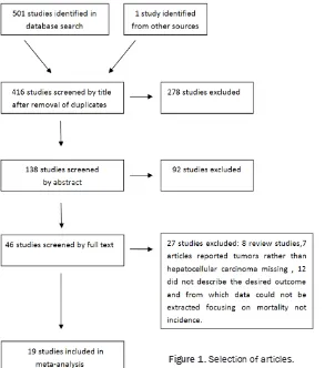
[image:2.612.91.384.70.402.2]examina-We used ORs with 95% CI to calculate associa-tions between CD133 and CD90 expression and various clinicopathological features of HCC including tumor differentiation, tumor stage, AFP level, tumor capsule, hepatitis (including HBV or HCV infection), cirrhosis, metastasis, and vascular invasion. We also utilized HRs with 95% CI to evaluate correlation between the overexpression of CSCs and survival out-comes. HRs with 95% CI of overall survival and disease-free survival were not directly found in some literature, however, these studies provid-ed Kaplan-Meier curves to describe survival outcomes. In these cases, the data were digi-tized and extracted using Engauge Digitizer 4.1 software to obtain the lnHR and variance by the methods provided by Tierney et al. [36] Heterogeneity was evaluated by Cochran w2 Q statistics and the I2 test which decided the use of either a fixed-effects or a random-effects model. We conclude from the study that hetero-geneity exists if the P-value is less than 0.05 or if the I2 is greater than 50%. The funnel plot of Begg’s or Egger’s was applied to assess the Figure 1. Selection of articles.
tion; (2) Immunohistochemical staining was used to detect the expression of CD133 and CD90; (3) The literature de- scribed the association of CD- 133 or CD90 overexpression with survival outcome or de- scribed the clinicopathologi- cal characteristics of liver neo-plasms; (4) Studies contained adequate data to estimate an odds ratio (OR) with 95% confi-dence interval (CI) of clinico-pathological parameters or a hazard ratio (HR) with 95% CI of OS or DFS. Meanwhile, stu- dies including less than 20 patients were excluded. Pati- ents with HCC, with BDTT, or those who had accepted TA- CE treatment were excluded.
Figure 1 displays the study screening process. To impro- ve reliability, two independent reviewers evaluated the rele-vant articles and resolved any disagreements by discussion.
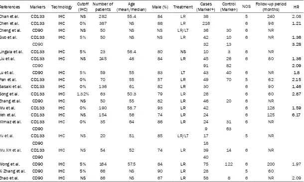
Footnotes: IHC, immunohistochemistry; NS, not specified; LR, liver resection; LT, liver transplantation; NR, not reported; NOS Newcastle-Ottawa Scale. The value of HR that could not be obtained directly from published data was calculated using the method described in the ‘Materials and methods’ section.
Zhao et al. Xi Zhang et al. Wong et al.
Wu XH et al. Yu et al.
Yilmaz et al.
[image:3.792.95.705.84.450.2]Yeh et al. Wu et al. Zhang et al. Song et al. Sasaki et al. Pan et al. Lu et al. Liu et al. Lingala et al. Guo et al. Cheng et al. Chen et al. Chan et al. References
Table 1. Characteristics of the articles included in this study
publication bias. Cochrane Review Manager version 5.2 and Stata statistical software ver-sion 12.0 were used to calculate statistics from relevant studies. For sensitivity analysis, we further examined the included studies by deleting one study each time.
Results
Description of studies
Based on the retrieval conditions, 501 total articles were searched and 1 study was obtained by analyzing the references of rele-vant articles. This analysis contained 1,981 patients. The smallest sample size of the in- cluded studies was 20 and the largest was 387. All positive cancer stem cell marker expression levels were detected by immunohistochemical
staining methods. According to the marker expression levels, the patients were divided into positive and negative groups. CD133 was detected as the CSC marker in 13 articles [10-11, 13-15, 17-19, 21-25] while CD90 was con-sidered as the CSC marker in 11 studies [12, 13, 15, 16, 20, 23-28]. Five studies utilized both CD90 and CD133 as CSC markers [13, 15, 23-25]. Fourteen articles reported Kaplan-Meier curves to assess the OS whereas only eight studies reported the DFS. Table 1 shows the main features of all studies in this analy- sis.
Association among CSC markers and clinico-pathological characteristics
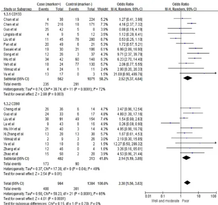
[image:4.612.91.519.70.472.2]cells with certain clinicopathological parame-ters including carcinoma with low degree of differentiation (pooled OR=2.52, 95% CI: 1.37-4.64, P=0.003, random-effect), advanced st- age of carcinoma (pooled OR=3.16, 95% CI: 1.31-7.66, P=0.01, random-effect), positive tu-
[image:5.612.92.518.70.374.2]mor capsule (pooled OR=0.47, 95% CI: 0.23-0.96, P=0.04, random-effect), and vascular thrombosis (pooled OR=1.81, 95% CI: 1.32-2.48, P=0.0002, fixed-effect). CD90-positive cells in HCC patients were also associated with biologically aggressive manifestations such as Figure 3. Association between stem cell markers and tumor grade.
Table 2. CD133 overexpression and clinicopathological characteristics in HCC
Clinicopathological characteristics Studies (n) Patients (n) Analytical modle Pooled OR (95% CI) P value Heterogeneity
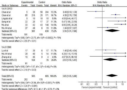
[image:5.612.92.528.430.602.2]poor tumor grading (pooled OR=2.14, 95% CI: 1.19-3.85, P=0.01, random-effect), advanced stage of carcinoma (pooled OR=2.53, 95% CI: 1.10-5.83, P=0.03, random-effect), and tumor size (pooled OR=1.96, 95% CI: 1.07-3.62, P=0.03).
[image:6.612.93.522.85.220.2]Overall for the entire analysis, HCCs with the assumed CSC markers were correlated with poor carcinoma differentiation (pooled OR= 2.31, 95% CI: 1.87-2.85, P<0.00001) (Figure 2) and advanced stage of carcinoma (pooled OR=3.05, 95% CI: 1.55-5.98, P=0.03) (Figure Table 3. CD90 overexpression and clinicopathological characteristics in HCC
Clinicopathological characteristics Studies (n) Patients (n) Analytical modle Pooled OR (95% CI) P value Heterogeneity
HBV (negative vs. positive) 3 202 FEM 1.54 (0.63, 3.75) 0.34 Q Chi 0.63 Cirrhosis (absent vs. present) 2 145 REM 0.37 (0,.01, 9.30) 0.55 Q Chi 0.01
AFP (absent vs. pres) 7 381 FEM 0.81 (0.45, 1.47) 0.49 Q Chi 0.451
Tumor differentiation (I+II vs. III+IV) 10 715 REM 2.14 (1.19, 3.58) 0.01 Q Chi 1.19, 3.58 Tumor stage (I+II vs. III+IV) 3 159 FEM 2.53 (1.10, 5.83) 0.03 Q Chi 1.10 Tumor size (I+II vs. III+IV) 5 271 FEM 1.96 (1.07, 3.62) 0.03 Q Chi 1.07 Encapsulation (absent vs. present) 3 189 REM 0.43 (0.11, 1.70) 0.23 Q Chi 0.11 Vascular invasion (absent vs. present) 3 365 REM 3.21 (0.30, 34.70) 0.34 Q Chi 0.30 Vascular thrombosis (absent vs. present) 2 295 REM 1.68 (0.09, 33.26) 0.73 Q Chi 0.09, 33.26 Footnotes: P<0.05 was considered to indicate statistically significant differences between CD90 expression and clinicopathological features; P>0.10 or I2>50% indicated the existence of heterogeneity; P<0.05 was representative of the significant publication bias; HBV hepatitis B virus, HCV hepatitis C virus, AFPα-fetoprotein, OR odds ratio, CI confidence interval, FEM fixed-effects model, REM random-effects model.
[image:6.612.92.520.266.633.2]3). Nevertheless, we did not find statistically significant associations between expression of CSC markers and HBV (pooled OR=1.15, 95% CI: 0.69-1.93, P=0.60, random-effect), HCV (pooled OR=0.79, 95% CI: 0.44-1.43, P=0.60, fixed-effect), cirrhosis status (pooled OR=0.97, 95% CI: 0.47-2.01, P=0.94, random-effect), tu- mor size (pooled OR=1.17, 95% CI: 0.72-1.90, P=0.53, random-effect), and high level of AFP (pooled OR=0.95, 95% CI: 0.65-1.39, P=0.80, random-effect) (Tables 2 and 3).
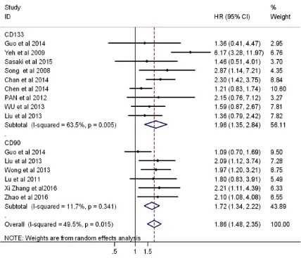
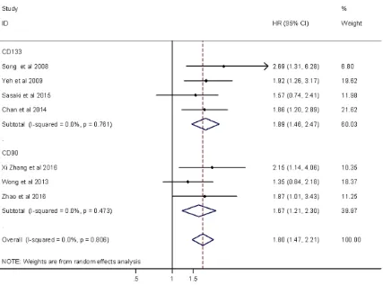
Correlation among CSC markers and survival outcomes in HCC patients
OS and DFS were used to assess the correla-tion between CSC markers and clinical charac-teristics of hepatocellular carcinomas. HRs with 95% CI, which were unmodulated, could be obtained from the Kaplan-Meier curves pre-sented in the articles. Regarding the HR of overall survival, we found diversity between CD133 and CD90 markers (Figures 4 and 5). In this subgroup, nine studies showed the rela-tionship between CD133-positive cells and
overall survival (pooled HR=1.96, 95% CI: 1.35-2.84, p<0.05) and there was little heterog- eneity (P=0.005 for the Q-test, I2=63.5% for the I2 test). On the other hand, six articles reported correlations between CD90 overex-pression and overall survival (pooled HR=1.72, 95% CI: 1.34-2.22, p<0.05) with no heteroge-neity (P=0.341 for the Q-test, I2=11.7% for the I2 test). The HR of DFS included four studies describing correlations in CD133 overexpres-sion and disease-free survival (pooled HR= 1.89, 95% CI: 1.46-2.47, P<0.05) with no het-erogeneity (P=0.761 for the Q-test, I2=0.0% for the I2 test). Three articles reported Kaplan-Meier curves for DFS in association with CD90. We calculated the correlation between high CD90 expression and disease-free survival (pooled HR=1.67, 95% CI: 1.21-2.30, p<0.05) with no heterogeneity (P=0.806 Q-test, I2= 0.0% I2 test).
Publication bias and sensitivity analysis
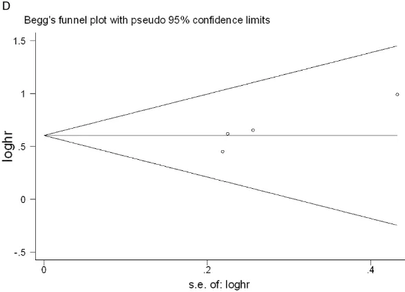
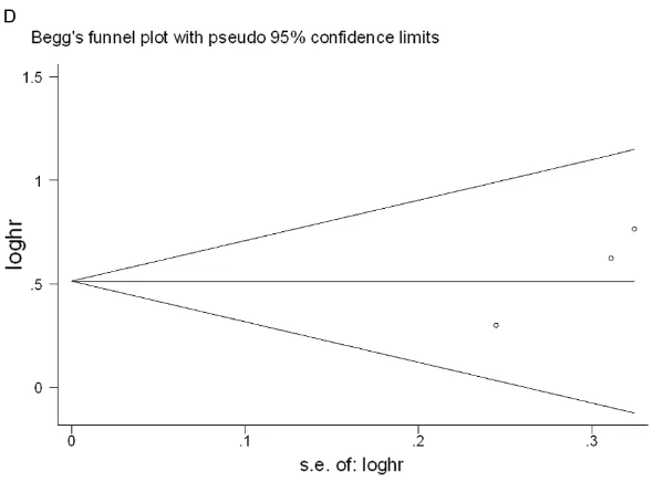
[image:7.612.94.521.68.385.2]the sample sizes of the included articles were gen-erally small and lacked ade-quate survival data was an important obstacle to more accurate calculations. In the case of survival out-come and CD133, the sh- apes of the Begg’s and Egger’s funnel plots reve- aled no clearly unsymmetri-cal patterns in the out-comes of overall survival (P=0.602 in Begg’s test; P=0.386 in Egger’s test) and DFS (P=0.089 and P=0.108, in Begg’s and Egger’s tests, respectively). For CD90, our study reve- aled no distinct publication bias for overall survival (P=0.707 in Begg’s test; P=0.156 in Egger’s test) or disease-free survival (P= 0.296 in Begg’s test; P= 0.08 in Egger’s test) (Fi- gures 6, 7 show the publi-cation bias plots for OS and DFS of CD133 and CD90). To judge the effect of every article on overall influence, we conducted a sensitivity analysis for our study to determine that the meta-analysis was not decided by an individual study. After the removal of each article one by one, the results were not different.
Discussion
that CSC markers, particularly CD133 and CD90, are highly correlated with clinicopatho-logical parameters and poor survival outcomes in HCC.
Chai S et al. [29] demonstrated CD133 as a tar-get of miR-142-3p in its capacity to offer can-cer-stem-cell-like features in liver cancer. Our findings support that CD133 overexpression is meaningfully correlated with low degree of dif-ferentiation carcinomas, advanced stage carci-nomas, vascular thrombosis, and tumor encap-sulation. Additionally, CD133 is considered a momentous factor for tumorigenesis and sur-vival outcome in HCC. Tang KH et al. [30] re- ported that CD133 accelerates tumor vasculo-genesis and stimulates self-renewal through the neurotensin/IL-8/CXCL1 signaling pathway, which influences the prognosis of hepatocellu-lar carcinoma patients. In our current study, we conclude that CD133 is closely correlated with poor survival outcomes. On the other hand, our results show that CD133 is not tightly associ-ated with hepatitis, cirrhosis, elevassoci-ated levels of AFP, tumor size, tumor number, metastasis, and vessel invasion. These results are mostly in agreement with previous research, with the exception of the tumor capsule [31]. Many fac-tors could explain this discrepancy. First, we included literature on the associations among CD133 overexpression and clinicopathological
already considered to play an important role in HCC tumor initiation and progression by ac- tivating self-renewal, invasion, and migration through the Notch and Wnt/β-catenin signaling pathways and has demonstrated enhanced tu- morigenicity, vascular invasion, and metastasis of liver cancer [34]. Our study demonstrates the statistical significance between CD90 over-expression and advanced tumor stage, poor tumor grading, and tumor size. Furthermore, high expression of CD90 was tightly correlated with poor DFS and OS. A previous study report-ed that CD90 overexpression has high speci- ficity and sensitivity in predicting poor differen-tiation in HCC [35]. Therefore, CD90 overex- pression may play a critical part in the assess-ment of clinical characteristics and survival outcomes which could serve as a new potential therapeutic target in hepatocellular carcino- ma.
However, there remained some unavoidable limitations. First, the sample size of the studies included was small. Furthermore, the patients were mostly Asian, as articles written in lan-guages other than English and Chinese were not included in this meta-analysis. Second, while we required positive detection of CSC markers via immunohistochemical staining, specific conditions such as different antibodies and scoring systems may have caused discrep-Figure 6. Egger’s and Begg’s publication bias plots for OS and DFS of CD133
(including A-D). (A) Egger’s publication bias plot for the OS of CD133. (B) Begg’s publication bias plot for the OS of CD133. (C) Egger’s publication bias plot for the DFS of CD133. (D) Begg’s publication bias plot for the DFS of CD133.
[image:9.612.90.383.74.285.2]ancies in this study since the cutoff value differed in those articles. Furthermore, we calculated the HRs of OS and DFS that were ob- tained from Kaplan-Meier curves as described by Ti- erney et al. [36] and this might have an influence on the accuracy of the results. In general, the heterogene-ity in our article could be accepted.
In summary, the results of our article indicate that positive expression of both CD133 and CD90 is significantly correlated with poor dif-ferentiation and advanced stage. Regarding survival outcomes, overexpression of both mar- kers was tightly associated with poor OS and poor DFS. Our findings suggest that CD133 and CD90 could be utilized as potential biomarkers for HCC which might help to subgroup high-risk HCC patients characterized by earlier recur-rence and poor overall survival after surgical or other treatments. We are looking forward to the potential of these new molecular-targeted ther-apies for hepatocellular carcinoma.
Acknowledgements
This work was supported by grants from the National Natural Science Foundation of China, grant number (81572888).
Disclosure of conflict of interest
None.
Abbreviations
HCC, Hepatocellular carcinoma; OS, Overall survival; DFS, Disease-free survival; CSCs, Cancer stem cells; OR, Odds ratio; CI, Con- fidence interval; BDTT, Bile duct tumor thrombi;
References
[1] Jemal A, Bray F, Center MM, Ferlay J, Ward E, Forman D. Global cancer statistics. CA cancer J Clin 2011; 61: 69-90.
[2] Liu M, Jiang L, Guan XY. The genetic and epigenetic alterations in human hepatocellu- lar carcinoma: a recent update. Protein Cell 2014; 5: 673-691.
[3] Chiba T, Zheng YW, Kita K, Yokosuka O, Saisho H, Onodera M, Miyoshi H, Nakano M, Zen Y, Nakanuma Y, Nakauchi H, Iwama A, Taniguchi H. Enhanced self-renewal capability in hepatic stem/progenitor cells drives cancer initiation. Gastroenterology 2007; 133: 937-950. [4] Ji J, Wang XW. Clinical implications of cancer
stem cell biology in hepatocellular carcinoma. Semin Oncol 2012; 39: 461-472.
[5] Rountree CB, Mishra L, Willenbring H. Stem cells in liver diseases and cancer: recent ad-vances on the path to new therapies. Hepatol-ogy 2012; 55: 298-306.
[6] Suetsugu A, Nagaki M, Aoki H, Motohashi T,
Kunisada T, Moriwaki H. Characterization of
CD133+ hepatocellular carcinoma cells as cancer stem/progenitor cells. Biochem Biophy Res Commun 2006; 351: 820-824.
[7] Ma S, Chan KW, Hu L, Lee TK, Wo JY, Ng IO,
Zheng BJ, Guan XY. Identification and charac
-terization of tumorigenic liver cancer stem/
[image:11.612.90.385.70.288.2]progenitor cells. Gastroenterology 2007; 132: 2542-2566.
Figure 7. Egger’s and Begg’s publication bias plots for OS and DFS of CD90 (including A-D). (A) Egger’s publication bias pot for the OS of CD90. (B) Begg’s publication bias pot for the OS of CD90. (C) Egger’s publication bias plot for the DFS of CD90. (D) Begg’s publication bias plot for the DFS of CD90.
TACE, Transcatheter arteri-al chemoembolization; HR, Hazard ratio; HBV, Hepatitis B virus; HCV, Hepatitis C virus; FEM, Fixed-effects model; REM, Random-ef- fects model; IHC, Immuno- histochemistry; NS, Not specified; LR, Liver resec-tion; LT, Liver transplanta-tion; NR, not reported; NOS, Newcastle-Ottawa Scale. Address correspondence to:
Dr. Jian Gao, Department of Gastroenterology and Hepa-
tology, Second Affiliated
[8] Sukowati CH, Anfuso B, Torre G, Francalnaci P, Croce LS, Tiribelli C. The expression of CD90/ Thy-1 in hepatocellular carcinoma: an in vivo and in vitro sutdy. PLoS One 2013; 8: e76830. [9] Zhu L, Zhang W, Wang J, Liu R. Evidence of
CD90+CXCR4+ cells as circulating tumor stem cells in hepatocellular carcinoma. Tumor Biol 2015; 36: 5353-5360.
[10] Chan AW, Tong JH, Chan SL, Lai PB, To KF. Ex-pression of stemness markers (CD133 and EpCAM) in prognostication of hepatocellular carcinoma. Histopathology 2014; 64: 935-950.
[11] Chen K, Li ZH, Jiang P, Zhang X, Zhang YJ, Jiang Y. CD44, CD133 and TF correlate with forma-tion of portal vein tumor thrombus and poor prognosis in patients with hepatocellular carci-noma. J Third Mil Med Univ 2014; 36: 1068-1073.
[12] Cheng BQ, Jiang Y, Li DL, Fan JJ, Ma M. Up-regulation of Thy-1 promotes invasion and metastasis of hepatocarcinomas. Asian Pac J Cancer Prev 2012; 13: 1349-1353.
[13] Guo Z, Li LQ, Jiang JH, Ou C, Zeng LX, Xiang BD. Cancer stem cell markers correlate with early recurrence and survival in hepatocellular carcinoma. World J Gastroenterol 2014; 20: 2098-2106.
[14] Lingala S, Cui YY, Chen X, Ruebner BH, Qian XF, Zern MA, Wu J. Immunohistochemical staining of cancer stem cell markers in hepatocellular carcinoma. Exp Mol Pathol 2010; 89: 27-35. [15] Liu LL, Li W, Li YJ. Expression of CD133 and
CD90 in hepatocellular carcinoma tissue and
their clinical significance. Acta Academiae Me -dicinae Qindao Universitatis 2013; 49: 393-396.
[16] Lu JW, Chang JG, Yeh KT, Chen RM, Tsai JJ, Hu RM. Overexpression of Thy1/CD90 in human hepatocellular carcinoma is associated with HBV infection and poor prognosis. Acta Histo-chem 2011; 113: 833-838.
[17] QX Pan, ZJ Su, CR Wang, JL Zhuang. Expres-sion of tumor stem cell markers EpCAM and CD133 in human primary hepatocellular carci-noma and their value in prognostic prediction. Tumor I 2012; 32: 628-633.
[18] Sasaki A, Kamiyama T, Yokoo H, Nakanishi K,
Kubota K, Haga H, Matsushita M, Ozaki M,
Matsuno Y, Todo S. Cytoplasmic expression of CD133 is an important risk factor for overall survival in hepatocellular carcinoma. Oncol Rep 2010; 24: 537-546.
[19] Song W, Li H, Tao K, Li R, Song Z, Zhao Q,
Zhang F, Dou K. Expression and clinical signifi -cance of the stem cell marker CD133 in hepa-tocellular carcinoma. Int J Clin Pract 2008; 62: 1212-1218.
[20] Zhang X, Mi LL, Wang QX. Clinical significance
and expression of CD90 in human
hepatocel-lular carcinoma. Chin J Diffic and Compl Cas
2011; 12: 911-913.
[21] Wu LM, Chen XX, Cheng CT. Prognostic signifi -cance of expression of CD133, VEGF and CD- 34 in hepatocellular carcinoma. Chin J Cancer Prev Treat 2013; 5: 37-41.
[22] Yeh CT, Kuo CJ, Lai MW, Chen TC, Lin CY, Yeh TS, Lee WC. CD133-positive hepatocellular carcinoma in an area endemic for hepatitis B virus infection. BMC Cancer 2009; 9: 324. [23] Yilmaz G, Akyol G, Cakir A, IIhan M. Investiga
-tion of diagnostic utility and expression profiles
of stem cell markers (CD133 and CD90) in he-patocellular carcinoma, small cell dysplasia, and cirrhosis. Pathol Res Pract 2014; 210: 419-425.
[24] Yu XH, Xu LB, Liu C, Zhang R, Wang J. Clinico-pathological characteristics of 20 cases of he-patocellular carcinoma with bile duct tumor thrombi. Digest Dis Sci 2011; 56: 252-259. [25] Wu XH, Wang SX, Cui DP, Li JK, Yang BM.
Ex-pression and significance of CD133 and CD90
in hepatocellular carcinoma. Chin J Hepatol 2011; 19: 376-377.
[26] Wong E, Srivastava S, Yeoh KG, Teh M,
Salto-Tellez M. Clinical and biological relevance of
Thy-1/CD90 protein overexpression in human hepatocellular carcinoma. Journal of Oncopa-thology 2013; 1: 1-9.
[27] Zhang X, Jiang P, Shuai L, Chen K, Li Z, Zhang Y, Jiang Y, Li X. miR-589-5p inhibits MAP3K8 and suppresses CD90+ cancer stem cells in hepatocellular carcinoma. J Exp Clin Canc Res 2016; 35:176-188.
[28] Zhao RC, Zhou J, Chen KF, Gong J, Liu J, He JY, Guan P, Li B, Qin Y. The prognostic value of combination of CD90 and OCT4 for hepatocel-lular carcinoma after curative resection. Neo-plasma 2016; 63: 288-298.
[29] Chai S, Tong M, Ng KY, Kwan PS, Chan YP, Fung TM, Lee TK, Wong N, Xie D, Yuan YF, Guan XY, Ma S. Regulatory role of miR-142-3p on the functional hepatic cancer stem cell marker CD133. Oncotarget 2014; 5: 5725-5735. [30] Tang KH, Ma S, Lee TK, Chan YP, Kwan PS,
Tong CM, Ng IO, Man K, To KF, Lai PB, Lo CM, Guan XY, Chan KW. CD133(+) liver tumor-initi-ating cells promote tumor angiogenesis, grow- th, and self-renewal through neurotensin/inter- leukin-8/CXCL1 signaling. Hepatology 2012; 55: 807-820.
[31] Zhong C, Wu JD, Fang MM, Pu LY. Clinico-
pathological significance and prognostic value
[32] Rege TA, Hagood JS. Thy-1 as a regulator of cell-cell and cell-matrix interactions in axon regeneration, apoptosis, adhesion, migration,
cancer, and fibrosis. Faseb J 2006; 20:
1045-1054.
[33] Yang ZF, Ho DW, Ng MN, Lau CK, Yu WC, Ngai P,
Chu PW, Lam CT, Poon RT, Fan ST. Significance
of CD90+ cancer stem cells in human liver cancer. Cancer Cell 2008; 13: 153-166. [34] Luo J, Wang P, Wang R, Wang J, Liu M, Xiong S,
Li Y, Cheng B. The notch pathway promotes the cancer stem cell characteristics of CD90+ cells in hepatocellular carcinoma. Oncotarget 2016; 7: 9525-9537.
[35] Liu R, Shen Y, Nan K, Mi B, Wu T, Guo J, Li M, Lv Y, Guo H. Association between expression of cancer stem cell markers and poor differentia-tion of hepatocellular carcinoma a meta-analy-sis. Medicine (Baltimore) 2015; 94: e1306.
[36] Tierney JF, Stewart LA, Ghersi D, Burdett S, Sydes MR. Practical methods for incorporating summary time-to-event data into meta-analy-sis. Trials 2007; 8: 16.
[37] Su R, Nan H, Guo H, Ruan Z, Jiang L, Song Y, Nan K. Associations of components of PTEN/ AKT/mTOR pathway with cancer stem cell markers and prognostic value of these bio-markers in hepatocellular carcinoma. Hepatol-ogy Research 2016; 46: 1380-1391.