Original Article
Effect of acupuncture on expression
levels of NF-κB, IL-1β, IL-6, and TNF-α in
rats with cerebral ischemia and reperfusion
Ying-Kui Si, Yang Yang, Hong Xu, Ya-Min Zhang, Su-Hui Chen, Hua Sun
Department of Traditional Chinese Medicine, Peking Union Medical College Hospital (PUMCH), Beijing, China
Received March 29, 2018; Accepted June 9, 2018; Epub September 15, 2018; Published September 30, 2018
Abstract: Objective: To explore the treatment effects of acupuncture at Baihui point and Zusanli point on the levels of nuclear factor-κB (NF-κB), interleukin -1β (IL-1β), interleukin -6 (IL-6), and tumor necrosis factor-α (TNF-α) in rats with cerebral ischemia and reperfusion. Methods: Sixty SD rats were randomly divided into three groups. The model group (n=20) was selected for cerebral ischemia and reperfusion, another group of rats were selected for acupunc-ture treatment following cerebral ischemia and reperfusion (n=20), and the control group of rats were fed normally and did not undergo cerebral ischemia (n=20). Results: Expression of IL-1β, IL-6, TNF-α, and NF-κB in the ischemia model group was increased compared to that in the control group. IL-1β expression was decreased in the acu -puncture group compared with ischemia model group (P < 0.05). In the acu-puncture group, IL-6 levels decreased, while in the model group IL-6 levels remained high (P < 0.05). TNF-α levels steadily increased in the acupuncture group, yet TNF-α maintained high expression in the model group (P < 0.05). NF-κB levels in the acupuncture group decreased gradually during treatment, while NF-kB sustained high expression levels without significant changes in the model group (P < 0.05). Conclusions: Acupuncture at Baihui point and Zusanli point effectively reduced the ex-pression levels of inflammatory factors in rats with cerebral ischemia and reperfusion, and demonstrated repairing effects on damaged brain tissue.
Keywords: Acupuncture, cerebral ischemia and reperfusion, NF-κB, IL-1β and IL-6
Introduction
Cerebrovascular disease is a global disease with a high incidence throughout the world. Ac- cording to statistics from Love et al. [1], the number of patients with cerebrovascular dis-eases has reached 1.6 million in 2016, most of which were middle aged and elderly people. The findings of Kamat et al. [2] showed that the incidence rate of cerebrovascular disease is increasing in young individuals. It is predict- ed that young and middle-aged patients will account for 35% of the overall cerebrovascular disease patients in the year 2030. Cerebrovas- cular diseases often occur suddenly and many patients become disabled and ultimately die due to an untimely response [3]. In the clinic, the most commonly applied method for the treatment of ischemic cerebrovascular disea- se is thrombolytic therapy with recombinant tis-sue plasminogen activator (rtPA) [4], but this method is treatment intensive and may cause intracranial hemorrhage and other
-cated pathogenesis, the treatment of cerebral ischemia in clinics has gradually begun to advo-cate the application of traditional Chinese med-icine acupuncture [9]. The goal of acupuncture is to improve the prognosis of the patients by reducing the damage caused by cerebral isch-emia through multiple channels and multiple targets [10]. Currently, there have been many studies [11-14] proving that acupuncture com -bined with Western medicine for the treatment of patients with cerebral ischemia has achieved remarkable results. However, due to the com -plicated structures and points of the human body, studies on the efficacy of acupuncture therapy for the treatment of cerebral ischemia and reperfusion are less reported.
Hwang IK et al. showed that electroacupunc-ture (EA) at ST36 (Zusanli) and GV20 (Baihui) enhanced cell proliferation and neuroblast dif-ferentiation in the rat dentate gyrus [15]. Jin Young Chuang et al. demonstrated that elec-troacupuncture (EA) at ST36 (Zusanli) and GV20 (Baihui) can ameliorate the reductions in proliferating cells and differentiated neuro-blasts in the dentate gyrus induced by type-2 diabetes without significantly reducing blood glucose levels with increasing BDNF levels [16].
Therefore, we established a rat model with ce- rebral ischemia and reperfusion to study acu-puncture at Baihui and Zusanli points to verify whether acupuncture treatment is applicable to patients with cerebral ischemia and reperfu-sion, and to further provide reliable reference and guidance for clinical practice in the future. Materials and methods
Animal
Sixty SD rats (30 male and 30 female) with body weights of 250~300 gram were provid- ed by the animal laboratory center of Central South University. Room temperature was 26°C and humidity 75%. 5 rats were fed in one cage. Modeling method and grouping
All rats were randomly divided into three groups. One group was used as the model group with cerebral ischemia and reperfusion (n=20), one group was treated with acupunc-ture following cerebral ischemia and reperfu-sion (n=20), and the final group served as the control group and did not undergo cerebral ischemia or acupuncture, and were maintained
on a normal diet (n=20). The rat model with cerebral ischemia and reperfusion was estab-lished by Mintorovitch et al. [17]. Briefly, intra -peritoneal injection of 10% chloral hydrate (0.3/100 g) was used for anesthesia. After dis-infection, the median neck skin was incised 2-3 cm and ligated the external carotid artery and cut it off. A suture was inserted from the cut side of the external carotid artery along the internal carotid artery until resistance was felt. The thread thrombus completely blocked the right middle cerebral artery entrance about 15 mm in. The silk thread above the cutting of the right neck general artery was ligated with about 2 cm of thread indwelled outside the rats’ body and the skin was sutured. Animals were anes -thetized after 2 hours of cerebral ischemia and the thread was slowly pulled out to the black mark and cut to achieve reperfusion.
Experimental method
At 24 hours after reperfusion, the rats in the acupuncture group received regular daily acu-puncture with disposable aseptic acuacu-puncture needles at Baihui and the left Zusanli points. The acupuncture lasted for 7 days. The pulse electrotherapy apparatus was delivered after needle placement so that the local muscle con-traction was used to assist the treatment. The parameters of the electrotherapy apparatus were set as a sparse dense wave with the fre-quencies of 2~100 Hz and intensity of 2 mA for 20 min. Rats in the model group were not given any treatment after cerebral ischemia and re- perfusion, and as a control were handled every time the rats in the acupuncture group were given treatment. The rats in the control group did not undergo cerebral ischemia and reperfu-sion, and were fed normally.
Western blotting
sodium dodecyl sulfate-polyacrylamide gel ele- ctrophoresis on 10% Tris-glycine gels. After in- cubation in blocking buffer and wash three times with Tris-buffered saline and Tween 20 (TBST) buffer (10 mM Tris-base, 100 mM NaCl, and 0.1% Tween 20; pH 7.5), blots were treat- ed with an anti-NF-κB p65 polyclonal antibody (1:1,000), in TBST buffer overnight. Blots were subsequently washed with TBST and incubated with a secondary horseradish peroxidase-con-jugated goat anti-mouse mAb for 1 hour. Blots
Of the 40 rats that underwent cerebral isch-emia and reperfusion, 36 surgeries were suc-cessful (90%). Of all the sucsuc-cessful rats, 17 were in the model group and 19 were in the acupuncture group. All rats in the control group were all alive.
IL-1β detection
[image:3.612.91.324.97.188.2]In the acupuncture group, IL-1β (71.64±20.84) on day 3 was significantly lower than the model Table 1. IL-1β levels in the control, acupuncture, and
model groups of rats
Control group
(n=20) group (n=19)Acupuncture Model group (n=17) 3 d 84.71±42.37 71.64±20.84□ 124.33±35.26□,☆ 5 d 125.16±34.67 96.27±29.33*,□ 160.51±21.16*,□,☆ 7 d 123.59±35.14Δ 85.26±32.92*,Δ,□ 168.74±8.21*,□,☆
F 0.01 3.65 53.06
P 0.99 0.03 < 0.01
[image:3.612.90.324.278.369.2]Note: *P < 0.05 compared with the IL-1β levels at 3 days. ΔP < 0.05 compared with the IL-1β levels at 5 days. □P < 0.05 compared with the IL-1β levels of control group. ☆P < 0.05 compared with the IL-1β levels of acupuncture group.
Table 2. IL-6 levels in the control, acupuncture, and model groups of rats
Control group
(n=20) Acupuncture group (n=19) Model group (n=17) 3 d 34.27±6.84 54.62±21.52□ 42.86±8.34□,☆ 5 d 35.02±5.59 39.26±6.85*,□ 42.63±7.86□,☆ 7 d 34.87±5.62 34.77±6.94*,Δ 41.86±8.13□,☆
F 0.09 8.30 0.07
P 0.92 < 0.01 0.93
Note: *P < 0.05 compared with the IL-6 levels at 3 days. ΔP < 0.05 compared with the IL-6 levels at 5 days. □P < 0.05 compared with the IL-6 levels of control group. ☆P < 0.05 compared with the IL-6 levels of acupuncture group.
were then washed, and the immunoreac-tive protein was detected using film expo-sed to enhanced chemiluminescence de- tection reagents.
ELISA
ELISA was adopted to detect the inflam -matory factors of IL-1β, IL-6, and TNF-α. Blood samples were collected at 3, 5, and 7 days following cerebral ischemia and reperfusion in all rats. Whole blood was centrifuged (13000×g for 15 minutes) and supernatants were collected to determine the level of TNF-α, IL-1β, and IL-6 in serum by available quantitative sandwich ELISA kits (R&D, USA). All use of ELISA kits was in strict accordance with the manufacturer’s protocols. The concentrations of the sam-ples were calculated according to the stan-dard curve. The serum TNF-α, IL-1β, and IL-6 levels are all expressed as ng/L.
Observation indexes
The expression levels of NF-κB, IL-1β, IL-6, and TNF-α on 3, 5, and 7 days following cerebral ischemia and reperfusion in all rats was examined, and data and variation were plotted.
Statistical method
Statistics software SPSS22.0 was used to analyze the data. All results are expressed by (mean ± standard deviation) and the data among 3 groups was compared by variance analysis. Paired t-tests were used to compare data at two different time points. P < 0.05 suggested that the differ-ence was statistically significant.
Results
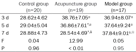
Cerebral ischemia and reperfusion results Table 3. TNF-α levels in the control, acupuncture, and
model groups of rats Control group
(n=20) Acupuncture group (n=19) Model group (n=17) 3 d 28.62±4.62 38.76±7.05□ 36.94±8.07□ 5 d 29.04±5.04 36.86±7.61*,□ 37.64±9.24□ 7 d 28.88±4.73 28.54±4.69*,Δ 37.84±9.01□,☆
F 0.04 12.99 0.05
P 0.96 < 0.01 0.95
Note: *P < 0.05 compared with the TNF-α levels at 3 days. ΔP < 0.05 compared with the TNF-α levels at 5 days. □P < 0.05 compared with the TNF-α levels of control group. ☆P < 0.05 compared with the
[image:3.612.90.322.458.548.2]group (124.33±35.26) and the control group (84.71±42.37), P < 0.05. In the control group on day 3, IL-1β (84.71±42.37) was also signifi -cantly lower than that of the model group (124.33±35.26), P < 0.05. On day 5, IL-1β in the acupuncture group was (96.27±29.33), which was significantly lower than that of the control group (125.16±34.67) and model group (160.51±21.16), all P < 0.05. IL-1β in the model group was significantly higher than that of the control group at day 5, P < 0.05. At day 7, the IL-1β in the acupuncture group was (85.26± 32.92), which was lower than that of the con- trol group (123.59±35.14), P < 0.05, and lower than that of the model group (168.74±8.21), P < 0.05 (Table 1).
IL-6 detection
As shown in Table 2, in the acupuncture group, IL-6 was (54.62±21.52) on day 3 which was
8.13), P < 0.05. On days 3, 5, and 7, the level of IL-6 in the control group and the model group did not change significantly (all P > 0.05), and decreased from relatively high to relatively low, and then decreased much lower in the acu-puncture group (P < 0.05).
TNF-α detection
[image:4.612.91.521.72.255.2]In the acupuncture group, TNF-α was (38.76± 7.05) on day 3 which was not significantly dif -ferent from that in the model group (36.94± 8.07), P > 0.05, yet was significantly higher than that of the control group (28.62±4.62), P < 0.05. TNF-α in the model group was signifi -cantly higher than that of the control group at day 3, P < 0.05. On day 5, TNF-α in the acu -puncture group was (36.86±7.61) which was significantly higher than that of the control group (29.04±5.04), P < 0.05, and lower than Figure 1. NF-κB protein levels in the control, acupuncture, and model groups of rats. Note: ΔP < 0.05 compared with the NF-κB levels at 3 days. #P < 0.05 compared with the NF-κB levels at 5 days. □P < 0.05 compared with the NF-κB levels of control group. *P < 0.05 compared with the NF-κB levels of model group.
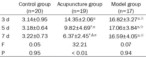
Table 4. NF-κB levels in the control, acupuncture, and model groups of rats
Control group
(n=20) Acupuncture group (n=19) Model group (n=17) 3 d 3.14±0.95 14.35±2.06□ 16.82±3.27□,☆ 5 d 3.18±0.64 9.82±4.69*,□ 17.06±3.84□,☆ 7 d 3.22±0.73 6.37±2.45*,Δ,□ 16.59±4.05□,☆
F 0.05 32.21 0.07
P 0.95 < 0.01 0.94
Note: *P < 0.05 compared with the NF-κB levels at 3 days. ΔP < 0.05 compared with the NF-κB levels at 5 days. □P < 0.05 compared with the NF-κB levels of control group. ☆P < 0.05 compared with the
NF-κB levels of acupuncture group.
[image:4.612.90.323.344.435.2]that of the model group (37.64±9.24), P < 0.05. TNF-α in the model group remained signifi-cantly higher than that of the control group at day 5, P < 0.05. On day 7, there was no sig- nificant difference between the acupuncture group (28.54±4.69) and the control group (28.88±4.73) in TNF-α levels, P > 0.05, and TNF-α in the acupuncture group was signifi -cantly lower than that of the model group (37.84±9.01), P < 0.05. On days 3, 5, and 7 the level of TNF-α in the control group and the model group did not change significantly (all P > 0.05) (Table 3).
NF-κB detection
The NF-κB levels of the rats in the acupunc-ture group was (14.35±2.06) which was signifi -cantly higher than that of the control group (3.14±0.95), P < 0.05, and lower than that of the model group (16.82±3.27), P < 0.05. NF-κB in the model group on day 3 was significantly higher than that of the control group, P < 0.05. On day 5, NF-κB in the acupuncture group was (9.82±4.69), which was significantly higher than that of the control group (35.02±5.59), P < 0.05, and was lower than that of the model group (17.06±3.84), P < 0.05. NF-κB in the mo-del group remained significantly higher than that of the control group, P < 0.05. At day 7, NF-κB in the acupuncture group was (6.37± 2.45), which was higher than that of the con- trol group (3.22±0.73), P < 0.05, and signifi -cantly lower than that of the model group (16.59±4.05), P < 0.05. On day 3, 5, and 7 the levels of NF-κB in the control group and the model group did not change significantly (all P > 0.05), and NF-κB in the acupuncture group decreased from relatively high to relatively low across time points (P < 0.05) (Figure 1; Table 4).
Discussion
Cerebral ischemia and reperfusion mainly cau- ses neuronal necrosis, vascular endothelial da- mage, and blood-brain barrier destruction [18]. The expression of inflammatory factors reflects both the injury and repair conditions of cere- bral ischemia and reperfusion more accurately. After damage of local neurons and glial cells in patients with cerebral ischemia, various cyto-kines are released to participate in the local inflammatory response of the injured cells [8, 19]. Relative signals are taken away from the brain by the blood directly to distal effector
organs. Therefore, the detection of relative sig-nals in the blood will reflect the local inflamma -tory damage of brain tissue, and the recovery progress of the inflammatory damage [20] can be measured through monitoring the inflamma -tory cytokines IL-1β, IL-6, and TNF-α [21]. At the earlier stages of cerebral ischemia and reper- fusion injury, the release of inflammatory cyto -kines in the damaged brain tissue mediates the inflammatory response. Due to the destruc -tion of the blood brain barrier, the inflammatory cells are transferred from the blood to the peri- phery to activate inflammatory factors in the peripheral immune system. Therefore, a large number of inflammatory factors are seen in rats with cerebral ischemia and reperfusion. NF-κB can be seen in all types of cells in the nervous system [22] and it has been demon -strated by Tabassum et al. [23] that its activa -tion enables an increase in apoptosis of cere-bral ischemic neurons. Our work aims to study the feasibility of treating cerebral ischemia and reperfusion with the acupuncture at Baihui and Zusanli points. Additionally, we attempt to pro-vide future guidance and reference for treat- ing patients in the clinic through the establish-ment of a rat model of cerebral ischemia and reperfusion, and the detection of rat inflamma -tory cytokines including IL-1β, IL-6, TNF-α, and NF-κB.
expression on day 3 reached a peak due to the severity of the injury. TNF-α is one of the most important effector molecules in the immune system and is directly involved in the inflamma -tory injury reaction. In this study there was no difference in the expression of TNF-α between the acupuncture group and the model group at the beginning of treatment, and the expression gradually decreased during the treatment and displayed no significant difference on day 7 between the acupuncture group and the con-trol group. This suggests that acupuncture at Baihui and Zusanli points is feasible for the treatment of cerebral ischemia and reperfu-sion. At the time of ischemia, NF-κB is activated in the nerve cells and the endothelial cells. This causes an interaction between the receptor on the cell surface and the inflammatory factors which amplifies brain tissue damage. In the treatment process of the acupuncture group, activation of NF-κB was reduced as were the infarct areas, which not only maintains immune system function and the normal structure of brain tissue, but also stabilizes the normal met-abolic function of the mitochondria and pre-vents apoptosis of neurons, reducing the dam-age caused by cerebral ischemia and reperfu-sion. In this study, NF-κB in the acupuncture group began to decrease gradually, which pro- ves that acupuncture treatment could effec-tively treat and inhibit brain injury due to cere-bral ischemia and reperfusion.
In summary, acupuncture at Baihui and Zusanli points can effectively reduce the expression level of inflammatory factors in rats with cere -bral ischemia and reperfusion, and positively impact repair of brain tissue damage. Due to the differences between rats and human, the results we observed in rats may not translate to humans. We will analyze patients with cerebral ischemia and reperfusion to further investigate the results of our experiment.
Acknowledgements
National Natural Science Foundation of China [grant number 8157150320].
Disclosure of conflict of interest
None.
Address correspondence to: Hua Sun, Department of Traditional Chinese Medicine, Peking Union
Me-dical College Hospital (PUMCH), No. Three No. 9, Dongdan, Dongcheng District, Beijing 100730, China. Tel: +86-138-01121322; E-mail: sunhuash- aa@163.com
References
[1] Love S and Miners JS. Cerebrovascular dis -ease in ageing and Alzheimer’s dis-ease. Acta Neuropathol 2016; 131: 645-658.
[2] Kamat P, Vacek J, Kalani A and Tyagi N. Homo -cysteine induced cerebrovascular dysfunction: a link to Alzheimer’s disease etiology. Open Neurol J 2015; 9: 9.
[3] Lahousse L, Tiemeier H, Ikram MA and Brus -selle GG. Chronic obstructive pulmonary dis-ease and cerebrovascular disdis-ease: a com- prehensive review. Respir Med 2015; 109: 1371-1380.
[4] Hartley A, Marshall DC, Salciccioli JD, Sikkel MB, Maruthappu M and Shalhoub J. Trends in mortality from ischaemic heart disease and cerebrovascular disease in Europe: 1980 to 2009. Circulation 2016; 133: 1916-26. [5] Nordestgaard LT, Tybjærg-Hansen A, Nordest
-gaard BG and Frikke-Schmidt R. Loss-of-func -tion muta-tion in ABCA1 and risk of Alzheimer’s disease and cerebrovascular disease. Alzheim-ers Dement 2015; 11: 1430-1438.
[6] Fang L, Gao H, Zhang W, Zhang W and Wang Y. Resveratrol alleviates nerve injury after cere-bral ischemia and reperfusion in mice by in- hibiting inflammation and apoptosis. Int J Clin Exp Med 2015; 8: 3219.
[7] Guo C, Yin Y, Duan J, Zhu Y, Yan J, Wei G, Guan Y, Wu X, Wang Y and Xi M. Neuroprotective ef-fect and underlying mechanism of sodium danshensu [3-(3,4-dihydroxyphenyl) lactic acid from Radix and Rhizoma Salviae miltiorrhizae = Danshen] against cerebral ischemia and re -perfusion injury in rats. Phytomedicine 2015; 22: 283-289.
[8] Saad M, Abdelsalam R, Kenawy S and Attia A. Montelukast, a cysteinyl leukotriene receptor-1 antagonist protects against hippocampal inju-ry induced by transient global cerebral isch-emia and reperfusion in rats. Neurochem Res 2015; 40: 139-150.
[9] Hua F, Tang H, Wang J, Prunty MC, Hua X, Say -eed I and Stein DG. TAK-242, an antagonist for toll-like receptor 4, protects against acute ce -rebral ischemia/reperfusion injury in mice. J Cereb Blood Flow Metab 2015; 35: 536-542. [10] Xu Q, Yang JW, Cao Y, Zhang LW, Zeng XH, Li F,
[11] Tan F, Fu W, Cheng N, Meng D and Gu Y. Ligus -trazine reduces blood-brain barrier permeabil-ity in a rat model of focal cerebral ischemia and reperfusion. Exp Ther Med 2015; 9: 1757-1762.
[12] Liu F, Jiang YJ, Zhao HJ, Yao LQ and Chen LD. Electroacupuncture ameliorates cognitive im-pairment and regulates the expression of apoptosis-related genes Bcl-2 and Bax in rats with cerebral ischaemia-reperfusion injury. Acupunct Med 2015; 33: 478-484.
[13] Gao HJ, Liu PF, Li PW, Huang ZY, Yu FB, Lei T, Chen Y, Cheng Y, Mu QC and Huang HY. Li -gustrazine monomer against cerebral isch-emia/reperfusion injury. Neural Regen Res 2015; 10: 832.
[14] Wu Z, Zou Z, Zou R, Zhou X and Cui S. Elec-troacupuncture pretreatment induces toler-ance against cerebral ischemia/reperfusion injury through inhibition of the autophagy pa- thway. Mol Med Rep 2015; 11: 4438-4446. [15] Hwang IK, Chung JY, Yoo DY, Yi SS, Youn HY,
Seong JK and Yoon YS. Effects of electroacu-puncture at Zusanli and Baihui on brain-de-rived neurotrophic factor and cyclic AMP re-sponse element-binding protein in the hip- pocampal dentate gyrus. J Vet Med Sci 2010; 72: 1431-1436.
[16] Chung JY, Yoo DY, Im W, Choi JH, Yi SS, Youn HY, Hwang IK, Seong JK and Yoon YS. Elec-troacupuncture at the Zusanli and Baihui acu-points ameliorates type-2 diabetes-induced reductions in proliferating cells and differenti-ated neuroblasts in the hippocampal dentate gyrus with increasing brain-derived neuro-trophic factor levels. J Vet Med Sci 2015; 77: 167-173.
[17] Mintorovitch J, Moseley M, Chileuitt L, Shimizu H, Cohen Y and Weinstein P. Comparison of diffusion-and T2-weighted MRI for the early detection of cerebral ischemia and reperfu-sion in rats. Magn Reson Med 1991; 18: 39-50.
[18] Engelhard K, Werner C, Eberspächer E, Bachl M, Blobner M, Hildt E, Hutzler P and Kochs E. The effect of the α2-agonist dexmedetomidine and the N-methyl-D-aspartate antagonist S(+)-ketamine on the expression of apoptosis-regu -lating proteins after incomplete cerebral isch-emia and reperfusion in rats. Anesth Analg 2003; 96: 524-31, table of contents.
[19] Yaidikar L and Thakur S. Arjunolic acid, a pen -tacyclic triterpenoidal saponin of terminalia arjuna bark protects neurons from oxidative stress associated damage in focal cerebral ischemia and reperfusion. Pharmacol Rep 2015; 67: 890-895.
[20] Zhao H, Wang R, Wu X, Liang J, Qi Z, Liu X, Min L, Ji X and Luo Y. Erythropoietin delivered via intra-arterial infusion reduces endoplasmic re-ticulum stress in brain microvessels of rats following cerebral ischemia and reperfusion. J Neuroimmune Pharmacol 2015; 10: 153-161. [21] Tao X, Sun X, Yin L, Han X, Xu L, Qi Y, Xu Y, Li H,
Lin Y and Liu K. Dioscin ameliorates cerebral ischemia/reperfusion injury through the down-regulation of TLR4 signaling via HMGB-1 inhi -bition. Free Radic Biol Med 2015; 84: 103-115.
[22] Li W, Suwanwela NC and Patumraj S. Curcu-min by down-regulating NF-kB and elevating Nrf2, reduces brain edema and neurological dysfunction after cerebral I/R. Microvasc Res 2016; 106: 117-127.
[23] Tabassum R, Vaibhav K, Shrivastava P, Khan A, Ahmed ME, Ashafaq M, Khan MB, Islam F, Saf -hi MM and Islam F. Perillyl alcohol improves functional and histological outcomes against ischemia-reperfusion injury by attenuation of oxidative stress and repression of COX-2, NOS-2 and NF-κB in middle cerebral artery occlu -sion rats. Eur J Pharmacol 2015; 747: 190-199.
[24] Li Q, Liu Y, Chen L. Effects of PPARγ agonist decreasing the IL-1β, IL-6 and TNF-α content in rats on focal cerebral ischemia-reperfusion in-jury. Biomedical Research 2017; 28: 9564-9566.
[25] Jiang YF, Liu ZQ, Cui W, Zhang WT, Gong JP, Wang XM, Zhang Y and Yang MJ. Antioxidant effect of salvianolic acid B on hippocampal CA1 neurons in mice with cerebral ischemia and reperfusion injury. Chin J Integr Med 2015; 21: 516-522.