Original Article
A method to induce high expression of human
lysozyme in the milk of transgenic mice
Yunyun Zheng1, Guihu Wang2, Jianfang Zhang1
1Department of Obstetrics and Gynecology, The First Affiliated Hospital of Air Force Medical University, Xi’an,
Shaanxi Province, China; 2National-Local Joint Engineering Research Center of Biodiagnostics and Biotherapy,
Xi’an Jiaotong University, Xi’an, Shaanxi Province, China
Received October 17, 2017; Accepted June 14, 2018; Epub September 15, 2018; Published September 30, 2018
Abstract: Objective: To investigate a low cost and efficient method to induce high expression of human lysozyme (hLYZ) in milk. Methods: a hybrid gene locus strategy was applied in this study, to construct a 50-kb bovine-αS1-casein-hLYZ hybrid gene locus, which was used as a mammary-gland-specific expression vector. The transgenic mice were generated. The expression of human lysozyme of the milk was measured by Western blot. The concentra -tion of rhLYZ was determined by Elisa method. Results: the expression of hLYZ of the milk samples from transgenic lines 15-4, 23-7, 23-11 could be detected by Western blot, while there was no expression in wild type mice. In the quantitative analysis by ELISA assay, the expression level of of hLYZ in the 3 lines was at the 5.35-6.06 g/l, while line 23-7 showed the highest expression level. Conclusion: Our transgenic mice carrying the bovine-αS1-casein-hLYZ hybrid gene locus represent a model system for the cost-effe
Keywords: Human lysozyme, bovine-αS1-casein, transgenic mice, hybrid gene locus
Introduction
Human lysozyme (hLYZ), also known as muram
-idase, can hydrolyze the mucopolysaccharide-β (1→4) glycosidic bonds of bacterial cell walls. It is an important non-specific defense factor in
the human body, participating in various
immu-nological reactions [1]. Human lysozyme is
present in a wide range of cells, tissues, and organs. Its concentration is about 0.5 mg/mL in human, which is 1500-4000 times higher than that in cow, sheep and goat [2]. Owing to its intrinsic antimicrobial activity, hLYZ plays a vital role in preventing intestinal diseases and boosting infant health [3, 4]. However, the resources are limited and the cost of tion is high. Therefore, its large-scale produc-tion for clinical use is desirable.
Transgenic farm animals that secrete
recombi-nant proteins into their milk, via efficient trans -gene expression, provide the opportunity to obtain high-quality and low-cost target protein [5]. In view of the utility of the mammary gland as a potential bioreactor, using of the
mamma-ry gland bioreactor system is not only a good new way to produce hLYZ, but also a process to avoid mastitis through the resisting against the growth of bacteria in dairy animals [6]. Maga et al. has successfully induced expression of hLYZ in the mammary gland of transgenic mice [6].
Shortly thereafter, guidance with the
bovine-α-S1-casein sequence in transgenic rabbits led to the successful expression of human interleu-kin-2 [7]. Furthermore, human protein C was expressed in the milk of transgenic pigs and reached the level of 1 g/L [8].
a 38.4-kb mouse whey acidic penomic-human tissue plasminogen activator (mWAP-htPA) hybrid gene locus was constructed, leading to htPA being expressed in the milk of transgenic mice at a level of 3.3 g/L [10]. A 37-kb mouse whey acidic penomic-human serum albumin (mWAP-hSA) hybrid gene locus was produced, and rhSA was expressed at a high level of 11.9 g/L in milk [11]. Therefore, using this method is feasible to obtain extremely high level expres-sion of target protein.
In the present study, regulatory elements of the
bovine-αS1-casein gene locus were chosen to
direct expression of the hLYZ genomic coding
sequence, and then a bovine-αS1-casein-hLYZ
hybrid locus was constructed. Using ELISA, we measured the expression level and calculated the single-copy locus expression.
Materials and methods
Gene clone
The BAC clone CH240-428L4 for the
bovine-αS1-casein gene locus and RP11-1143G9
(GenBank No. AC020656) containing the hLYZ gene locus were purchased from the BACPAC Resources Center of American the Children’s Hospital. The gap-repair method used was based on the Red recombinant system encod-ed by the pKD46 vector (GenBank No. AY048746.1) [12].
Construction of the pBR322-gap-repair vector
T1-T6, six 400-500-bp homologous arms, were
amplified by PCR with BAC as a template, which
were then individually subcloned into the pMD18-T vector. T1 was located in the region 9
kb upstream of the bovine-αS1-casein start
codon. T2 was located in the region just
upstream of the bovine-αS1-casein start
codon. T1 and T2 were used for gap repair of
the 5-kb 5’-flanking region of the
bovine-αS1-casein gene locus. T5 was located in the region
9 kb upstream of the bovine-αS1-casein stop
codon, while T6 was located in the region just upstream of this codon. T5 and T6 were used
for gap repair of the 9-kb 5’-flanking region of the bovine-αS1-casein gene locus. T3 was
located in the region just downstream of the hLYZ start codon, while T4 was located in the region just upstream of the hLYZ stop codon. T3 and T4 were two arms used for gap repair of
the whole 20-kb hLYZ genomic sequence from ATG to TGA. All six homologous arms were ed together in the following order: T1 was ligat-ed to T2 via an Swa I site, T3 was ligatligat-ed to T4 via a Pme I site, while T5 was ligated to T6 via an Hpa I site through PCR connection between T2 and T3 because between T1 and T6 homolo-gous with each arm sequence spell non-trace
enzyme sites connection, there were no suit
-able enzyme loci between T4 and T5 arms, only
with homology themselves EcoR I connection.
All enzyme sites were native to the
bovine-αS1-casein or hLYZ sequence and the six arms were seamlessly ligated, with no base pairs being introduced or deleted. After ligation, the six arms were cut out from the pMD18-T vector by
PvuI enzyme digestion and then cloned into the
PvuI site of pBR322.
Successive three-step gap repair
For the first step of gap repair, DH10b bacteria containing the bovine-αS1-casein BAC insert
were transfected with the pKD46 plasmid. Then, the transformants carrying the
bovine-αS1-casein BAC insert and pKD46 were incu -bated in 5-ml SOB cultures with ampicillin/ chloramphenicol double-resistance LB liquid medium at 30°C and 220 r/min, after OD600 reached at of 0.20-0.25, 1 mol/L L-arabinose was added. This mixture was then incubated for 45-60 min until an OD600 reached at 0.45-0.50. After that it was centrifuged at 4000 r/ min for 15 min. The bacterial sediment was col-lected using cold aseptic water, and then elec-trocompetent was made after concentrating 100-fold and washing three times with ice-cold 10% glycerol. Electroporation was performed using a Cell-Porator (Gbico) with a voltage booster and 0.15-cm chambers according to the manufacturer’s instructions. This process
featured 25 μl cells and 100 ng pBR322-gap-repair vector, which was linearized by Hpa I to dissociate the homologous arms T5 and T6. The shocked cells were supplemented with 1 ml of SOC, incubated for 1 h at 37°C, and then half of the mixture was spread onto an LB plate
containing 50 μg/ml tetracycline.
In the second step, the gap repair was initiated
with the plasmid resulting from the first step.
The 9-kb 3’region of the pBR322-gap-repair
electro-Figure 1. A sketch of the three-step gap-repair method for construction of the bovine-αS1-casein-hLYZ hybrid gene locus. First, homologous arms T5 and T6 were dissociated by Hpa I to catch the 9-kb 3’-flanking region of the bovine-αS1-casein. Second, homologous arms T3 and T4 were dissociated by Pme I to catch the 4.3-kb hLYZ genomic coding sequence from ATG to TGA. Third, homologous arms T1 and T2 were dissociated by SwaI to catch the 8.6-kb 5’-flanking region of the casein. The final bovine-αS1-casein-hLYZ hybrid gene locus was released from the pBR322 vector by Pvu I digestion and used for subsequent microinjection.
porated into competent DH-
10β bacteria containing the
hLYZ BAC insert and pKD46 plasmid.
In the third step, the gap repair was initiated with the plasmid resulting from the second step. The pBR322-gap-repair vector-9-kb 3’-5-kb hLYZ was
linearized by Swa I to dissoci-ate the homologous arms T1 and T2, and then
electropor-ated into competent DH10β
bacteria containing the bov-
ine-αS1-casein BAC insert and
pKD46 plasmid once again. Other procedures were the
same as in the first step of gap
repair.
Six homologous arms of T1-T6, 400-500 bp in length, which
were amplified by PCR with
BAC as a template, were sea- mlessly ligated together for cloning into the PBR322 vec-tor to construct the succes-sive three-step gap-repair
vec-tor. In the first step,
homol-ogous arms T5 and T6 were dissociated by HpaI digestion,
and the linearized vector was
electroporated into compe-tent cells containing the bo-
vine-αS1-casein BAC clone
and pKD46 plasmid. The plas-mid pKD46 expressed the Red system under the control of a well-regulated promoter to avoid unwanted recombina-tion events under
non-induc-ing conditions. The 9-kb 3’-fl-anking region of
[image:3.612.91.369.72.605.2]codon TGA was gap-repaired. In the third step, started from the second step, the arms T1 and T2 were dissociated by SwaI digestion, and electroporated into competent cells containing
the bovine-αS1-casein BAC clone and pKD46 plasmid once again; then, the 8.6-kb 5’-flanking region of bovine-αS1-casein was gap-repaired. Lastly, the bovine-αS1-casein -9-kb 3’-f-4.3-kb
hLYZ-8.6-kb 5’-f was obtained. The three frag-ments were then seamlessly ligated, which resulted in the successful construction of bo-
vine-αS1-casein-hLYZ; the bovine-αS1-casein
gene locus was exactly replaced by the 4.3-kb hLYZ genomic coding sequence from the start
codon to the stop codon. The
bovine-αS1-casein-hLYZ hybrid gene locus was proven to
be accurate by enzyme digestion and sequence
analysis. The three-step gap-repair process is shown in Figure 1.
Generation of transgenic mice
The 50-kb bovine αS1-casein-hLYZ hybrid gene
locus was released from the pBR322 vector by
PvuI digestion and purified by agarose gel elec -trophoresis. The DNA was microinjected into
fertilized C57BL/6 mouse eggs, and the eggs
were reimplanted into pseudopregnant fe- males. The mice were housed at the transgenic mouse facility. Total genomic DNA was pre-pared from a short segment of mouse tail to check for integration of the injected DNA. PCR of tail DNA was performed with the primer pair mouse-F and mouse-R (mouse-F, 5’-CTGTGA- CTACAGGTGTGTACCAC-3’; mouse-R, 5’-TCATAG- CAGATACATAGGCTGATG-3’) corresponding to
the bovine-αS1-casein and hLYZ gene. As a
result, a PCR product of 630 bp was generated after the following schedule: 94°C for 4 min, and then 30 cycles of 94°C for 30 s, 58°C for 30 s, and 72°C for 1 min. In case of integration
of the bovine αS1-casein-hLYZ hybrid gene
locus construct. Southern blot analysis of
SacI-HF and NcoI-SacI-HF double-enzyme-digested tail
DNA was performed in accordance with the standard procedure, following the protocols provided in the DIG High Prime DNA Labeling and Detection Starter Kit II. The probe used to check for integration of the construct was a 630 bp fragment located between intron 1 and
intron 3 of hLYZ, which was amplified by the specific primer pair mouse-F and mouse-R
(mouse-F, located in intron 1 of hLYZ; mouse-R, located in intron 3 of hLYZ), and then labeled with digoxigenin.
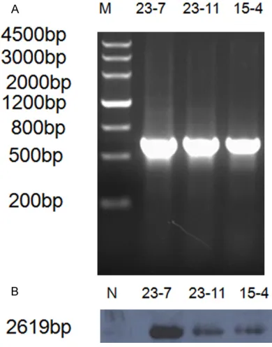
The bovine-αS1-casein-hLYZ construct was
released from the pBR322 vector by PvuI diges-tion and microinjected into C57BL/6 mouse eggs, resulting in three transgenic mouse lines. These three lines appeared to inherit the trans-gene stably over several trans-generations, so they were used in a breeding program to follow the inheritance and expression of the transgene from generation to generation. Integration of
the final hybrid gene locus was confirmed by
PCR and Southern blot analysis. A 630-bp PCR
product was generated from the
bovine-αS1-casein-hLYZ integrated unit (Figure 2A). The mouse genomic DNA was digested with EcoRI and probed with an hLYZ probe, resulting in the
identification of a 2619 kb band representing
[image:4.612.91.286.74.322.2]the fragment from intron 1 to intron 3 of the hLYZ genomic sequence (Figure 2B). As indi-cated by the homologous BLAST, there was no similarity between all introns of the hLYZ gene
and the mouse counterpart no similarity can be found all introns of the hLYZ gene and the mouse counterpart.
Determination of transgene copy numbers
The integration copy number for each transgen-ic mouse strain was determined according to
rice was used as a positive control. Western blotting was performed in accordance with standard protocols. After blotting, the mem-brane was blocked for 1 h in Tris-buffered saline (pH 7.5) containing 0.2% Tween-20 and 5% bovine serum albumin. The membrane was then incubated for 1 h with a primary monoclo-nal rabbit anti-hLYZ antibody diluted at 1:500 in 0.2% Tween 20 and 1% Protifar in TBS (pH 7.5). Then, the membrane was washed with TBS containing 0.2% Tween-20 for 1 h at room temperature, after which it was incubated with a goat anti-rabbit horseradish peroxidase-con-jugated secondary antibody. Enhanced chemi-lumin-escence detection was then performed using Na-luminol and p-coumaric acid.
The concentration of rhLYZ was determined using an AssayMax Human Lysozyme
ELISA kit (milk) (Assaypro, Brooklyn, NY) was used according to the manufacturer’s protocol. The activity of rhLYZ was calculated relative to the standard curve of known hLYZ con- centrations.
Primers
All the primers were listed in Table 1. Primers
J1F and J1R were used to achieve the first step of PCR identification of the 3’
bovine-αS1-casein gene fragment. The primers J2F and J2R were used to achieve the second step of PCR
identification of the hLYZ gene fragment. The
primers J3F and J3R were used to achieve the
third step of PCR identification of the 5’bovine-αS1-casein gene fragment. The primers
mouse-Table 1. Primers used in this study
No Primer sequences product (bp)Length of T1F 5’-GCCCGATCGGCCTTGTGGCTCGAATCTTCTAG-3’
T1R 5’-GCATTTAAATGCACTGGCAATTTCTTGGTAA-3’ 332 T23F 5’-GCATTTAAATATTGACGCTTCTCTATTC-3’
T23R 5’-TGGCGATCGGTTTAAACATCTATTCACTCAAAG-3’ 551 J1F 5’-TCTGAGGGACTCCACAGTTATG-3’
J1R 5’-TAACACATACCCACTGCTCCTG-3’ 680 J2F 5’-AAATATTGACGCTTCTCTATTCCTC-3’
J2R 5’-TTCCTTTATCGGGTATCTCTGG-3’ 627
J3F 5’-GGAACTCTAGGAGTCAAACGTG-3’
J3R 5’-TGGCGATCGGTTTAAACATCTATTCACTCAAAG-3’ 876 Mouse-F 5’-CTGTGACTACAGGTGTGTACCAC-3’
Mouse-R 5’-TCATAGCAGATACATAGGCTGATG-3’ 630
our previous report [9]. The mo- use-F and mouse-R primers were used to assess the Ct val-ues of the hLYZ gene; both prim-ers were located in intron 3 of hLYZ.
Analysis of hLYZ expression
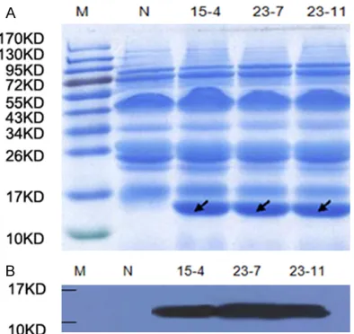
[image:5.612.90.369.86.267.2]For SDS-PAGE, milk samples were diluted (1:40) in phos-phate-buffered saline at equal volumes (10 µl), which were loaded under reducing condi-tions onto a 10% SDS/PAGE gel. The milk of wild-type mice was used as a negative control, and hLYZ standard from transgenic
Figure 3. Expression of hLYZ in the milk of transgenic mice. A. Milk samples were applied to SDS-PAGE and then stained for total protein. M protein marker, N
[image:5.612.93.290.290.476.2]F and mouse-R were used for the identification
of transgenic mice. Results
High hLYZ expression measured by Western blot
To evaluate the expression of hLYZ, milk was collected from three female transgenic mouse lines at day 12 of lactation. The band of about 16 KD was illustrated in SDS-PAGE analysis of milk samples from transgenic lines 15-4, 23-7,
23-11, consisting of a significant amount of
hLYZ (Figure 3A). In western blot analysis, com-pared with wild-type mouse milk, a unique pro-tein band of 16 kDa was observed in the milk of three transgenic mice. There was no band of 16 kDa in the negative control sample (Figure 3B). High hLYZ expression measured by ELISA The expression level of hLYZ in milk was deter-mined by total antigen assay ELISA. Human
lysozyme standard was diluted to 6.0, 3.0, 1.5,
zyme in milk was built. The mammary gland has
been considered as a potential bioreactor for the expression of recombinant proteins. The results show that the high expression of
recom-binant proteins requires optimized constructs, featuring an optimized integration locus. In pre -vious study, rhLZ was expressed in the milk of the transgenic animal using 23 kb of a
bovine-αS1-casein regulatory element, which resulted
in rhLYZ being expressed at concentrations from 0.25 to 0.07 g/L [13]. Therefore,
lyso-zymes were chosen in this study. In addition,
transgenic goats were established by standard pronuclear microinjection, which led to rhLYZ expression in milk at a level of 0.27 g/L [14]. In another study, the milk from transgenic mice could produce hLYZ at the level of 1.4 g/L [15], which was achieved by cloning the genomic sequence of hLYZ into the commercial expres-sion vector pBC1. Moreover, transgenic cattle expressing rhLYZ in milk were produced at a level of 25.96 mg/L [16]. The hybrid construct
of goat β-casein fused to rhLYZ gDNA seemed to be an optimized construct, as the rhLYZ was
expressed at a desirable level in transgenic livestock. It was reported that 0.026 g/L of rhLYZ appeared in transgenic cow milk [17]. In contrast, rhLYZ was expressed at a very low level in transgenic cloned pigs, namely, 0.32 mg/L [18]. To replace the genomic sequence of human lactoferrin (hLF) in the hLF BAC clone
with that of hLYZ, which is optimized for the hLYZ expression vector, five transgenic mouse
lines were generated, four of which expressed rhLYZ at levels from 1.2 to 1.76 g/L [19]. Our expression cassette was set based on the hybrid gene locus strategy works, which
[image:6.612.89.374.73.231.2]includ-Figure 4. A standard graph for the determination of hLYZ concentration by ELISA. Human lysozyme standard dilution into six gradient. X-axis: standard density, Y-axis: absorption values reflecting luminosity.
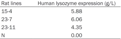
Table 2. Expression of human lysozyme in
transgenic mouse milk. Expression levels represent the values of milk pools from female mice at day 12 of lactation for each transgenic line
Rat lines Human lysozyme expression (g/L)
15-4 5.88
23-7 6.06
23-11 4.35
N 0.00
N: negative control.
0.75, 0.375, 0.187, 0.093,
and 0.000 μg/ml, and ELISA standard curve fitting equa -tion was used CurveExpert
software analysis, finally got
the data analysis (Figure 4). The expression level of of hLYZ in the 3 lines was at the 4.35-6.06 g/l (Table 2), while line 23-7 showed the highest expression level.
Discussion
[image:6.612.91.289.355.419.2]lyso-ed both the expression level and the proportion of high productive transgenics. A 50 kb mWAP-hLF hybrid gene locus was constructed and an extremely high level expression of rhLF (16.7-29.8 g/L) was found in the milk [9]. A 38.4 kb mWAP-htPA hybrid gene locus was constructed, which gave high level expression of rhtPA attained to 3.3 g/L [10]. A chimeric mWAP-hLYZ vector was constructed and the expression of rhLYZ in transgenic mice attained to 35 g/L [20].
In our study, a bovine-αS1-casein gene was
used as a promoter to drive the expression of
hLYZ, while bovine-αS1-casein itself is the
endogenous high expression protein in
rumi-nants. After bovine-αS1-casein gene was intro -duced into the murine germline, transgene expression occurred in all transgenic mice, and
was confines to the lactating mammary gland.
The highest levels of expression were obtained
with a transgene containing 14.2 kb of 5’flank -ing sequence, which was 20 mg/ml [21]. High expression of GM-CSF (Granulocyte Macro- phage-Colony Stimulating Factor) could be
induced in transgenic mice by a
bovine-αS1-casein gene [22]. The above research showed
that the bovine-αS1-casein could not only guide
the expression of exogenous gene in bovine mammary epithelial cell, but also guides expression in transgenic mice, rabbits or sheep, which may be related to the conservation of the expression regulation of milk protein gene.
Moreover, the bovine-αS1-casein gene is very
similar to the mouse casein gene, which is pos-sibly originated from the same ancestor.
Therefore, the bovine-αS1-casein was chosen
as the regulatory element to guide the expres-sion of hLYZ in mammary gland. Red homolo-gous recombination system was used to replace part of the genome, which was used to build a milk target protein hybrid locus. In the
current study, a bovine-αS1-casein-LYZ hetero
-zygous locus containing a relatively intact bovine-αS1-casein gene upstream and a con -trol sequence downstream was established.
When the established bovine-αS1-casein-hLYZ heterozygous locus were transferred by the
microinjection method and integrated into
fer-tilized egg cells in mice, the mouse mammary
gland bioreactor was achieved. 631 pieces of
mouse fertilized eggs were transplanted to 23
receptors in rat. Among 23 offspring mice, PCR
and Southern blot identification revealed three
transgenic mice, yielding a positive rate of 13.0%, which proved that the hybrid loci could
be consolidated in mice, and the efficient expression of lysozyme could be obtained. Our current study established a heterozygous locus that was integrated into fertilized egg cells of
mice, which need less transgenic mice. The reasons should be: (i) the rearing environment caused abnormal female fertility; (ii) the super-ovulation of false pregnant mothers was not
good; (iii) the toxicity of the heterozygous locus; and (iv) the large size of the
bovine-αS1-casein-hLYZ hybrid gene fragment would cause prob-lems in gene integration. The amount of rhLYZ
in milk was also quantified by ELISA, which
showed that three transgenic mouse lines had a high level of expression. This proved that the
bovine-αS1-casein BAC vector provided consis -tent expression, and our expression cassette based on the hybrid gene locus strategy
func-tioned efficiently. The expression level of hLYZ
reached as high as 6.06 g/L, while another cases reached at 5.88 g/L and 4.35 g/L. There was no abnormal phenomenon in transgenic mice, which meant that our hybrid loci are safe. In the process of breeding the F1 generation of transgenic mice, 50 F2 mice were obtained, of
which 22 (44%) were genetically modified, con -forming to the Mendelian law of inheritance. It proved that the stable genetic traits of trans-genic mice could be obtained.
In conclusion, the results of this study show that the mammary gland expression vector
with the bovine-αS1-casein-hLYZ heterozygous
locus can be expressed in mice and there was no effect on normal protein expression in mouse milk. As the milk of dairy cows is easy to be obtained and continuously available, the use of mammary gland bioreactor system of dairy cows may provide a new way to produce
hLYZ and transfer the benefits of human milk to
cow milk.
Disclosure of conflict of interest
None.
References
[1] Maidment C, Dyson A, Beard J. A study into measuring the antibacterial activity of lyso-zyme-containing foods. Nutr Food Sci 2009; 39: 29-35.
[2] Yu Z, Li W, Brunk UT. 3-Aminopropanal is a ly-sosomotropic aldehyde that causes oxidative stress and apoptosis by rupturing lysosomes. APMIS 2003; 111: 643-52.
[3] Lonnerdal B. Nutritional and physiologic signifi -cance of human milk proteins. Am J Clin Nutr 2003; 77: 1537S-1543S.
[4] Huang JM, Wu LY, Yalda D, Adkins Y, Kelleher SL, Crane M, Lonnerdal B, Rodriguez RL, Huang N. Expression of functional recombi-nant human lysozyme in transgenic rice cell culture. Transgenic Res 2002; 11: 229-239. [5] Ebert KM, DiTullio P, Barry CA, Schindler JE,
Ayres SL, Smith TE, Pellerin LJ, Meade HM, Denman J, Roberts B. Induction of human tis-sue plasminogen activator in the mammary gland of transgenic goats. Biotechnology (N Y) 1994; 12: 699-70.
[6] Maga EA, Cullor JS, Smith W, Anderson GB, Murray JD. Human lysozyme expressed in the mammary gland of transgenic dairy goats can inhibit the growt of bacteria that cause masti-tis and the cold-spoilage of milk. Foodborne Pathog Dis 2006; 3: 384-92.
[7] Buhler T, Bruyere T, Went DF, Stranzinqer G, Burki K. Rabbit beta-casein promoter directs secretion of human interleukin-2 into the milk of transgenic rabbits. Biotechnology (N Y) 1990; 8: 140-3.
[8] Park JK, Lee YK, Lee P, Chunq HJ, Kim S, Lee HG, Seo MK, Han JH, Park CG, Kim HT, Kim KS, Kim JH, Lee HT. Recombinant human erythro-poietin produced in milk of transgenic pigs. Biotechnology 2006; 122: 362-371.
[9] Shi G, Chen H, Wu X, Zhou Y, Liu Z, Zheng T, Huang P. A mWAP-hLF hybrid gene locus gave extremely high level expression of human lac-toferrin in the milk of transgenic mice. Trans-genic Res 2009; 18: 573-582.
[10] Zhou Y, Lin Y, Wu X, Xiong F, Lv Y, Zheng T, Huang P, Chen H. The high-level expression of human tissue plasminogen activator in the milk of transgenic mice with hybrid gene locus strategy. Mol Biotechnol 2012; 50: 137-144. [11] Wu X, Lin Y, Xiong F, Zhou Y, Yu F, Deng J, Huang
P, Chen H. The extremely high level expression of human serum albumin in the milk of trans-genic mice. Transtrans-genic Res 2012; 21: 1359-66.
[12] Datsenko KA, Wanner BL. One-step inactiva-tion of chromosomal genes in escherichia coli K-12 using PCR products. Proc Natl Acad Sci U S A 2000; 97: 6640-6645.
[13] Maga EA, Anderson GB, Murray JD. The effect of mammary gland expression of human lyso-zyme on the properties of milk from transgenic mice. J Dairy Sci 1995; 78: 2645-2652. [14] Maga EA, Shoemaker CF, Rowe JD, BonDurant
RH, Anderson GB, Murray JD. Production and processing of milk from transgenic goats ex-pressing human lysozyme in the mammary gland. J Dairy Sci 2006; 89: 518-524.
[15] Yu Z, Meng Q, Yu H, Fan B, Yu S, Fei J, Wang L, Dai Y, Li N. Expression and bioactivity of re-combinant human lysozyme in the milk of transgenic mice. J Dairy Sci 2006; 89: 2911-2918.
[16] Yang P, Wang J, Gong G, Sun X, Zhang R, Du Z, Liu Y, Li R, Ding F, Tang B, Dai Y, Li N. Cattle mammary bioreactor generated by a novel pro-cedure of transgenic cloning for large-scale production of functional human lactoferrin. PLoS One 2008; 3: e3453.
[17] Yang B, Wang J, Tang B, Liu Y, Guo C, Yang P, Yu T, Li R, Zhao J, Zhang L, Dai Y, Li N. Character-ization of bioactive recombinant human lyso -zyme expressed in milk of cloned transgenic cattle. PLoS One 2011; 6: e17593.
[18] Tong J, Wei H, Liu X, Hu W, Bi M, Wang Y, Li Q, Li N. Production of recombinant human lyso-zyme in the milk of transgenic pigs. Transgenic Res 2011; 20: 417-9.
[19] Liu S, Li X, Lu D, Shanq S, Wang M, Zheng M, Zhang R, Tang B, Li Q, Dai Y, Li N. High-level expression of bioactive recombinant human lysozyme inthe milk of transgenic mice using a modified human lactoferrin BAC. Transgenic Res 2012; 21: 407-14.
[20] Wu X, Lin Y, Xi Y, Shao Z, Zhou Y, Liu F, Chen H. The development of transgenic mice for the ex-pression of large amounts of human lesozyme in milk. Biotechnol Lett 2014; 36: 1197-1202. [21] Rijnkels M, Kooiman PM, de Boer HA, Pieper
FR. Organization of the bovine casein gene lo -cus. Mamm. Genome (in press). Mamm Ge-nome 1997; 8: 148-52.