Elsevier Editorial System(tm) for Materials
Letters
Manuscript Draft
Manuscript Number: MLBLUE-D-18-00536R1
Title: Pectin coatings on titanium alloy scaffolds produced by additive
manufacturing: promotion of human bone marrow stromal cell proliferation
Article Type: Short Communication
Keywords: Biomaterials; Thin films; Biomimetic
Corresponding Author: Dr. Timothy E.L. Douglas, PhD
Corresponding Author's Institution: Lancaster University
First Author: Timothy E.L. Douglas, PhD
Order of Authors: Timothy E.L. Douglas, PhD; Ute Hempel; Jagoda Zydek;
Alina Vladescu; Krzysztof Pietryga; Julia Kaeswurn; Maria Buchweitz;
Roman Surmenev; Maria Surmeneva; Cosmin Cotrut; Andrey Koptyug; Elzbieta
Pamula
Abstract: Ti6Al4V is a popular biomaterial for load-bearing implants for
bone contact, which can be fabricated by additive manufacturing
technologies. Their long-term success depends on their stable anchoring
in surrounding bone, which in turn depends on formation of new bone
tissue on the implant surface, for which adhesion and proliferation of
bone-forming cells is a pre-requisite.. Hence, surface coatings which
promote cell adhesion and proliferation are desirable.
2018-01-23
Dear Prof. Boccaccini,
Thank you for giving us the opportunity to submit a revision of our manuscript MLBLUE-D-18-00536
"Pectin coatings on titanium alloy scaffolds produced by additive manufacturing: promotion of
human bone marrow stromal cell proliferation"
We have prepared a point-by-point response to the reviewer comments (below). We have altered
the manuscript according to the suggestions of the reviewer and marked changes with the “Track
Changes” function.
Thank you for your attention and we look forward to hearing from you.
Yours sincerely,
Timothy E.L. Douglas
Point-by-point response to reviewer comments
Reviewer #1: Fig. 2c, statistical analysis should be performed. The labels for scale bars of Fig. 2 should be clearer.
AUTHORS: We have performed statistical analysis as recommended by the reviewer and modified the scale bars.
For coatings on implant, the adhesive strength is important. It is would be interesting to test and compare the adhesive strength of C-ALP and A-ALP on Ti6Al4V implant.
It is not clear how thick are the coatings.
AUTHORS: The reviewer is correct to point out the importance of adhesive strength and that the thickness of the coatings is unclear.
We would have liked to have tested adhesive strength. Unfortunately the Ti6Al4V substrate is rough and the coating is uneven, as shown on the optical microscopy and SEM images (Figures 2a and 2b). Hence, due to lack of a clear methodology to calculate adhesive strength and coating thickness directly, we chose to perform alternative tests, namely stability tests. We incubated the coatings upon incubation in an aqueous environment and analyse release of ALP and pectin. The results are shown in Figures 2d and 2e in the revised version.
Highlights
Ti6Al4V discs were prepared by additive manufacturing (EBM) Discs were coated with pectins from citrus (C) and apple (A)
Coatings also contained the enzyme alkaline phosphatase (ALP)
Coatings promoted proliferation of human bone marrow stromal cells (hBMSC)
1
2
3
4
5
6
7
8
9
10
11
12
13
14
15
16
17
18
19
20
21
22
23
24
25
26
27
28
29
30
31
32
33
34
35
36
37
38
39
40
41
42
43
44
45
46
47
48
49
50
51
52
53
54
55
56
57
58
59
60
61
62
63
64
65
1
Pectin coatings on titanium alloy scaffolds produced by additive manufacturing: promotion of human bone marrow stromal cell proliferation
Timothy E.L. Douglas1,2*, Ute Hempel3, Jagoda Żydek4, Alina Vladescu5,6, Krzysztof Pietryga4, Julia A.H.
Kaeswurm7, Maria Buchweitz7, Roman A. Surmenev6, Maria A. Surmeneva6, Cosmin M. Cotrut6,8, Andrey V. Koptyug9, Elżbieta Pamuła4
1
Engineering Dept., Lancaster University, United Kingdom, 2Materials Science Institute (MSI), Lancaster University, United Kingdom, 3Institute of Physiological Chemistry, Technische Universität Dresden, Germany,
4Dept. Biomaterials, AGH University of Science and Technology, Kraków, Poland, 5National Institute for
Optoelectronics, Dept. Advanced Surface Processing and Analysis by Vacuum Technologies, Romania,
6Physical Materials Science and Composite Materials Centre, National Research Tomsk Polytechnic University,
Russia, 7Analytical Food Chemistry, Faculty of Chemistry, University of Stuttgart, Germany, 8University
Politechnica of Bucharest, Romania, 9Sports Tech Research Centre, Mid Sweden University, Sweden. *Email: t.douglas@lancaster.ac.uk
1
2
3
4
5
6
7
8
9
10
11
12
13
14
15
16
17
18
19
20
21
22
23
24
25
26
27
28
29
30
31
32
33
34
35
36
37
38
39
40
41
42
43
44
45
46
47
48
49
50
51
52
53
54
55
56
57
58
59
60
61
62
63
64
65
2
AbstractTi6Al4V is a popular biomaterial for load-bearing implants for bone contact, which can be fabricated by additive manufacturing technologies. Their long-term success depends on their stable anchoring in surrounding bone,
which in turn depends on formation of new bone tissue on the implant surface, for which adhesion and proliferation of bone-forming cells is a pre-requisite.. Hence, surface coatings which promote cell adhesion and
proliferation are desirable.
Here, Ti6Al4V discs prepared by additive manufacturing (EBM) were coated with layers of pectins,
calcium-binding polysaccharides derived from citrus (C) and apple (A), which also contained alkaline phosphatase (ALP), the enzyme responsible for mineralization of bone tissue.
Adhesion and proliferation of human bone marrow stromal cells (hBMSC) were assessed. Proliferation after 7 days was increased by A-ALP coatings and, in particular, by C-ALP coatings. Cell morphology was similar on
1
2
3
4
5
6
7
8
9
10
11
12
13
14
15
16
17
18
19
20
21
22
23
24
25
26
27
28
29
30
31
32
33
34
35
36
37
38
39
40
41
42
43
44
45
46
47
48
49
50
51
52
53
54
55
56
57
58
59
60
61
62
63
64
65
3
1. IntroductionThe titanium alloy Ti6Al4V is a popular biomaterial for load-bearing implants for bone contact which can be
fabricated by additive manufacturing technologies [1]. The adhesion and proliferation of bone-forming cells are pre-requisites for formation of new bone tissue on the implant surface, which guarantees implant stability and
long-term success. Hence, surface coatings which promote cell adhesion and proliferation are desirable. Coatings containing certain polysaccharides have improved cell adhesion and proliferation [2-4].
Pectins are a family of complex, anionic, calcium-binding polysaccharides found in the primary cell wall and intercellular regions of higher plants, composed primarily of linear D-galactopyranosyluronic acids joined via α(14) glycosidic linkages (Homogalacturonan: HG). These are either partially methyl-esterified, acetylated or
both [19]. In the Rhamnogalacturonan–I (RG-I) the linear galacturonic acid (GalA) chain is disturbed by α-(1,2)-linked L-rhamnose (L-Rha) units. Depending on plant source and isolation method, L-Rha residues are
substituted with neutral saccharide side chains. The RG-II fragment is a substituted galacturonan being composed of a partially methyl esterified GalA backbone.
Commercial pectins from apple and citrus pomace are extracted under hot acidic conditions and therefore, many regions containing high proportions of neutral sugars are hydrolysed. Thus, extracted pectins are mainly
composed of HG containing free or genuine esterified carboxylic groups that are more acid-stable. They form hydrogels and are thus widely applied in viscous, hydrated foods such as jams. They are also inexpensive and widely available and have been applied as scaffolds for tissue regeneration [5]. Pectic RG-I nanocoatings have
been used to tailor surface properties of tissue culture polystyrene (TCPS) [6, 7] and titanium [8]. However,
pectin coatings on Ti6Al4V remain relatively unexplored.
The enzyme alkaline phosphatase (ALP) plays an important role in hard tissue mineralization. ALP catalyses hydrolysis of organic phosphate monoesters to yield inorganic phosphates. ALP has been used as a coating
material on Ti6Al4V surfaces [9, 10], which has improved the osteogenic response.
Here, Ti6Al4V discs prepared by additive manufacturing (electron beam melting, EBM) were coated with pectins derived from citrus (C) and apple (A) and ALP. Coatings were characterized physicochemically and cell
1
2
3
4
5
6
7
8
9
10
11
12
13
14
15
16
17
18
19
20
21
22
23
24
25
26
27
28
29
30
31
32
33
34
35
36
37
38
39
40
41
42
43
44
45
46
47
48
49
50
51
52
53
54
55
56
57
58
59
60
61
62
63
64
65
4
2. Materials and methods
All materials, including alkaline phosphatase (P7640) and BCA Assay (BCA1) were acquired from
Sigma-Aldrich, unless stated otherwise. Pectins C (degree of esterification (DE) 35%, Galacturonic acid content (GalC) 86%) and A (DE 34%, GalC 74%) were obtained from Herbstreith & Fox KG Pektin-Fabriken, D. Rough
Ti6Al4V discs of diameter 2 cm were prepared using additive manufacturing techniques in an ARCAM EBM A2 machine as described previously [1]. Ti6Al4V discs and 0.8% (w/w) C (degree of esterification (DE) 35%,
Galacturonic acid content (GalC) 86%) and A (DE 34%, GalC 74%) pectin solutions were autoclaved (121ºC, 15 minutes). Sterile-filtered ALP solution (1.6% (w/v)) and pectin solution were mixed 1:1 (v/v). 250 µl of this
solution was spread on Ti6Al4V and allowed to air-dry under sterile conditions.
Morphologies of uncoated and coated samples were examined by digital optical microscopy (KEYENCE
VHX-5000) and scanning electron microscopy (SEM, TableTop 3030PLUS, Hitachi).
Surface contact angles were determined using a drop shape analysis system (DSA 10Mk2, KRÜSS). UHQ water
droplets (approximate volume 0.2 µl) were deposited on sample surfaces. Images captured by video camera were analyzed to calculate the contact angle. Results (30 drops per sample) were expressed mean ± standard deviation.
To test coating stability, uncoated and coated samples were immersed in 4 ml ddH2O for 1 and 15 h. ALP release
was quantified by the BCA Assay according to the manufacturer’s instructions. Pectin release was quantified by
the method of van den Hoogen et al. [11].
For cell experiments, hBMSC from two different donors (ethical approval granted by ethics committee of
Technische Universität Dresden, No. 466112016).were seeded at a density of 7000 cells/cm². Cells were seeded onto the samples in 400 µl of cell culture medium (DMEM with 10% heat-inactivated fetal calf serum, and
antibiotics (penicillin and streptomycin). After 2 h the medium was filled up to 4 ml and culture proceeded at 37°C in a humidified CO2 incubator. Proliferation was assessed by the MTS assay. Cells were treated with 10%
dye solution in DMEM for 2 h. The formed formazan amount was measured photometrically at 490 nm. Analyses were performed 24 h and 7 days after seeding. Statistical significance was analyzed by one-way
ANOVA and Bonferroni post-test (prism graph pad software). Cell morphology was assessed after 24 h. Cells were fixed with 4% paraformaldehyde and stained with Alexa488-phalloidine to visualize F-actin cytoskeleton
(green fluorescence) and with DAPI to stain the nuclei (blue fluorescence). The images (three from each sample) were taken with Axiophot microscope (Zeiss) using a digital camera and Axiovision software. Focusing of cells
1
2
3
4
5
6
7
8
9
10
11
12
13
14
15
16
17
18
19
20
21
22
23
24
25
26
27
28
29
30
31
32
33
34
35
36
37
38
39
40
41
42
43
44
45
46
47
48
49
50
51
52
53
54
55
56
57
58
59
60
61
62
63
64
65
5
3. Results and Discussion
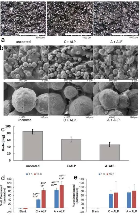
The presence of A-ALP and C-ALP coatings was confirmed by optical microscopy (Figure 2a) and SEM (Figure 2b). Coatings lowered contact angle, and A-ALP coatings were more hydrophilic than C-ALP coatings (Figure
2c). ALP release was more pronounced from C-ALP coatings (Figure 2d) and increased from 1 to 15 h. No differences in pectin release from A-ALP and C-ALP coatings after 1 and 15 h were observed (Figure 2e). Cells
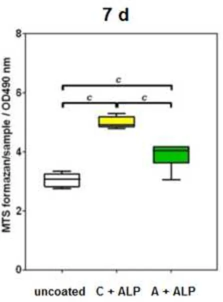
retained viability and proliferated over 24 h and 7 days (Figure 3a). Proliferation was significantly higher on C-ALP coatings than on A-C-ALP coatings after 24 h, and higher than on both uncoated samples and A-C-ALP coatings
after 7 days. A-ALP coatings were significantly superior to uncoated samples after 7 days. Cells on all substrates displayed a spread morphology and distinct, well organized F-actin fibers, characteristic for good adhesion
(Figure 3b).
Several authors have reported that polysaccharides, including pectins, have promoted adhesion and proliferation
of cells [12]. Coatings of RG-I derived from plant pectins on tissue culture polystyrene have promoted proliferation of primary osteoblasts and osteoblast-like MC3T3-E1 cells [13]. Conversely, other studies by the
same group (Gurwaszka K et al) on different RG-I preparations on titanium surfaces revealed no positive effect or, in some cases, an inhibitory effect of the coatings on the proliferation of SaOS-2 osteoblast-like cells [8]. It is
possible that the effect on proliferation is cell-type dependent; in this study, hBMSC were used, while Gurwaszka K and colleagues used primary osteoblasts, MC3T3-E1 and SaOS-2 cells. The reasons for the
stimulatory effect of pectin coatings on proliferation, and the differences between the C-ALP and A-ALP coatings remain unclear. It can be speculated that the differences in GalC (C: 86%, A:74%) may play a role, as
may the differences in wettability. Pectin-coated samples were considerably more hydrophilic than uncoated surfaces (Figure 2c). A stimulatory effect of ALP on cell proliferation is not ruled out. The morphology of
hBMSC did not seem to be affected by the C-ALP and A.ALP coatings (Figure 3b). This seems to be in agreement with previous work by Svava et al, where coatings of different RG-I preparations did not influence
morphology of SaOS-2 cells [7]. Since the A-ALP and C-ALP coatings improve cell proliferation, future work should focus on the ability of these coatings to promote osteogenic differentiation of hBMSC.
1
2
3
4
5
6
7
8
9
10
11
12
13
14
15
16
17
18
19
20
21
22
23
24
25
26
27
28
29
30
31
32
33
34
35
36
37
38
39
40
41
42
43
44
45
46
47
48
49
50
51
52
53
54
55
56
57
58
59
60
61
62
63
64
65
6
hBMSC proliferation after 7 days was increased by A-ALP coatings and, in particular, by C-ALP coatings. Cell
morphology was similar on coated and uncoated samples.
5. Acknowledgement
Era-Net Rus Plus program: project “Fabrication and investigation of new hybrid scaffolds with the controlled porous hierarchy for bone tissue engineering” (Intelbiocomp).
6. References
[1] Surmeneva M, Chudinova E, Syrtanov M, Koptioug A, Surmenev R. Investigation of the HA film deposited
on the porous Ti6Al4V alloy prepared via additive manufacturing. 3Rd International Youth Conference on Interdisciplinary Problems of Nanotechnology, Biomedicine and Nanotoxicology (Nanobiotech 2015). 2015;98.
[2] Bierbaum S, Douglas T, Hanke T, Scharnweber D, Tippelt S, Monsees TK, et al. Collageneous matrix coatings on titanium implants modified with decorin and chondroitin sulfate: characterization and influence on
osteoblastic cells. J Biomed Mater Res A. 2006;77:551-62.
[3] Wolf-Brandstetter C, Lode A, Hanke T, Schamweber D, Worch H. Influence of modified extracellular
matrices on Ti6AL4V implants on binding and release of VEGF. Journal of Biomedical Materials Research Part A. 2006;79A:882-94.
[4] Rother S, Salbach-Hirsch J, Moeller S, Seemann T, Schnabelrauch M, Hofbauer LC, et al. Bioinspired Collagen/Glycosaminoglycan-Based Cellular Microenvironments for Tuning Osteoclastogenesis. Acs Applied
Materials & Interfaces. 2015;7:23787-97.
[5] Moreira HR, Munarin F, Gentilini R, Visai L, Granja PL, Tanzi MC, et al. Injectable pectin hydrogels
produced by internal gelation: pH dependence of gelling and rheological properties. Carbohydrate Polymers. 2014;103:339-47.
[6] Gurzawska K, Svava R, Syberg S, Yu YH, Haugshoj KB, Damager I, et al. Effect of nanocoating with rhamnogalacturonan-I on surface properties and osteoblasts response. Journal of Biomedical Materials Research
Part A. 2012;100A:654-64.
[7] Svava R, Gurzawska K, Yu YH, Haugshoj KB, Dirscherl K, Levery SB, et al. The structurally effect of
1
2
3
4
5
6
7
8
9
10
11
12
13
14
15
16
17
18
19
20
21
22
23
24
25
26
27
28
29
30
31
32
33
34
35
36
37
38
39
40
41
42
43
44
45
46
47
48
49
50
51
52
53
54
55
56
57
58
59
60
61
62
63
64
65
7
Materials Research Part A. 2014;102:1961-71.
[8] Gurzawska K, Svava R, Yu YH, Haugshoj KB, Dirscherl K, Levery SB, et al. Osteoblastic response to pectin nanocoating on titanium surfaces. Materials Science & Engineering C-Materials for Biological Applications.
2014;43:117-25.
[9] Berendsen AD, Smit TH, Hoeben KA, Walboomers XF, Bronckers AL, Everts V. Alkaline
phosphatase-induced mineral deposition to anchor collagen fibrils to a solid surface. Biomaterials. 2007;28:3530-6.
[10] Schouten C, van den Beucken JJ, de Jonge LT, Bronkhorst EM, Meijer GJ, Spauwen PH, et al. The effect of
alkaline phosphatase coated onto titanium alloys on bone responses in rats. Biomaterials. 2009;30:6407-17. [11] van den Hoogen BM, van Weeren PR, Lopes-Cardozo M, van Golde LMG, Barneveld A, van de Lest CHA.
A microtiter plate assay for the determination of uronic acids. Analytical Biochemistry. 1998;257:107-11. [12] Gurzawska K, Syaya R, Jorgensen NR, Goffredsen K. Nanocoating of Titanium Implant Surfaces with
Organic Molecules. Polysaccharides Including Glycosaminoglycans. Journal of Biomedical Nanotechnology. 2012;8:1012-24.
1
2
3
4
5
6
7
8
9
10
11
12
13
14
15
16
17
18
19
20
21
22
23
24
25
26
27
28
29
30
31
32
33
34
35
36
37
38
39
40
41
42
43
44
45
46
47
48
49
50
51
52
53
54
55
56
57
58
59
60
61
62
63
64
65
8
7. Figure captions
Figure 1. Schematic description of coating of Ti6Al4V discs with pectin and ALP.
Figure 2. a) optical microscopy images of bare Ti6Al4V disc (uncoated), Ti6Al4V coated with C + ALP and
Ti6Al4V coated with A + ALP. Scale bar: 1000 µm. b) SEM images of bare Ti6Al4V disc (uncoated), Ti6Al4V coated with C + ALP and Ti6Al4V coated with A + ALP. Scale bars: 500 µm (top row), 100 µm (bottom row).
c) Contact angle measurements on bare Ti6Al4V disc (uncoated), Ti6Al4V coated with C + ALP and Ti6Al4V coated with A + ALP. Differences between all groups were statisitically significant (p<0.001 in all cases). d)
Results of BCA Assay. Significances: *:p<0.05;***:p<0.001. A1: significant relative to A + ALP after 1 h
incubation. A15: significant relative to C + ALP after 15 h incubation. C1: significant relative to C + ALP after
1 h incubation. C15: significant relative to C + ALP after 15 h incubation. e) Results of pectin assay. No
statistically significant differences were observed between any sample groups. Error bars show standard
deviation.
Figure 3. a) MTS assay 24 h (left) and 7 days (right) after seeding of hBMSC on bare Ti6Al4V disc (uncoated), Ti6Al4V coated with C + ALP and Ti6Al4V coated with A + ALP. a and c indicate significant differences