EFFECT OF CO-ADMINISTRATION OF ZINC AND SELENIUM IN CCl
*,1
Amol Gurav,
1Mondal, D. B.,
1Kumar,
1
Division of Medicine, Indian Veterinary Research Institute, Izatnagar, Bareilly, U.P, India
2
Division of Pathology, Indian Veterinary Research Institute, Izatnagar, Bareilly, U.P, India
2
Central Avian Research Institute, Izatnagar, Bareilly,
ARTICLE INFO ABSTRACT
Effect of zinc and selenium alone and in combination was evaluated against carbon tetrachloride induced hepatotoxicity. Rats were divided into 6 groups with group I and II as healthy and diseased control.
therapeutic agents respectively. Heapatotoxicity was induced in rats by giving @ 2 ml CCl olive oil/kg (1:1; v/v) body weight orally twice a week for 4 weeks. At the
biochemical alterations revealed that levels of ALT, AST and ALP increased significantly in group II as compared to healthy control whereas
groups which does not vary statistical
LPO as well as significant decrease in SOD, GSH and Catalase in liver tissue. LPO value of group VI reduced significantly as compared to group IV and V which reveals better antiperoxidation act zinc and selenium in combination as compared to their actions alone. Therapeutic regimen of group VI reflected best SOD and Catalase activity as compared to group IV and V. Hepatocytes of group IV and V showed mild to moderate degree of fatty change
cells and revealed score 1
changes with mild fibrous tissue proliferation between lobules as compared to group IV and V with score of 1. Co
model.
Copyright © Amol Gurav, et al., This is an open access article distributed under the Creative Commons Attribution License, which permits unrestricted use, distribution, and reproduction in any medium, provided the original work is properly cited.
INTRODUCTION
Hepatobiliary dysfunctions occur in number of acute and chronic clinical conditions, including drug induced hepatotoxicity, infectious diseases, congenital or neoplastic diseases, metabolic disorders, degenerative process, vascular injury, autoimmune diseases and even blunt trauma. Liver is major organ for detoxification process (Tamburro, 1979) of xenobiotic compounds such as CCl4, cadmium, arsenic etc. These compounds are predominantly oxidized by microsomal mixed function oxidase (MFO) system along with nicotinamide adenine dinucleotide phosphate
phosphate (NADPH-CYP) reductase enzyme system in liver (Zimmerman, 1999). Liver injury involves co
peroxidative damage of cell. A deficiency of hepatic L-glutathione (GSH) and its antioxidant par
radicals species may contribute to the progression of liver disease. Oxidative stress is known to up regulate production of inflammatory cytokines. Mitochondrial oxidative damage plays an important role in the etiology of numerous oxidati stress–mediated clinical conditions; one possible protective strategy would be to enrich tissue mitochondria with antioxidants thereby limiting mitochondrial oxidative damage,
*Corresponding author: Amol Gurav, Division of Medicine, Veterinary Research Institute, Izatnagar, Bareilly, U.P, India
ISSN: 0975-833X
Article History:
Received 31st July, 2013
Received in revised form 12th August, 2013
Accepted 04th September, 2013
Published online 23rd October, 2013
Key words:
Heapatotoxicity, CCl4,
Zinc and Selenium.
RESEARCH ARTICLE
ADMINISTRATION OF ZINC AND SELENIUM IN CCl
4INDUCED HEAPATOTOXICITY
Kumar, M.
2Monalisa Sahoo,
1Vijayakumar
Indian Veterinary Research Institute, Izatnagar, Bareilly, U.P, India
Division of Pathology, Indian Veterinary Research Institute, Izatnagar, Bareilly, U.P, India
Central Avian Research Institute, Izatnagar, Bareilly, U.P, India
ABSTRACT
Effect of zinc and selenium alone and in combination was evaluated against carbon tetrachloride induced hepatotoxicity. Rats were divided into 6 groups with group I and II as healthy and diseased control. Group III, IV, V and VI received silymarin, zinc, selenium and zinc + selenium as therapeutic agents respectively. Heapatotoxicity was induced in rats by giving @ 2 ml CCl olive oil/kg (1:1; v/v) body weight orally twice a week for 4 weeks. At the
biochemical alterations revealed that levels of ALT, AST and ALP increased significantly in group II as compared to healthy control whereas these levels were significantly reduced in all treatment groups which does not vary statistically from healthy group. Group II showed significant increase in LPO as well as significant decrease in SOD, GSH and Catalase in liver tissue. LPO value of group VI reduced significantly as compared to group IV and V which reveals better antiperoxidation act zinc and selenium in combination as compared to their actions alone. Therapeutic regimen of group VI reflected best SOD and Catalase activity as compared to group IV and V. Hepatocytes of group IV and V showed mild to moderate degree of fatty changes with mild infiltration of inflammatory cells and revealed score 1-2 by histopathological scoring (HPS). Group VI showed mild degree fatty changes with mild fibrous tissue proliferation between lobules as compared to group IV and V with score of 1. Co-administration of zinc and selenium revealed synergistic hepatoprotective action in rat model.
This is an open access article distributed under the Creative Commons Attribution License, which permits unrestricted use, distribution, and reproduction in any medium, provided the original work is properly cited.
number of acute and chronic clinical conditions, including drug induced hepatotoxicity, infectious diseases, congenital or neoplastic diseases, metabolic disorders, degenerative process, vascular injury, autoimmune diseases and even blunt trauma. Liver is major organ for detoxification process (Tamburro, 1979) of , cadmium, arsenic etc. These compounds are predominantly oxidized by microsomal mixed function oxidase (MFO) system along with hosphate-cytochrome CYP) reductase enzyme system in liver (Zimmerman, 1999). Liver injury involves co-lateral oxidative/
peroxidative damage of cell. A deficiency of hepatic glutathione (GSH) and its antioxidant partners and or free
radicals species may contribute to the progression of liver disease. Oxidative stress is known to up regulate production of inflammatory cytokines. Mitochondrial oxidative damage plays an important role in the etiology of numerous oxidative mediated clinical conditions; one possible protective strategy would be to enrich tissue mitochondria with antioxidants thereby limiting mitochondrial oxidative damage,
Division of Medicine, Indian Research Institute, Izatnagar, Bareilly, U.P, India
cellular injury and the initiation and progression of disease (Fariss et al., 2005). Considering increasing evidences (Ohta
et al., 2006) that reactive oxygen species (ROS) are important
mediators in liver injury, effective therapy of hepatobiliary diseases requires disease directed interventions with the aim at elimination of causative factors, reducing hepatic inflammation, minimizing fibrosis, controlling complications and initiating hepatic regeneration. Using antioxidants as supplements to protect cellular structures against oxidative stress and lipid peroxidation in liver damage is of paramount importance. The present study aims to investigate the protective effect of zinc and seleniu
combination on rat liver damage induced by CCl
MATERIALS AND METHODS
Animals
Wistar male Albino rats of around 150 to 200 gm body weight were procured from Laboratory Animal Resource Section (LARS) of Indian Veterinary Res
Bareilly (UP) and were housed in laboratory animal shed of medicine division. Rats were maintained under standard managemental condition and acclimatized for 1 week before the start of the actual experiment. They were fed balanced ration obtained from feed technology unit, IVRI and fresh,
International Journal of Current Research
Vol. 5, Issue, 10, pp.3179-3183, October,2013
INTERNATIONAL
INDUCED HEAPATOTOXICITY
Vijayakumar, H. and
3Pange, A. D.
Indian Veterinary Research Institute, Izatnagar, Bareilly, U.P, India
Division of Pathology, Indian Veterinary Research Institute, Izatnagar, Bareilly, U.P, India
U.P, India
Effect of zinc and selenium alone and in combination was evaluated against carbon tetrachloride induced hepatotoxicity. Rats were divided into 6 groups with group I and II as healthy and diseased Group III, IV, V and VI received silymarin, zinc, selenium and zinc + selenium as therapeutic agents respectively. Heapatotoxicity was induced in rats by giving @ 2 ml CCl4 + 2 ml
olive oil/kg (1:1; v/v) body weight orally twice a week for 4 weeks. At the end of trial, serum biochemical alterations revealed that levels of ALT, AST and ALP increased significantly in group II these levels were significantly reduced in all treatment ly from healthy group. Group II showed significant increase in LPO as well as significant decrease in SOD, GSH and Catalase in liver tissue. LPO value of group VI reduced significantly as compared to group IV and V which reveals better antiperoxidation action of zinc and selenium in combination as compared to their actions alone. Therapeutic regimen of group VI reflected best SOD and Catalase activity as compared to group IV and V. Hepatocytes of group s with mild infiltration of inflammatory 2 by histopathological scoring (HPS). Group VI showed mild degree fatty changes with mild fibrous tissue proliferation between lobules as compared to group IV and V with inistration of zinc and selenium revealed synergistic hepatoprotective action in rat
This is an open access article distributed under the Creative Commons Attribution License, which permits unrestricted use,
cellular injury and the initiation and progression of disease . Considering increasing evidences (Ohta 2006) that reactive oxygen species (ROS) are important mediators in liver injury, effective therapy of hepatobiliary diseases requires disease directed interventions with the aim at elimination of causative factors, reducing hepatic inflammation, minimizing fibrosis, controlling complications patic regeneration. Using antioxidants as supplements to protect cellular structures against oxidative stress and lipid peroxidation in liver damage is of paramount importance. The present study aims to investigate the protective effect of zinc and selenium either alone or in combination on rat liver damage induced by CCl4.
MATERIALS AND METHODS
Wistar male Albino rats of around 150 to 200 gm body weight were procured from Laboratory Animal Resource Section (LARS) of Indian Veterinary Research Institute (IVRI), Bareilly (UP) and were housed in laboratory animal shed of medicine division. Rats were maintained under standard managemental condition and acclimatized for 1 week before the start of the actual experiment. They were fed balanced ation obtained from feed technology unit, IVRI and fresh,
clean drinking water was offered ad libitum. Animals were provided 12 hours light and dark cycle. The temperature and relative humidity were maintained at 18 ± 3oC and 55 ± 5% respectively.
Experimental procedure
Rats were divided into six groups with 6 rats (n=6) in each group. Group I was healthy control, group II was diseased control receiving CCl4. Hepatotoxicity was induced in rats by
giving CCl4 along with olive oil (1:1; v/v) @ 2 ml CCl4 + 2 ml olive oil/kg body weight orally twice a week for four weeks (Doi et al.,1991). All groups has received hepatotoxic dose except healthy control group. Group III received silymarin (5 mg/kg b. wt p.o), group IV received zinc (500 ppm of zinc chloride ad lib through drinking water), group V treated with selenium (0.1 ppm of selenium ad lib through drinking water) and group VI received zinc and selenium as per same dosage as described earlier. All the antioxidants were given for 28 days period. Rats were sacrificed at the end of experiment (day 28) under light chloroform anaesthesia then blood and liver samples were collected for various biochemical, oxidative stress indices and histopathological examination.
Serum biochemical profile
Biochemical parameters viz. alanine aminotranseferase (ALT) and aspartate aminotransferase (AST) (Reitman and Frankle, 1957), alkaline phosphatase (ALP) (pNPP method), serum total protein and albumin (Verley, 1980), serum bilirubin (Zilva and Pannal, 1979) and serum glucose (Kaplan, 1984) were estimated using standard test kits (Span Diagnostic Ltd. India).
Liver oxidative profile
Lipid peroxidation (LPO) (Ohkawa et al., 1979), catalase (CAT) (Cohen et al., 1970), superoxide dismutase (SOD) (Marklund et al., 1974) and glutathione (GSH) (Sedlak and Lindsay, 1968) were estimated in 10% liver homogenate.
Protein content in liver homogenates was determined by the
Lowry method.
Histopathological examination
Liver from all groups were processed for histopathological examination as per standard procedures (Culling, 1963). The pathological changes of fatty liver and degeneration of hepatocytes were graded (score) as normal (0), mild degree (1), moderate degree (2), severe degree (3).
Statistical analysis
Data was subjected to statistical analysis using ANOVA (Snedecor and Cochran, 1994) and Tukey multiple comparison post hoc test at P<0.05 level of significance.
RESULTS AND DISCUSSION
Serum biochemical alterations revealed that levels of ALT, AST and ALP increased significantly in Gr II at the tune of 156.5±20.1, 338.5±10.7 and 288.0±10.9 respectively as compared to healthy and other treatment groups (Table 1). The increased activities of liver enzymes such as ALT, AST and
ALP in the serum of CCl4 induced rats indicate damage to hepatic cells (Wolf, 1999). Damage to the cell integrity of the liver by CCl4 is reflected by an increase in the activity of AST, which is released into circulation after cellular damage. In CCl4 mediated toxicity increased permeability of the hepatocyte membrane and cellular leakage causes high levels of ALP in serum which is an ectoenzyme of the hepatocyte plasma membrane (Paduraru et al., 1996). Muriel and Escobar (2003) reported that due to repeated doses of CCl4 there is leakage of enzymes like AST and ALT across hepatocellular membrane and in blood indicating liver dysfunction and cellular injury. The findings of our study concur with the above reports. The levels of ALT, AST and ALP in all treatment groups were significantly reduced as compared to Gr II which does not vary statistically significant from healthy group. It shows the hepatoprotective effect of all the treatment regimens to prevent the possible hepatocellular injury. The value of serum ALT and ALP of Gr VI does not vary significantly as compared to Gr IV and Gr V but it shows insignificant variation from same groups.
The ALT level in group IV (Zinc) was reduced to 58.9±1.9 level which may be attributed to the anti-peroxidative effect of the zinc in CCl4 induced liver injury (Dhawan and Goel, 1995). The reduction in level of liver enzymes in Gr V showed the efficacy of selenium in preventing hepatocellular injury due to CCl4. Selenium acts as a free radical scavenger due to presence of selenoprotein-P in plasma membrane (Burk and Hill, 1993). Group VI showed reduction in ALT value (54.7 ± 3.2) which was non-significantly lower as compared to either Gr IV (60.3±2) or Gr V (58.9 ± 1.9) alone. The above effect may be due to synergistic action of zinc and selenium in reducing the hepatotoxicity (Jihen et al., 2009). In contrast the levels of ALP in groups III, V and VI were considerably higher than the healthy control group which may be due to concurrent increase in ALP values by CCl4 induced nephrotoxicity. Significant reduction in total protein (4.4 ± 0.9) and albumin (2.9 ± 0.1) in Gr II as compared to all the groups except Gr III (silymarin) was found, which may be due to decreased synthesis of protein in liver of rats due to long term treatment with CCl4 (Patel et al.,2010). Manjunatha et al. (2008) reported the decreased total protein levels in rats treated with CCl4 @ 0.1 ml/kg i.p for 14 days, which supports our study.
failure of antioxidant defence mechanisms to prevent the formation of excessive free radicals (Shenoy et al., 2001). These findings were in agreement with findings of Doi and co-workers (1991) where they found that rats given 2 ml CCl4 with 2 ml olive oil per kg body weight orally produced hepatotoxicity and revealed increased LPO levels in liver of exposed rats. However, there was significant decrease in the values of LPO in all treatment groups as compared to Gr II which reveals hepatoprotective actions of all treatment regimens. LPO value of Gr VI (3.96±0.18) reduced significantly as compared to Gr IV and V which reveals better antiperoxidation action of Zinc and Selenium in combination as compared to their actions alone. Dhawan and Goel (1995) also revealed the synergistic action of zinc and selenium.
Modi et al. (2006) found that zinc (5mg/kg, p.o) and N- acetylcysteine (10mg/kg i.p) prevented lead acetate induced
lipid peroxidation. Similarly Tupe et al. (2010) found that zinc @ 30mg/kg in diet significantly protected against butyl hydroperoxide induced increased lipid peroxidation.
The level of SOD in Gr II decreased significantly as compared to all groups but there was no significant difference in all treated groups with the healthy control group revealing efficacy of antioxidants as an hepatoprotectant. Therapeutic regimen in Gr VI reflected increased SOD activity as compared to Gr IV and V revealing zinc and selenium in combination restored better SOD activity as compared to selenium and zinc alone. These findings on SOD were in agreement with findings of Jihen et al. (2009) who reported that zinc and selenium significantly improved SOD level in cadmium induced toxicity. Combined effect of zinc and selenium found to be much better than their individual effect on SOD because selenium and zinc are well known essential elements and cofactors of antioxidant enzyme such as copper, zinc – superoxide dismutase (CuZn SOD) which dismutates O2
into H2O2 and selenoenzyme Glutathione peroxidase, which
catalyzes H2O2 into H2O. Zinc significantly improved SOD
level, metallothionein and GSH level in oxidative stress of liver (Cabre et al., 2001). The level of GSH decreased
[image:3.595.57.533.75.169.2]significantly in Gr II as compared to all other groups. The level of GSH increased significantly in all treatment groups as compared to Gr II which reveals its antioxidant potential.GSH level of Gr VI (2.9±0.14) restored well as compared to Gr IV (2.7±0.3) and GR V (2.8±0.3). Zinc and selenium are believed to interact with GSH affecting its antioxidant activity. The levels of GSH were also found to be correlated well with dietary zinc status (Oztuk et al., 2003). The level of catalase decreased significantly in Gr II in comparison to all other groups. The catalase value was best restored in Gr VI as compared to Gr IV and V which again reveals better antioxidant potential of zinc and selenium in combination. These findings were similar with the findings of Jihen et al., (2009) who reported that selenium and zinc when given in drinking water for 35 days significantly improved catalase activity in liver. Histopathological appearance of liver of rats in Gr I were normal with no specific pathological changes and had a grade 0 by histopathological scoring system (HPS) (Fig.1).
Fig. 1. Histopathological appearance of liver of rats
[image:3.595.61.533.219.301.2]The cords of hepatocytes were well preserved, cytoplasm was vacuolated, and sinusoids well demarcated. Chandan et al. (2008) and Rai et al. (2001) reported the same observations in
Table 1. Biochemical profile of rats serum receiving various treatments against CCl
4 induced hepatotoxicity (Mean ± SE)
Groups
I (Healthy control) ( CClII
4)
III (CCl
4+silymarin
IV (CCl
4+Zn)
V (CCl
4+Se)
VI (CCl
4+Zn+Se)
ALT (IU/L) 41.9 ±5.3a 156.5 ±20.1b 68.49 ± 11.3 a 58.9 ±1.9a 60.34 ±2.01a 54.07 ± 3.2a
AST (IU/L) 161.2 ±16.2a 338.5 ±10.7c 230.7 ±35.6b 194.7±10.7ab 185.1±12.9ab 179.1±19.5ab
ALP (IU/L) 165.4± 16.8a 288.0±10.9b 203.8±10.8ab 183.9±2.8a 235.9±18.8ab 196.1± 5.8ab
Total Protein (g/dl) 6.9± 1.1d 4.4± 0.96a 5.07± 0.9ab 5.5± 0.12bc 5.8± 0.17bc 6.01± 0.5bc
Albumin (g/dl) 4.5 ±0.1e 2.9 ±0.14a 3.2± 0.12ab 3.5 ± 0.13bc 3.9 ±0.2cd 3.8± 0.23cd
Glucose (mg/dl) 63.8±3.8abc 120.6±2.5e 70.0 ±7.4abcd 44.3±3.4a 70.8±9.4abcd 52.2± 5ab
Total Billirubin (mg/dl) 0.38 ±0.03a 0.55 ±0.06b 0.33± 0.07a 0.31 ±0.01a 0.38± 0.04ab 0.37± 0.02a
Values bearing different superscripts in the same column differ significantly (p≤0.05)
Table 2. Oxidative stress indices in rat liver tissue receiving various treatment against CCl4 induced hepatotoxicity
(Mean ± SE)
Groups LPO (nmoles MDA/mg Protein) SOD (U/mg Protein) Catalase (U/mg Protein) GSH (µmoles/ mg Protein)
I (Healthy control) 3.2± 0.15a 3.2± 0.14b 9.2± 0.3ef 3.01±0.2c
II (CCl
4) 8.9± 0.4
d 1.6±0.2a 4.7±0.4a 1.7±0.12a
III (CCl
4+silymarin) 5.1± 0.2
c 2.7 ±0.1b 6.7±0.2bc 2.5±0.2b
IV (CCl
4+Zn) 5±0.12
c 2.8±0.2b 6.2±0.12b 2.7±0.3bc
V (CCl
4+Se) 4.8±0.3
c 2.7±0.14 b 7.1±0.3bc 2.8±0.3bc
VI (CCl
4+Zn+Se) 3.96±0.2
b 2.96±0.3 b 7.8±0.2cde 2.9±0.14c
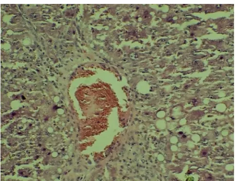
[image:3.595.327.538.549.710.2]mice and rat liver respectively. Hepatocytes of Gr II revealed severe fatty changes and necrosis associated with fibrosis. It was characterized by cytoplasmic vacuolations of variable sizes putting the nucleus to adjacent areas. Liver tissues revealed grade 3 by HPS (Fig.2).
[image:4.595.40.287.123.310.2]Fig. 2. Liver tissues revealed grade 3 by HPS
Fig. 3. moderate fatty changes and mild infiltration of inflammatory cells with HPS score of 2
[image:4.595.313.552.309.496.2]Fig. 4. mild to moderate degree fatty changes with mild infiltration of inflammatory cells
[image:4.595.43.285.339.529.2]Fig. 5. mild to moderate degree fatty changes with mild infiltration of inflammatory cells
Fig. 6. mild degree fatty changes with mild fibrous tissue proliferation between lobules
Figures. Histopathology (Light micrographs) of livers of Group I,II,III,IV,V and VI. (HE x 400)
[image:4.595.42.286.567.759.2]proliferation between lobules. Liver tissues revealed grade 1 by HPS. The above findings also showed better hepatoprotective potential of zinc and selenium in combination as compared to their actions alone. In conclusion, co-administration of Zinc and Selenium as antioxidant had better synergistic hepatoprotective action in CCl4 induced
hepatotoxicity in rats.
REFERENCES
Burk, R. F. and Hill, K. E. 1993. Regulation of selenoproteins.
Ann. Rev. Nutr., 13:65-81.
Chandan, B.K., Saxena, A.K., Shukla, S., Sharma, N., Gupta, D.K., Singh, K., Suri, J., Bhadauria, M. and Qazi, G.N. 2008. Hepatoprotective activity of Woodfordia fruticosa flowers against carbon tetrachloride induced hepatotoxicity. J. Ethnopharmacol., 119(2): 218-224. Cabre, M., Camps, J., Ferre, N., Paternain, J. L. and Joven, J.
2001. The antioxidant and hepatoprotective effect of zinc are related to hepatic cytochrome P450 depression and metallothionein induction in rats with experimental cirrhosis. Int. J. Vita. Nutr. Res., 71:229-236.
Cohen, G., Dembiec, D. and Marcus, J. 1970. Measurement of catalase activity in tissues extracts. Anal. Biochem., 34, 30-38.
Culling, C. F. A. 1963. Handbook of histopathological techniques (including museum techniques). 2nd edition. Butterworth. London. pp 230-248.
Dhawan, D. and Goel, A. 1995. Further evidence for zinc as a hepatoprotective agent in rat liver toxicity. Exp. and Mol.
Pathol., 63: 110-117.
Doi, K., Kurabe, S., Shimazu, N. and Inagaki M. 1991. Systemic histopathology of rats with CCl4 induced hepatic cirrhosis. Lab. Ani., 25: 21-25.
Fariss, M. W., Chan, C. B., Patel, M., Van Houten, B. and Orrenius, S. 2005. Role of mitochondria in toxic oxidative stress. Mol. Interv., 5:94–111.
Jihen, E. H., Imed, M., Fatima, H. and Abdel-hamid, K. 2009. Preventive effect of selenium (Se) and zinc (Zn) on cadmium (Cd) toxicity in the liver of the rat: Effects on oxidative stress. Eco. Environ. Safety, 72:1559-1564. Kaplan, L. A. 1984. Carbohydrate and metabolites in clinical
chemistry: Theory, analysis and correlation. Kaplan, L. A. and Pesce, A. J., Eds. C. B. Mosby Toranto pp 1032-1040. Manjunatha, B. K. and Vidya, S. M. (2008). Hepatoprotective
activity of Vitex trifolia against carbon tetrachloride induced hepatic damage. Ind. J. Pharm. Sci., 70(2):241-5. Marklund, S. and Marklund, G. 1974. Involvement of
superoxide anion radical in the auto-oxidation of pyrogallol and a convenient assay for superoxide dismutase. Biochem.
J., 47:469-474.
Modi, M., Kaul, R. K., Kanran, G. M. and Flora, S. J. S. 2006. Co-administration of zinc and n-acetyl cysteine prevents arsenic induced tissue oxidative stress in male rats. J.
Trace Ele. Med. Bio. 20:197-204.
Muriel, P. and Escobar, Y. 2003. Kupffer cells are responsible for liver cirrhosis induced by carbon tetrachloride. J. App.
Toxicol., 23(2):103-108.
Ohkawa, H., Ohishi, N. and Yagi, K. 1979. Assays of peroxides in animal tissues by thiobarbituric acid reaction.
Analytical Biochem., 95:351-358.
Ohta, Y., Imai, Y., Matsura, T., Kitagawa, A. and Yamada, K. 2006. Preventive effect of neutropenia on carbontetrachloride induced hepatotoxicity in rats. J. of
Appl. Tech., 26: 178-186.
Ozturk, A., Baltaci, A. K., Mogulkoc, R., Oztekin, E., Sivrikaya, A., Kurtoglu, E., Kul, A., 2003. Effects of zinc deficiency and supplementation on malondialdehyde and glutathione levels in blood and tissues of rats performing swimming exercise. Biol. Trace Elem. Res., 94: 157-166. Paduraru, I., Saramet, A., Danila, G. H., Nichifor, M., Jerca, L.
and Iacobovici. A.1996. Antioxidant action of a new flavonic derivative in acute carbon tetrachloride intoxication. Eur. J. Drug Metab. Pharmacokinet., 21 : 1-6. Patel, N., Joseph, C., Corcoran, G. B. and Ray, S. D. 2010. Silymarin modulates doxorubicin induced oxidative stress, Bcl-xL and p53 expression while preventing apoptotic and necrotic cell death in the liver. Toxicol. Appl. Pharma., 245:143-152.
Rai, M., Pai Mirabel, R. S. M., Sabitha, P. and Pai, P. 2001. Hepatoprotective influence of selenium in experimental liver cirrhosis. Kuwait Med. J., 33(4):333-336.
Rao, K. S. and Mishra, S. H. 1997. Anti-inflammatory and hepatoprotective activities of Sida rhombifolia Linn. Ind. J.
Pharma., 29:110-116.
Reitman, S. and Frankel, S. 1957. Calorimetric determination of serum glutamic oxalacetic transminase and serum glutamic pyruvic transaminase. Am. J. Clin. Pathol., 28: 56-63.
Sedlak, J. and Lindsay, R. H. 1968. Estimation of total, protein bound and non-protein sulfhydril groups in tissues with Ellman’s reagent. Anal. Biochem., 25(1):192-205.
Shenoy, K.A., Somayaji, S. N. and Bairy, K. L. 2001. Hepatoprotective effects of Ginkgo biloba against carbon tetrachloride induced hepatic injury in rats. Ind. J.
Pharmacol., 33:260–266.
Snedecor, G.W. and Cochran, W.G. 1994. Statistical methods, 8th edn. Iowa state university press, USA.
Tamburro, C. H. 1979. Chemical hepatitis: pathogenesis, detection and management. Med. Clin. North Am., 63: 545-566.
Tupe, R. S., Tupe, S. G., Tarwadi, K. V. and. Agte, V. V. 2010. Effect of different dietary zinc levels on hepatic antioxidant and micronutrients indices under oxidative stress conditions. Metbol., 59 (11):1603–1611.
Verley, H., 1980. Practical clinical biochemistry, 5th edition, William Heinemann publisher, London, pp 550-555. Wolf, P. L. 1999. Biochemical diagnosis of liver diseases. Ind.
J. Clin. Biochem., 14:59-65.
Zilva, J.F. and Pannall, P.R. 1979. Clinical chemistry in diagnosis and treatment. 4th Edn., Lloyd-Luke, London, UK. pp: 348-352.
Zimmerman, H. J. 1999. Drug-induced liver disease, in: Schiff, E. R., Sorrell, M. F., Maddrey, W. C. (Eds.). Schiff’s Diseases of the Liver, eighth ed. Lippincott-Raven, Philadephia, pp. 973-1064.