A Mathematical Model for Inter-Cellular Inductive
Notch Signaling
Jeyaraman Srividhya
The Biocomplexity Institute, Indiana University, Bloomington, USA Email: [email protected]
Received August 14,2012; revised September 15, 2012; accepted September 23, 2012
ABSTRACT
In vertebrate limb, a group of specialized epithelial cells called Apical Ectodermal Ridge (AER) form at the boundary of dorsal and ventral limb ectoderm. Recent experiments suggest that AER forms at the boundary of Fringe expressing and Fringe non-expressing cells by a specific type of receptor-ligand interaction called as inductive signaling, involving the transmembrane proteins Notch, Serrate and Delta. Experiments conducted on Drosophila wing disc have shown that Fringe inhibits the binding ability of Serrate ligand to Notch and enhances that of Delta to Notch. Although several of the signaling elements have been identified experimentally, it remains unclear how the inter-cellular interactions can give rise to such a boundary of specialized cells. Here we present an ordinary differential equation (ODE) model in-volving Delta→Notch and Serrate→Notch interactions between juxtaposed Fringe expressing and Fringe nonexpressing cells. When simulated in a compartmentalized set up, this model gives rise to high Notch levels at the boundary of Fringe expressing and Fringe non-expressing cells.
Keywords: Delta-Notch Signaling; Apical Ectodermal Ridge; Compucell3D; Boundary Formation; Inductive Signaling
1. Introduction
Notch signaling is one of the highly conserved signaling pathways in animal kingdom and plays a vital role in determining the fate of developing tissues [1]. Notch receptor and its ligands Delta, Serrate and Lag/Jagged (DSL ligands) are transmembrane proteins that have large extracellular domains with Epidermal Growth Factor (EGF) like repeats. Notch signaling is activated when the Notch receptor of one cell binds with a Notch ligand of the neighboring cell, which is called as trans-activation. Notch activation within the same cell is termed as cis- activation. When Notch receptor accepts a ligand from the neighboring cell, a multi-step cleavage leads to the formation of an active intracellular domain called as Notch intracellular domain (NICD). NICD further acti-vates several other transcription factors required for dif-ferentiation and other important transformations [1].
Notch signaling in developing tissues can be catego-rized into three types: 1) Lateral inhibition: Here equi- valent cells in a population acquire different fate due to Notch signaling resulting in one cell with high Notch and the neighboring cell with low Notch forming a checker board pattern; 2) Lineage decisions: An asymmetric cell division occurs where cell progenies acquire different levels of Notch resulting in differentiated cell types, and 3) Induction: Here specialized cells are formed at the boundary of two non-equivalent cell populations that
express different genes [2,3]. Inductive signaling of Notch plays a vital role in Drosophila wing disc forma-tion and limb formaforma-tion in vertebrates [4,5]. In chick embryos, at HH (Hamburger-Hamilton) [6] stage 16, an inductive signaling is believed to occur at the bending of the lateral plate mesoderm across the somites 15 - 18, to give rise to a growing limb bud. This inductive signaling gives rise to a specialized type of cells called Apical Ectodermal Ridge (AER) which secretes the morphogens belonging to the Fibroblast Growth Factor family (Figure 1). The AER runs along the anteroposterior axis of the
[image:1.595.355.494.601.710.2]embryo and forms at the boundary of the dorsal and ven- tral ectoderm. Several genes are expressed in the dorsoventral domains before the AER formation giving the ectoderm a dorsal and ventral identity. Wnt-7a gene
expressed in the dorsal ectoderm influences the dorsal fate of the ectoderm and the underlying mesenchyme [7,8]. Engrailed En-1 is expressed in the ventral ecto-derm and has been shown to influence the ventral cell fate [9]. In Drosophila, the gene fng, which is expressed only in the dorsal ectoderm is believed to be directly in-volved in the formation of the wing boundary [10,11]. Similarly in chick, an analogous gene has been identified as Radical fng or rfng which is expressed only at the dorsal ectoderm [9,12].
Recent experimental work by Rodriguez-Esteban et al. [9] and Laufer et al. [12] in chick limb, has derived po-tential parallels between chick AER formation and Dro-sophila wing disc boundary formation. Laufer et al. [12] suggest the existence of evolutionarily conserved paral-lels between vertebrate AER and Drosophila wing margin. Although AER forms at the boundary of dorsal and ven- tral ectoderm, dorsal and ventral identity does not seem to affect its formation. Experiments on Drosophila show that the juxtaposition of Fringe expressing and nonex-pressing cells seems to be necessary and sufficient for the boundary formation [13]. Several other genes like Vg (vestigial), Ap (apterous) operate upstream of Fringe in the Fringe-expressing cells [14,15]. In Fringe-nonex-pressing cells, expression of Fringe is suppressed or in- hibited by the gene Engrailed (En).
Although experiments in Drosophila have shed light on several elements of the signaling mechanism, a com-plete understanding of this inductive signaling is still unclear. One way of verifying the core signaling is through mathematical modeling involving both inter- and intra-cellular signaling. In this short report, we propose a
mathematical model using ordinary differential equation (ODE) to describe the inductive Notch signaling leading to boundary formation. In order to couple the Notch sig- naling at the inter-cellular level, we use multiple com- partment ODEs. Using our model we show the formation of boundary in 1D dimensional arrangement of cells which qualitatively agrees with the higher Notch levels at the AER in chick limb. We finally discuss the potential application of the model and its future extensions in into 2D and 3D spatial scales.
2. The Model
To build the model we first consider two non-equivalent cell populations of the same cell type juxtaposed in a 1D line: Fringe expressing (dorsal) (2 pink cells in Figure 2)
and Fringe non-expressing (ventral) (2 blue cells in Fig-
ure 2). Each cell has its own Notch signaling network.
We model Notch signaling in each cell using three vari- ables namely, Notch (N), Serrate (S) and Delta (D). Fringe (F) is set as a constant in dorsal cells and absent in ventral cells. Although experiments have shown that the dorsal and ventral identity is not related to the presence of Fringe, in this paper, we will refer Fringe-expressing cells as dorsal and Fringe non-expressing cells as ventral respectively. For the current model, we have not consid-ered the molecular level interaction of Notch and its ligands to form NICD since we believe that biochemistry does not matter greatly for a phenomenological repre- sentation. The model is based on the putative core mecha- nism as follows:
Dorsal cells express Serrate and Notch.
Ventral cells express Notch and Delta.
In the dorsal cells, activation of Notch through Serrate is prevented by Fringe: Fringe modifies the Notch ligand through glycosylation and decreases Notch’s affinity to Serrate.
In the ventral cells activation of Notch through Delta is not strong in the absence of Fringe.
At the boundary, dorsal Serrate is able to activate the Notch in ventral cells without being inhibited by Fringe and ventral Delta is able to activate the Notch in the dorsal cells which is enhanced by Fringe.
The resulting high Notch activity at the boundary, in turn activates the transcription of the ligands Serrate and Delta possibly forming a positive feedback loop [16].
The positive feedback loop sustains the high Notch activity as well as the high ligand activity at the boun- dary.
The above mechanism is illustrated in Figure 2. The
mechanism is cast into the following set of differential equations.
d d
i
synN nc i nc i degN i
N
k f D f F f S g F k
t N
(1)
dd
i
synS S i degS i
S
k Z f N k S
t
(2)
dd
i
synD D i degD i
D
k Z f N k D
t
(1)
0,1, 2,3 k k k , m m m
i f x a x gb b x
Here Ni, Di, Siand Fi represent levels of active Notch,
Delta, Serrate and Fringe proteins in cell i, f and g are monotonously increasing and decreasing functions re-spectively. f and g vary between 0 and 1. ksyn and kdeg are
synthesis rate and degradation rate constant of each of the species. Z represents the factor responsible for the differential expression of Serrate and Delta in dorsal and ventral compartments respectively. Z can be considered similar to a cofactor required for the transcription of a specific gene. There is experimental evidence for restric-tion of Serrate expression in dorsal compartment in dro-sophila, however, the expression of Delta is unclear [16]. In order to mathematically represent differential expres-sion, we consider Zs = 1 for dorsal cells and Zs = 0 for
ventral cells. There is evidence of complimentary ex-pression of Serrate and Delta in chick neural tube [12]. Dnc indicates the activity of Delta in neighboring cells. In
a 1D line of cells
1 1
2nc i i
D D D
and in a 2D arrangement, it is the average taken over the immediate neighbors and given as:
1 nc i i D D r
The same principle applies for Serrate also. We do not co
3. Results
iable Model
uced to a one variable nsider cell division since cell cycle times are much greater than the time scale of the inter-cellular dynamics and hence we assume it does not have an effect.
3.1. One-Var
The proposed model can be red
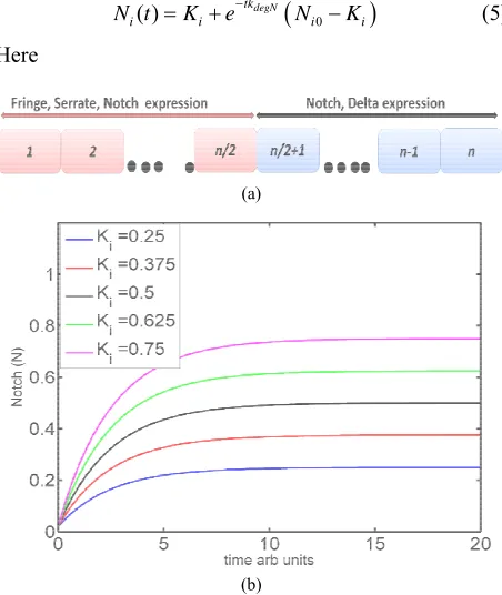
system by making the variables Serrate and Delta con-stants along with Fringe. We consider n cells arranged in a line in a 1D representation (Figure 3(a)). The initial
value of Fringe (F) and Serrate (S) is non-zero for cells from 1 to n/2 and zero from n/2+1 trough n. Similarly the initial value of Delta (D) is zero for cells 1 to n/2 and non-zero for cells n/2+1 to n. The initial value of Notch (N) is low but non-zero throughout the cell arrangement. The boundary condition is non-periodic. The dynamics of Notch inside each cell is reduced as follows:
, , ,
Ni ksynNi Si1 Di1 Si1 Di1 kdegNNi (4)
where the factor Φi represents all the terms in th
i (5)
Here
e paren-thesis in Equation (1). Since Serrate (S) and Delta (D) are constants, the factor Φi will also be a constant depending
upon the value of S and D in the neighboring cells. This results in a simple stable system with steady states Ni = 0, Ki and solution:
(
N t) degN
0
tki Ki e Ni K
(a)
(b)
[image:3.595.310.536.409.677.2]
1, 1, 1, 1
i synN i i i i i degN K k S D S D kThe simulation of various solution curves of N(t is given
3.2. Four-Compartment ODE Model
(1)-(3) is
odel, w
)
in Figure 3(b). The steady state of N is simply Ki, which
implies that the steady state level of Notch depends on the ratio of its synthesis and degradation rate. The pa-rameter Kidepends on the Serrate and Delta activities of
the neighboring cells. For each cell, the value of Φi is
different. This results in a series of coupled system with different Ki s, eventually giving rise to different steady
state in each of the cells. Although the dynamics of the single cell is less interesting, when coupled as a four cell system, the two cells at the boundary results in a steady state that has a higher Notch activity when compared to the cells in the either side of the line of cells. We illus-trate this in the following section.
The three-variable model given in Equations
more complex as it involves the dynamics of Serrate and Delta. The most important aspect of this model is that a positive feedback is induced by the active Notch over the synthesis of the ligands Serrate and Delta. Unlike the previously known model of lateral inhibition [17] where active Notch has been shown to inhibit Delta in the same cell, in this model active Notch activates Delta and Ser-rate in the same cell. There is experimental support sug-gesting a possible positive feedback due to active Notch in Drosophila [16]. The individual dynamics of the sys-tem is simple, however when the cell interacts with its neighbors, interesting pattern emerges which is explained in the numerical simulations as follows.
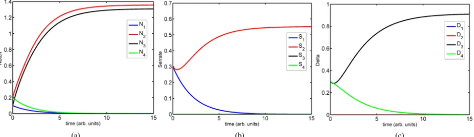
In order to construct a multi-compartment ODE m e consider the model in Equations (1)-(3) where we have 4 compartments representing four cells i = 1, 2, 3, 4. The initial conditions are set such that compartments 1 and 2 have non-zero levels of Fringe, Serrate and Notch, while compartments 3 and 4 have non-zero levels of Delta and Notch. In each compartment
1 1
2i i
i nc
D D
D
and
1 1
2i i
i nc
S S
S
(See Figure 4). We have simulated the four compartment
[image:4.595.374.460.84.153.2]model using Systems Biology Workbench (SBW) [18]. The results of the simulation of the ODE are given in
Figure 5. In Figure 5 we show the time evolution of the
variables N1··· N4, S1 ··· S4, and D1 ··· D4. We can see
that in Figure 5(a), the concentrations of N1 and N4 are
zero where as N2 and N3 which are at the boundary, are
high. Similarly the concentrations of D1, D2 and D4 are
zero while D3 is high (Figure 5(c)). The concentrations
of S1, S3 and S4 are zero while S2 has a non-zero high
value (Figure 5(b)). The parameters of the simulation are
[image:4.595.312.534.401.508.2]given in Supplementary material SBW jarnac code. Since this model involves differentially expressed genes it makes analytical analysis of the large scale model ex-tremely complex. However, as shown above the three- variable model can be reduced to one variable model whose dynamics is described in previous section. Exten-sion of this model into spatial 2D and 3D arrangement of cells is underway.
Figure 4. Four compartment ODE model set up with Notch signaling in each compartment. Dnc and Snc indicate the
averaged concentrations from neighboring cells as in the ODE simulations.
(a) (b) (c)
[image:4.595.61.538.567.705.2]. Discussions
absence of dynamics of Fri4
In the recent years, significant progress has been made in
xpressing
fine-graine
inductive Notch sig-na
formation. Our model has some limitations such as
nge and 2) absence of bio-
he Center; Data-Generating in Developmental Toxicity in
U. Lendahl, “Notch
Signaling: Sim lity in Function,”
Development, pp. 3593-3612.
understanding both the origin and molecular nature of the signals controlling patterning of the dorsoventral limb axis and AER formation. Inductive signaling of Notch has not been well understood despite its critical role in Drosophila wing disc as well as vertebrate limb forma-tion. In addition to this, differential expression of fng and serrate genes has been a hurdle for mathematical model-ing efforts. In this short report, we propose an ODE based compartmental mathematical model to describe inductive Notch signaling involved in the boundary formation at the dorsoventral limb axis. Our model is phenomenol-ogical and hence does not involve any Notch-related biochemical reactions. This qualitative approach allows us to derive the following conclusions:
Boundary of specialized high Notch-e cells is formed due to the interaction of two cell populations with differential gene expressions: In real biological systems, differential gene expression patterns are pro- grammed in the developmental protocols and hence they need to be considered as such. To interpret this differential gene expression mathematically, we sup-pressed the dynamics of the relevant variable in the respective compartments. However, this approximation limits the application of global analysis of the model.
The positive feedback loop at the boundary cells fur-ther maintains high Notch levels by activating tran-scription of more Serrate and Delta: In the experi-ments on chick limb AER formation, initially Serrate expression is observed throughout the dorsal side and then restricts only to the AER [9,12]. To simulate this observation, the model requires a positive feedback loop from Notch to Serrate and Delta formation, eventually creating a boundary which expresses high Serrate and Delta in addition to Notch. However, this result is left to be shown experimentally.
This form of model can only account for d patterns of cell specialization: Our model explains the interactions only between one nearest neighbor. How- ever, there may be long range interactions, which are not accounted in this model.
This is the first attempt to model
ling giving rise to boundary formation in developing tissues. An added advantage of this approach is that this can be extended into 2D arrangement of cells as well as into any agent based modeling approaches potentially leading to a multi-scale model. We are currently making efforts to incorporate this model in a cell-based modeling environment in a 2D and 3D spatial arrangement as well as parameter search that can show this behavior. Our model is the first to represent differential gene expression mathematically and is able to simulate the boundary
chemical reactions involving Notch-Delta ligand forma- tion. Nevertheless, this model presents a versatile frame- work on which further extensive models can be built.
5. Acknowledgements
We would like to acknowledge the EPA grant—T
1)
Texas-Indiana Virtual STAR vitro and in silico Models of
Embryonic Stem Cells and Zebrafish.
REFERENCES
[1] E. R. Andersson, R. Sandberg andplicity in Design, Versati Vol. 138, No. 17, 2011, doi:10.1242/dev.063610
[2] N. Haines and K. D. Irvine, “Glycosylation Regulates Notch Signalling,” Nature Reviews Molecular Cell Biol- ogy, Vol. 4, No. 10, 2003, pp. 786-797.
[3] S. J. Bray, “Notch Signalling: A Simple Pathway Be- comes Complex,” Nature Reviews Molecular Cell Biol- ogy, Vol. 7, No. 9, 2006, pp. 678-689.
doi:10.1038/nrm2009
[4] S. S. Blair, “Limb development: Marginal Fringe Bene- fits,” Current Biology, Vol. 7, No. 11, 1997, pp. R686- R690. doi:10.1016/S0960-9822(06)00356-3
[5] K. D. Irvine and T. F. Vogt, “Dorsal—Ventral Signaling in Limb Development,” Current Opinion in Cell Biology, Vol. 9, No. 6, 1997, pp. 867-876.
doi:10.1016/S0955-0674(97)80090-7
[6] V. Hamburger and H. L. Hamilton, “A Series of Normal Stages in the Development of the Chick Embryo,” De-
velopmental Dynamics, Vol. 195, No. 4, 1992, pp. 231-
272. doi:10.1002/aja.1001950404
[7] B. A. Parr and A. P. McMahon, “Dorsalizing Signal Wnt-7a Required for Normal Polarity of D-V and A-P Axes of Mouse Limb,” Nature, Vol. 374, No. 6520, 1995, pp. 350- 353. doi:10.1038/374350a0
[8] R. D. Riddle, et al., “Induction of the LIM Homeobox Gene Lmx1 by WNT6a Establishes Dorsoventral Pattern in the Vertebrate Limb,” Cell, Vol. 83, No. 4, 1995, pp. 631-640. doi:10.1016/0092-8674(95)90103-5
[9] C. Rodriguez-Esteban, et al., “Radical Fringe Positions the Apical Ectodermal Ridge at the Dorsoventral Bound- ary of the Vertebrate Limb,” Nature, Vol. 386, No. 6623, 1997, pp. 360-366. doi:10.1038/386360a0
[10] T. Klein and A. M. Arias, “Interactions among Delta, Serrate and Fringe Modulate Notch Activity during Dro- sophila Wing Development,” Development, Vol. 125, No. 15, 1998, pp. 2951-2962.
[11] C. Rauskolb, T. Correia and K. D. Irvine, “Fringe-De- pendent Separation of Dorsal and Ventral Cells in the Drosophila Wing,” Nature, Vol. 401, No. 6752, 1999, pp. 476-480. doi:10.1038/46786
Bud Ectoderm Regulates Apical Ectodermal Ridge For- mation,” Nature, Vol. 386, No. 6623, 1997, pp. 366-373. doi:10.1038/386366a0
[13] K. D. Irvine and E. Wieschaus, “Fringe, a Boundary- Specific Signaling Molecule, Mediates Interactions be- tween Dorsal and Ventral Cells during Drosophila Wing Development,” Cell, Vol. 79, No. 4, 1994, pp. 595-606. doi:10.1016/0092-8674(94)90545-2
[14] M. Milan and S. M. Cohen, “A Re-Evaluation of the Con- tributions of Apterous and Notch to the Dorsoventral Lineage Restriction Boundary in the Drosophila Wing,”
Development, Vol. 130, No. 3, 2003, pp. 553-562.
doi:10.1242/dev.00276
[15] S. Koelzer and T. Klein, “Regulation of Expression of Vg and Establishment of the Dorsoventral Compartment Boundary in the Wing Imaginal Disc by Suppressor of Hair-
less,” Developmental Biology, Vol. 289, No. 1, 2006, pp. 77-90. doi:10.1016/j.ydbio.2005.10.008
[16] J. F. de Celis and S. Bray, “Feed-Back Mechanisms Af-
attern Formation by Lateral Inhibi- fecting Notch Activation at the Dorsoventral Boundary in the Drosophila Wing,” Development, Vol. 124, No. 17, 1997, pp. 3241-3251.
[17] J. R. Collier, et al., “P
tion with Feedback: A Mathematical Model of Delta- Notch Intercellular Signalling,” Journal of Theoretical
Biology, Vol. 183, No. 4, 1996, pp. 429-446.
doi:10.1006/jtbi.1996.0233
[18] H. M. Sauro, et al., “Next Generation Simulation Tools:
37670
The Systems Biology Workbench and BioSPICE Integra- tion,” OMICS: A Journal of Integrative Biology, Vol. 7, No. 4, 2003, pp. 355-372.
doi:10.1089/1536231033226