University of Warwick institutional repository: http://go.warwick.ac.uk/wrap
This paper is made available online in accordance with
publisher policies. Please scroll down to view the document
itself. Please refer to the repository record for this item and our
policy information available from the repository home page for
further information.
To see the final version of this paper please visit the publisher’s website.
Access to the published version may require a subscription.
Author(s): Richard C. AMEY, Anna ATHEY-POLLARD, Claire BURNS,
Peter R. MILLS, Andy BAILEY and Gary D. FOSTER
Article Title: PEG-mediated and
Agrobacterium
-mediated transformation
in the mycopathogen
Verticillium fungicola
Year of publication: 2002
Link to published version:
Richard C. AMEY1, Anna ATHEY-POLLARD1, Claire BURNS1, Peter R. MILLS2, Andy BAILEY1
and Gary D. FOSTER1*
"School of Biological Sciences,University of Bristol,Woodland Road,Bristol BS8 1UG,UK.
#Horticulture Research International,Wellesbourne,Warwick CV35 9EF,UK. E-mail:Gary.Foster!bristol.ac.uk
Received 27 May 2001 ; accepted 10 September 2001.
Verticillium fungicola, a severe mycopathogen of the cultivated mushroomAgaricus bisporus, was successfully
transformed using both PEG-mediated andAgrobacterium-mediated techniques. PEG-mediated co-transformation was successful with hygromycin B resistance (hph),uidA (β-glucuronidase GUS), and green fluorescent protein (GFP) genes. Agrobacterium-mediated transformation was successful with thehphgene. Transformation frequencies of up to 102 transformants perµg DNA and 4068 transformants per 10&conidia were obtained for PEG-mediated and
Agrobacterium-mediated transformation respectively. Expression of integrated genes in co-transformants was stable after 18 months of successive sub-culturing on non-selective medium, and following storage atk80mC in glycerol. Molecular analysis of PEG-mediated transformants showed integration of the transforming genes into the target genome. Molecular analysis ofAgrobacterium-mediated transformants showed integration of transforming DNA as single copies within the target genome. Co-transformants exhibited symptoms of disease in inoculation experiments and were at least as virulent as the wild-type fungus. GFP and GUS expression were observedin-vivowith the GFP-tagged strain showing great potential as a tool in epidemiological and host-pathogen interaction studies. The development of transformation systems forV.fungicolawill allow in-depth molecular studies of the interaction of this organism with A.bisporus.
I N T R O D U C T I O N
The mycoparasiteVerticillium fungicola var.fungicola is the causal agent of the disease dry bubble in the cultivated white button mushroom Agaricus bisporus. Of all the pathogens infecting mushrooms, fungal diseases are responsible for the greatest losses to the mushroom industry, with V. fungicola posing the greatest threat. The process by which V. fungicola infects the mushroom is not well understood (Calonje et al. 1997, 2000) as the fungus : fungus interaction is complex. Specialised penetration structures observed by Dragtet al. (1996) and direct penetration play a role in the infection process as does the action of extra-cellular enzymes produced by the pathogen.
To date control ofV.fungicolainvolves two distinct approaches : (1) traditional control consisting of strict hygiene, cultural practices and manipulation of en-vironmental conditions (Jeffries & Young 1994), the
* Corresponding author.
ultimate aim of which is preventing disease outbreaks ; and (2) application of approved fungicides when disease risk is high (Fletcher, White & Gaze 1986). Disease control in late flushes is difficult to achieve using hygiene alone, and the only available compound for control, Prochloraz manganese (Aventis), does not give 100 % control and is being phased out due to environmental concerns over resistance build-up and high residue levels in mushroom farm waste (Dragtet al. 1996).
The mushroom industry is in need of novel and effective control methods for V. fungicola. The de-velopment of an efficient transformation system may advance studies of the pathogen and may ultimately lead to a broader range of control strategies. A transformation system could be used to elucidate genes and associated pathways responsible for V. fungicola pathogenicity, by analysing gene function and ex-pression. Indeed, a number of studies have shown that complementation and disruption of putative patho-genicity genes gives a good indication of their
in-DOI : 10.1017\S0953756201005251 Printed in the United Kingdom.
PEG-mediated and
Agrobacterium
-mediated transformation in
R. C. Amey and others 5
volvement in infection within plant systems (Talbotet al. 1993, Bowyeret al. 1993, Roheet al. 1995). This may also be applied to the infection of mushrooms. Transformation of V. fungicola may allow further studies involving targeted gene disruptions, reporter gene fusions and insertional mutagenesis by restriction enzyme mediated integration (REMI) and T-DNA tagging.
To date studying and monitoring of individual V. fungicolaisolates in mushroom houses has been difficult due to an inability to visualize the pathogen in-vivo. This may be improved by transforming V. fungicola with selectable markers and reporter genes such as β -glucuronidase (GUS) and green fluorescent protein (GFP). This may allow the epidemiology ofV.fungicola, and the molecular basis of its interaction with A. bisporusto be examined further.
Until recently, PEG-mediated transformation of protoplasts has been the method of choice for fila-mentous fungi. However, an established method for plant transformation byAgrobacterium tumefacienshas been further developed for use in yeast (Bundocket al. 1995), and a number of filamentous fungi (de Grootet al. 1998, Goukaet al. 1999, Covertet al. 2001, Mullins et al. 2001). It was decided to investigate both these methods for the transformation ofV.fungicola.
Here PEG-mediated co-transformation ofV. fungi -colaprotoplasts using vectors conferring resistance to hygromycin B, GUS activity and GFP activity, is described. Transformation of V. fungicola to hygro-mycin B resistance using Agrobacterium-mediated transformation is also demonstrated. Integration of transforming DNA is studied by Southern hybrid-ization, with the stability and properties of vector DNA integration being investigated and discussed. Virulence tests demonstrate the efficacy of the transformant strains as tools for pathogen detection and epidemiology in vivo.
M E T H O D S
Fungal strains
All experiments were withVerticillium fungicolaisolate CBS 440n34 (Centraalbureau voor Schimmelcultures, Utrecht), a standard infective strain ofAgaricus bisporus originally isolated from a diseased mushroom, which was stored as mycelium at k80mC in 30 % glycerol. For experimental purposes cultures were grown in potato dextrose broth (PDB, Oxoid), malt extract agar (MEA : malt extract broth, Oxoidj2 % agar), or on potato dextrose agar (PDA : PDBj2 % agar) at 24m.
Protoplast preparation
A 500µl spore suspension (1i10' spores ml−") was
inoculated into PDB and incubated at 20mwith constant shaking (150 rev min−") for 2 d. Fungal cells were
harvested by centrifugation (3600g, 30 min) and washed twice in 10 ml 0n6KCI. Fungal cells were
re-suspended in either 10 ml 20 mg ml−" Novozyme 234
(Interspex Products, USA) in SCS (1sorbitol ; 20 m sodium citrate), or 10 ml β-D-glucanase (Interspex Products, USA) and driselase (Interspex Products, USA) both at 10 mg ml−" in SCS and shaken
(75 rev min−") at room temperature. Protoplast release
was monitored by light microscopy. When 90 % protoplasting was achieved the protoplasts were filtered through sterile Miracloth (Calbiochem, USA) and centrifuged (2000g, 10 min), washed twice in 10 ml SCS, and washed once more in 10 ml STC2 (1 sorbitol ; 10 mTris-HCl ; 100 mCaCl#, pH 7n5). The protoplasts were then adjusted to a concentration of 2i10%protoplasts ml−"in STC2 and stored on ice until
required.
Transformation
The transformation vector pAN7-1 (Puntet al. 1987) contains the hygromycin B phosphotransferase re-sistance gene (hph) fromEscherichia coli regulated by anAspergillus nidulanspromoter (gpdA) and terminator (trpC). Vector pST28 (from Paul Bowyer, IACR, Long Ashton) contains the hph gene regulated by the A. nidulans trpC promoter and terminator. The vector pNOM102 contains a β-glucuronidase gene (uidA) from E. coli regulated by an A. nidulans promoter (gpdA) and terminator (trpC). The vector pGPDGFP contains a GFP gene regulated by an A. nidulans promoter (gpdA) and terminator (trpC). For fungal transformation plasmid DNA was prepared using the Qiagen midi-prep kit.
Transformation of protoplasts was carried out essentially as described by Hargreaves and Turner (1992), with selection using 5 ml Czapek-Dox agar amended with 1 sorbitol and a final overall hygro-mycin B concentration of 200µg ml−".
Agrobacterium-mediated transformation was carried out with plasmid vector pBIN7.1, built essentially as described by de Groot et al. (1998). The plasmid pBIN7.1 was constructed by digesting pAN7.1 with BglII andHindIII. This resulted in a 3201 bp fragment containing a gpdA promoter, hph gene and trpC terminator. The fragment was inserted into pBIN19 (Bevan 1984) between the left and right border sequences as described by de Grootet al. (1998).
100µl of 3n2i10% and 3n2i10& conidia ml−" were
mixed with 100µl (8n4i10'cells)A.tumefaciensstrain LBA1126 containing pBIN7.1, and plated out on cellophane disks on induction media (IM) (Hooykaas, Roobol & Schilperoort 1979). Control plates not amended with acetosyringone were included for each conidial concentration plated. After 2 d incubation, the cellophane disks with germinating conidia were trans-ferred to cefotaxime (200µg ml−") and hygromycin B
(550µg ml−") selection medium to kill the Agrobac
amended with hygromycin B (550µg ml−") to maintain
selection. After a further 4 d, remaining colonies were sub-cultured on to hygromycin B and cefotaxime selection plates as before, and on to control plates without hygromycin B, to ensure that viableVerticillium fungicola colonies had been transferred. Colonies that grew strongly on hygromycin B plates were selected for further analysis.
Analysis of transformants
Expression of GUS
GUS expression was determined fluorometrically as follows : 2 ml molten PDA containing 2µ methyl-umbelliferyl β-D-glucuronide (MUG, Melford) was pipetted into each well (16 mm diam) of a flat-bottomed 24-well micro-titre plate. 23 wells were inoculated with a 5 mm plug of putative hygromycin B-resistant\ GUS-expressingVerticillium fungicolaco-transformants. The remaining well was inoculated with wild-type CBS 440-34V.fungicola. After incubation at room temperature for 3 d GUS activity was assessed using a long wavelength UV emitter. One good GUS-expressing colony (designated 440GUS) was selected for further analysis.
Histochemical visualization of GUS activity in isolates potentially transformed with the GUS gene, and in Agaricus bisporus infected with a GUS trans-formant, was carried out as follows : Potentially transformed mycelium or 1 mm sections of infectedA. bisporus were placed on microscope slides. These were treated with 500µl 10 m sodium phosphate buffer (pH 7n5) supplemented with 20µl 5-bromo-4-chloro-3-indolyl glucuronide (X-gluc) and incubated overnight at room temperature in the dark. The slides were then examined macro- and microscopically.
Expression of GFP
Putative GFP-expressing colonies were identified using a LEICA DMLB fluorescence microscope fitted with an I3 blue filter set. One good GFP-expressing colony (designated 440GFP) was selected for further analysis.
Confirmation of transformation by PCR
Isolation of genomic DNA for analysis of transformants was essentially carried out as described by Keon & Hargreaves (1998). PCR was carried out with genomic DNA for PEG-mediated co-transformants and Agro -bacterium-mediated transformants. Primers designed to amplify part of thehphgene were used for amplification
(Hygl-5h-GCGTGGATATGTCCTGCGGG-3h and
Hyg2-5h-CCATACAAGCCAACCACGG-3h). The fol-lowing protocol was used in PCR (94m2 min ; 30 cycles of 94m30 s, 50m30 s, 72m30 s ; 72m4 min). The same primers and protocol were used with pAN7.1 as a template to obtain a probe for use in Southern analysis.
Confirmation of transformation by Southern analysis
Southern analysis was carried out on each transformant (440GUS, 440GFP, and five Agrobacterium-mediated transformants) and wild-type genomic DNA. 10µg samples of genomic DNA were digested with ap-propriate restriction enzymes. Restriction fragments were separated by electrophoresis, blotted on to Hybond-N membrane (Amersham Pharmacia Biotech) and fixed by UV cross-linking. Probes were radio-labelled with$#P-dCTP using the rediprimeTMII random
prime labelling system (Amersham Pharmacia Biotech). Following hybridization X-ray film was exposed to the filter for 5 h.
Virulence testing of co-transformants
Mushroom compost spawned with Agaricus bisporus mycelium (Amycel 2100) was used to assess the efficacy of 440GFP and 440GUS strains as tools inV.fungicola epidemiology. The spawn-run compost was placed in 10-inch pots and cased. The pots were incubated in the dark at 17mwith 85 % relative humidity (RH) and were inoculated with 5000 spores in 100 ml sterile distilled water of their respectiveV.fungicolastrains, 12 d after casing (before pinning was visible). The treatments were A : 440GFP, B : 440GUS, C : CBS 440-34 wild type and D : Sterile distilled water. The pots were arranged in a randomized block design in the growth chamber and were assessed periodically for expression of disease symptoms and yield at each flush. Four replicates of each treatment were used.
R E S U L T S
Analysis of PEG-mediated transformants
To enable effective transformation to be investigated the intrinsic resistance of Verticillium fungicola to hygromycin B was established.V.fungicola mycelium and conidia of the strain used grew on CDA at concentrations up to but not including 50µg ml−"
hygromycin B. With PDA and malt extract agar (MEA : malt extract broth, Oxoidj2 % agar) the minimum inhibitory concentration was higher than that for CDA, with 200µg ml−" and 200µg ml−" hygromycin B
needed to suppress growth respectively (data not shown). Protoplasting was equally efficient with Novo-zyme 234 and a blend of driselase and glucanase, giving between 15 000 and 20 000 protoplasts ml−".
TheV.fungicolaisolate was co-transformed with the plasmids pAN7-1 or pST28 in conjunction with either pNOM102 or pGPDGFP via PEG-mediated trans-formation of protoplasts. The same isolate was also transformed using the plasmid pBIN7.1 via Agro -bacterium-mediated transformation.
R. C. Amey and others 7
A
B
C
D
[image:5.612.313.543.51.227.2]1 2 3 4 5 6
Fig. 1. β-glucuronidase activity detected by a MUG assay. MUG plate assay assessing transformants for the presence of
[image:5.612.72.293.52.217.2]β-glucuronidase (GUS) activity. Wells A6, B5-6, C6 and D4 are all positive for GUS activity as indicated by fluorescence. Well D1 is wild-typeVerticillium fungicolaCBS 440-34. D4 is transformed with pAN7-1 and pNOM102. All other positives (A6, B5, B6 & C6) are transformed with pST28 and pNOM102.
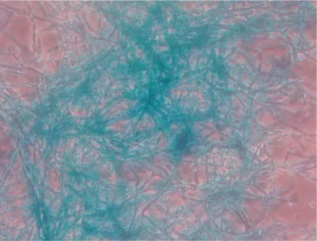
Fig. 2.β-glucuronidase activity can be detected using X-gluc as a substrate. Photomicrograph of histochemical visu-alization of GUS activity within GUS-tagged Verticillium fungicola(440GUS) grown in liquid culture (i10).
on CDA without antibiotic selection followed by CDA amended with 500µg ml−" hygromycin B.
Trans-formants were stored atk80min glycerol stocks and remained stable upon subsequent sub-culture. Single spore colonies of the selected transformants were used for further analysis.
Transformation rates were extremely variable with zero to 102 transformants per µg DNA. Co-trans-formation efficiency varied depending upon the plasmid used.
From 57 pAN7-1\pNOM102 and 102 pST28\
[image:5.612.312.545.297.434.2]pNOM102 transformants, 23 were selected at random and assessed forβ-glucuronidase expression using the MUG microtitre plate assay (Fig. 1). Five of the transformants assessed showed activity. One of these
Fig. 3.Verticillium fungicolaco-transformed with pGPDGFP and pAN7-1 expresses strong fluorescence. Photomicrograph of GFP-tagged mycelium grown in liquid culture and observed microscopically with UV light (i100).
Fig. 4. PEG-mediated transformation, with transforming DNA integrating randomly and in varying copy number into the target DNA. (a)–(b) Genomic DNA of 440GUS (a) and 440GFP (b) was digested withEcoRI (lanes 1 and 4),KpnI (lanes 2 and 5) andSalI (lanes 3 and 6) and was probed with a 600 bphphfragment obtained by PCR using pAN7-1 as a template. Single bands of varying size in lanes 1 and 3 indicate single copy random integration of the pAN7-1 DNA into the genome of the GUS co-transformant. Lane 2 did not resolve successfully. Multiple bands of varying sizes in lanes 4–6 indicate multi-copy, multi-locus random integration of the pAN7-1 DNA into the genome of the GFP co-transformant. (c) 440GUS genomic DNA digested withEcoRI,KpnI and BamHI in lanes 7–9 respectively, was probed with a 2000 bp NcoI pNOM102 fragment. Single bands of varying sizes in lanes 7–9 indicate single copy random integration of the pNOM102 transforming DNA into the target genome. (d) 440GFP genomic DNA digested withEcoRI,PstI andKpnI in lanes 10–12 respectively was probed with a 1 kbEcoRI pUCGFP fragment. Single bands in lanes 10–12 of varying sizes indicate single copy random integration of the pGPDGFP transforming DNA into the target genome. Selected molecular weight size markers are indicated.
[image:5.612.68.297.335.509.2]Fig. 5.Transforming DNA integrating randomly and in single copy number into the target DNA. A 550 bpPvuII-NcoI fragment from pBIN7.1. was used to probe theAgrobacterium-mediated transformants. (a) Lanes 1–5.HindIII digests of genomic DNA from fiveAgrobacterium-mediated transformants selected at random ; Lane 6. Wild-typeVerticillium fungicola440-34 genomic DNA.HindIII cuts once in the transforming DNA and once in the genomic DNA. The differing band sizes indicate integration at random loci, and the single bands show single integration events. (b) Lanes 7–11.EcoRI digests of genomic DNA from fiveAgrobacterium-mediated transformants selected at random ; Lane 12. Wild-typeV. fungicola440-34 genomic DNA.EcoRI cuts twice in the transforming DNA (once in the 550 bp region used as a probe), and once in the genomic DNA. Cutting the two sites within the vector generates the 2n4 kb band indicated. This band would be expected to have a greater intensity as it has 441 bp homologous to the probe, compared to 109 bp for the bands of variable size. The presence of different sized bands shows random integration, and two bands confirm single integration.
methods is successful. One transformant, designated 440GUS (pAN7-1 & pNOM102) was selected for further molecular analysis and was checked for histo-chemical localization of GUS activity (Fig. 2).
Twelve putative GFP transformants were obtained using plasmids pAN7-1 and pGPDGFP. These were assessed visually for GFP (Fig. 3) as described above. Three transformants expressed GFP, with one (440GFP) being selected for further molecular analysis. This particular isolate was chosen for comparison of pAN7-1 integration in the GFP co-transformant with the GUS co-transformant under separate transform-ation events. Fluorescence was not observed in the wild-type strain.
After initial PCR analysis to check for the presence of transforming DNA, Southern hybridisation showed that the probes for the hygromycin B resistance, β -glucuronidase and GFP genes did not hybridise to digested wild-type 440-34 genomic DNA. The probe for the hygromycin B gene hybridised to one or more fragments in digested DNA of the PEG-mediated transformed strains, thus confirming integration of the plasmid into the genomes of the transformants (Figs 4a–b). 440GUS (Fig. 4a) shows single bands when examined with a probe for the hygromycin B resistance gene, indicating single copy integration. 440GFP (Fig. 4b) shows multiple bands when screened with a probe for the hygromycin B resistance gene, indicative of multi-copy, multi-locus integration. Although difficult to determine accurately, it is estimated that three to four copies of the hygromycin B resistance gene are present in 440GFP. The GUS probe and the GFP probe hybridised to one fragment in the GUS (Fig. 4c) and GFP (Fig. 4d) transformants respectively, con-firming single copy integration of the respective plasmids into the genomes of these transformants. The
different hybridisation patterns for the hygromycin B-resistant transformants indicates that integration can occur singly, and in multiple copies for protoplast transformation.
Analysis ofAgrobacterium-mediatedtransformants
Hygromycin B-resistant transformants were generated usingA.tumefaciensstrain LBA1126 transformed with the vector pBIN7-1. This was mixed withV.fungicola strain 440-34 conidia and transformation was induced by incubation on induction medium. 4068 putative transformants were obtained from 8n6i10& conidia. Putative hygromycin B-resistant transformants were sub-cultured on to minimal medium amended with 550µg ml−" hygromycin B. Five transformants were
selected at random for further analysis. Growth of transformants was exhibited at hygromycin B concen-trations up to 2 mg ml−".
R. C. Amey and others 9
A
[image:7.612.67.299.51.414.2]B
Fig. 6.Isolate 440GFP as a tool to trackVerticillium fungicola infection in vivo in Agaricus bisporus. (a) Infection of A. bisporus by the V. fungicola GFP-tagged transformant (440GFP) resulting in a necrotic lesion on the mushroom cap. (b) Microscopic examination of the spotting symptom shown in Figure 6.a and visualised using UV light. TheV.fungicola green fluorescent hyphae can be seen clearly against the host (mushroom) tissue (i10).
must have resulted in functional copies of the gene to enable the fungus to grow on selection medium.
Assessment of virulence of co-transformants
As hypothesised by Joneset al. (1999), it is important to know whether the constitutive Aspergillus nidulans promoters place an undesirable metabolic burden on the transformant, and to determine whether the transformants have altered in their ability to cause disease in theAgaricushost as a result of integration of transforming DNA within the genome. Initial com-parison of growth rates of the co-transformants showed similarity with that of the wild-type. We then compared the co-transformants with the wild-type fungus in virulence tests onA.bisporus. Disease impact of the two strains was evaluated by measuring crop yield and assessment of symptom expression. It was observed that 440GUS and 440GFP were as virulent as the
wild-type, showing similar reductions in yield (results not presented) and the three main symptoms ofV.fungicola infection (cap spotting, stipe blow-out and dry bubble).
GUS and GFP expression of co-transformants in Agaricus bisporus
To observe GUS activity, infected mushrooms were observed following the X-gluc staining as described above. Although the GUS transformant gave blue colouration, histochemical localization of GUS activity within Verticillium fungicola mycelium in vivo within the infected mushroom proved impossible due to the high background of GUS activity generated by the host (mushroom) tissue. This may indicate that the value of the GUS transformant to investigate direct interactions withAgaricus bisporusis limited.
GFP expression of V. fungicola in infected mush-rooms was observed as described earlier. Hyphae of the co-transformant 440GFP were barely distinguishable from the host under incident light. In contrast, 440GFP exhibited distinct green fluorescence when observed with ultra-violet light, allowing it to be clearly dis-tinguished from its host (Fig. 6).
D I S C U S S I O N
fungicola. Random T-DNA tagging would certainly be one possibility for further investigation. Transformants could be examined for altered pathogenicity, and the cause of the changed phenotype ascertained by search-ing for the integration site of the T-DNA in the target genome. Protoplast-mediated transformation displays reduced value in this situation due to the unpredictable number of gene copies entering the target genome, possibly causing more than one phenotypic change.
TheuidA gene has proved to be useful in the study of fungal biomass of Cladosporium fulvum and Lepto -sphaeria (Oliver et al. 1993), root colonisation by Fusarium oxysporum (Eparvier & Alabouvette 1994), infection of Bipolaris sorokiniana(Lijeroth, Jansson & Schafer 1993) and biocontrol ofSclerotinia sclerotiorum byConiothyrium minitans(Joneset al. 1999). However, the GUS transformant has limited application as a tool for studying infection of Agaricus bisporus. This was due to the backgroundβ-glucuronidase production of A. bisporus making it difficult to distinguish between the host and pathogen. However, it may remain useful for studies ofV. fungicola biomass and epidemiology studies in compost and casing experiments. In contrast, the GFP strain may prove to be an invaluable tool in studying the interaction ofV.fungicolawithA.bisporus and may also advance the study of the epidemiology of the disease and the spread through both host and compost. The possession of hygromycin-B resistant, GFP-expressing and GUS-expressing isolates will allow selective isolation, quantification andin vivotracking of the transformants from, and in mushrooms and compost, especially as no reduction in virulence was observed. This will allow experiments to be carried out into the survival, growth, epidemiology and host colonisation ofV.fungicola.
The establishment of these transformation systems for V. fungicola and validation of GUS and GFP reporter genes allows further information to be obtained on the fungus : fungus interaction betweenV.fungicola and its hostA.bisporusby promoter : reporter fusions, REMI and T-DNA tagging of mutants. Targeted disruption of genes with potential roles in pathogenicity will be investigated.
A C K N O W L E D G E M E N T S
The authors would like to thank Steve Lincoln (Horticulture Research International, Wellesbourne) for technical support and supplying isolate CBS 440n34, and Neil Willoughby (Horticulture Research International, Wellesbourne) for help and advice with mushroom cultivation. This work was partly funded by the UK Ministry of Agriculture Fisheries and Food (MAFF) and The Royal Society.
R E F E R E N C E S
Bae, Y. S. & Knudsen, G. R. (2000) Cotransformation ofTrichoderma
harzianum with β-glucuronidase and green fluorescent protein
genes provides a useful tool for monitoring fungal growth and activity in natural soils.Applied and Environmental Microbiology
66: 810–815.
Bevan, M. (1984) Binary Agrobacterium vectors for plant trans-formation.Nucleic Acids Research22: 8711–8721.
Bowyer, P., Clarke, B. R., Lunness, P., Daniels, M. J. & Osbourn, A. E. (1993) Host range of a plant pathogenic fungus is determined by a saponin detoxifying enzyme.Science287: 371–374. Bundock, P., den Dulk-Ras, A., Beijersbergen, A. & Hooykas, P. J. J.
(1995) Trans-kingdom T-DNA transfer from Agrobacterium
tumefaciens to Saccharomyces cerevisiae. EMBO Journal 14:
3206–3214.
Calonje, M., Garcia Mendoza, C., Galan, B. & Novaes-Ledieu, M. (1997) Enzymic activity of the mycoparasiteVerticillium fungicola
on Agaricus bisporus fruit body cell walls. Microbiology 143:
2999–3006.
Calonje, M., Garcia Mendoza, C., Perez Cabo, A., Bernardo, D. & Novaes-Ledieu, M. (2000) Interaction between the mycoparasite
Verticillium fungicolaand the vegetative mycelial phase ofAgaricus
bisporus.Mycological Research104: 988–992.
Covert, S. F., Kapoor, P., Lee, M., Briley, A. & Nairn, C. J. (2001)
Agrobacterium tumefaciens-mediated transformation ofFusarium
circinatum.Mycological Research105: 259–264.
de Groot, M. J. A., Bundock, P., Hooykaas, P. J. J. & Beijersbergen, A. G. M. (1998)Agrobacterium tumefaciens-mediated transform-ation of filamentous fungi.Nature Biotechnology16: 839–842. Dragt, J. W., Geels, F. P., de Bruijn, W. C. & van Griensven,
L. J. L. D. (1996) Intracellular infections of the cultivated
mush-roomAgaricus bisporusby the mycoparasiteVerticillium fungicola
var.fungicola.Mycological Research100: 1082–1086.
Eparvier, A. & Alabouvette, C. (1994) Use of ELISA and GUS-transformed strains to study competition between pathogenic and non-pathogenicFusarium oxysporumfor root colonisation.
Bio-control Science & Technology4: 35–47.
Fletcher, J. T., White, P. F. & Gaze, R. H. (1986)Mushrooms : pest and disease control. Intercept, Newcastle Upon Tyne.
Gouka, R. J., Gerk, C., Hooykaas, P. J. J., Bundock, P., Musters, W., Verrips, C. T. & de Groot, M. J. A. (1999) Transformation of
Aspergillus awamoribyAgrobacterium tumefaciens-mediated
hom-ologous recombination.Nature Biotechnology17: 598–601. Hargreaves, J. & Turner, G. (1992) Gene transformation in plant
pathogenic fungi. InMolecular Plant Pathology(S. J. Gurr, M. J. McPherson & D. J. Bowles, eds) : 79–97. Oxford University Press, Oxford.
Hooykaas, P. J. J., Roobol, C. & Schilperoort, R. A. (1979) Regu-lation of the transfer of Ti-plasmids ofAgrobacterium tumefaciens.
Journal of General Microbiology110: 99–109.
Inglis, P. W., Araga4o, F. J. L., Fraza4o, H., Magalha4es, B. P. & Valadares-Inglis, M. C. (2000) Biolistic co-transformation of
Metarhizium anisopliae var. acridum strain CG423 with green
fluorescent protein and resistance to glufosinate ammonium.FEMS
Microbiology Letters191: 249–254.
Jeffries, P. & Young, T. W. K. (1994)Interfungal Parasitic Relation-ships. CAB International, Wallingford.
Jones, E., Carpenter, M., Fong, D., Goldstein, A., Thrush, A., Crowhurst, R. & Stewart, A. (1999) Co-transformation of the sclerotial mycoparasiteConiothyrium minitanswith the hygromycin B resistance andβ-glucuronidase markers.Mycological Research
103: 929–937.
Keon, J. & Hargreaves, J. (1998) Isolation and heterologous expression of a gene encoding 4-hydroxyphenylpyruvate dioxy-genase from the wheat leaf-spot pathogen,Mycosphaerella
gramini-cola.FEMS Microbiology Letters161: 337–343.
Lijeroth, E., Jansson, H. B. & Schafer, W. (1993) Transformation of
Bipolaris sorokinianawith the GUS gene and use for studying
fungal colonisation of barley roots.Phytopathology83: 1484–1489. Mullins, E. D., Chen, X., Romaine, P., Raina, R., Geiser, D. M. & Kang, S. (2001) Agrobacterium-mediated transformation of Fusarium oxysporum: an efficient tool for insertional mutagenesis and gene transfer.Phytopathology91: 173–180.
R. C. Amey and others 11
infected plant tissues. Molecular Plant–Microbe Interactions 6: 521–525.
Punt, P. J., Oliver, R. P., Dingemanse, M. A., Pouwels, P. H. & van den Hondell, C. A. M. J. J. (1987) Transformation ofAspergillus based on the hygromycin resistance marker fromEscherichia coli. Gene56: 117–124.
Rohe, M., Gierlich, A., Hermann, H., Hahn, M., Schmidt, B., Rosahl, S. & Knogge, W. (1995) The race-specific elicitor,NIP1,
from the barley pathogen, Rhynchosporium secalis, determines avirulence on host plants of the RRS1 resistance genotype.EMBO
Journal14: 4168–4177.