http://www.scirp.org/journal/ojem ISSN Online: 2332-1814
ISSN Print: 2332-1806
Total Hip Replacement in Young Adults Less
Than Fifty Year Old: Our Experience
Adonis Magoumou, Narcisse Dabiré, Yassir El Andaloussi, Said Abdallah, Amine Belmoubarik,
Rachid Ait-Mouha Karim Ahed, Nabil Omari, Ahmed Reda Haddoun, Driss Bennouna,
Mohamed Moujtahid, Mohamed Nechad, Mustapha Fadili
Department of Orthopedic Traumatology, Wing 4, Chu Ibn Rochd, Casablanca, Morocco
Abstract
Background: We report a retrospective study of 47 total prosthesis of hip put in place among the young adult of less than fifty years in the service of Trauma-tology and Orthopedics at the Wing IV of the CHU Ibn Rochd of Casablanca, over a period from January 2008 to March 2014, with an average rate of decline of 42 months. Results: The deliberate group of 43 patients (47 hips including 4 bilateral) consisted of 26 women, either 60, 47% and 17 men, either 39, 53%. The average age of our patients was 36 years. In our series, the predominant eti-ology has been conducted for the indication of the hip replacement was the aseptic osteonecrosis of the femoral head which has represented 31.91%. With regards to the choice of implants, 85% of PTH were non-cemented, the ce-mented PTH has been used in 15% of cases. The Friction Torque met-al/polyethylene was used in all cases. The functional outcome was appreciated by the rating of Merle of Aubigne before and after arthroplasty. The average rate of decline of 42 months, and the results were generally satisfactory. They were excellent in 27.78% of cases, very good in 19.44% of cases and good in 44.44% of cases. The results on the Pain were remarkable: the rating of the pain going 2.8 in pre-operative at 5.6 at the latest decline. The results on the walking and mo-bility were also good. It was noted as a complication: 4 cases of prosthetic dislo-cation, 1 case of aseptic descellement bipolar, 2 cases of early sepsis on hard-ware, 3 cases of paralysis of the sciatic external popliteal and 4 cases of peripro-thetiques ossification. In total, 5 total prosthesis of hip were occasions. The young age does not seem to be an obstacle to the prosthetic surgery. Conclusion: However, a major question remains concerning the distant future of these prostheses in young active subjects, because they will be submitted for many years in a job which can cause the descellement of prosthetic parts.
Keywords
Total Hip Replacement, Young Subjects, Hip Disease, Casablanca
How to cite this paper: Magoumou, A., Dabiré, N., El Andaloussi, Y., Abdallah, S., Belmoubarik, A., Ait-Mouha K. Ahed, R., Omari, N., Haddoun, A.R., Bennouna, D., Moujtahid, M., Nechad, M. and Fadili, M. (2017) Total Hip Replacement in Young Adults Less Than Fifty Year Old: Our Experience. Open Journal of Emergency Medicine, 5, 43-74.
https://doi.org/10.4236/ojem.2017.52006
Received: November 21, 2016 Accepted: June 23, 2017 Published: June 26, 2017
Copyright © 2017 by authors and Scientific Research Publishing Inc. This work is licensed under the Creative Commons Attribution International License (CC BY 4.0).
1. Introduction
The goal of total hip replacement is to replace the worn surfaces of the ball-and- socket joint. The number of total prosthesis of hip (PTH) implanted by year is in perpetual increase in the industrialized countries but also in Morocco, the quali-ty of life is better because of the increase in the life expectancy and the availabil-ity of prostheses in the public hospitals since the introduction of the medical as-sistance scheme (RAMED).
The indication by excellence of the total hip remains the coxarthrose, this tra-ditional indication limited the practice of this technique to the elderly, but the excellent results of this technique have pushed the orthopaedic surgeons to the practice in subjects of more and more young people.
However, the implantation on young patients increases the risk of iterative occasions. The pre-operative planning is therefore paramount in the choice of the surgical technique and the choice of implants.
Through this retrospective study of 43 patients (47 total prosthesis of hip) collected at the service of orthopaedic traumatology wing IV of the Center Hos-pitalier Universitaire Ibn Rochd of Casablanca, we want to relate our experience in the total hip replacement in the subject of less than 50 years, while comparing our results in the light of different data in the literature.
2. Materials and Methods
Our study is concerned with the reviewing of 43 patients aged less than 50 years, having benefited from 47 total prosthesis of hip between January 2008 and March 2014 in the service of orthopaedic traumatology wing IV of the Universi-ty Hospital Center (CHU) Ibn Rochd of Casablanca.
The clinical information, para-clinical scalable and have been collected from the records of the sick and collected by the means of a farm, a copy of which is shown below. We have studied the data pre-therapeutic, the surgical act and suites post-operation. Our criteria of inclusions were: Patients who benefited from a total hip aged less than 50 years of age at the time of arthroplasty, auto-nomous patients, decline of minimum 6 months post-operative is required. Our criteria of non-inclusions were: age greater than 50 years at the time of the total hip replacement, psychomotor retardation, neuropsychiatric disorders, Co- morbidities serious: dysimmunitary field, decompensated heart disease.
3. Ethics
Patients or their relatives gave informed consent to be part of the study.
4. Results
Epidemiological Study -Age
-Sex
In our series we note a predominance of women: 26 patients were women (ei-ther 60, 47%); 17 patients were men (ei(ei-ther 39, 53%). With a sex-ratio male/female of 0.65.
-Operated Side
The right hip was operated in 24 cases, either 55, 81% and the left hip in 15 cases, either 34, 88%.
Four of our patients (9.3%) have benefited from a bilateral PTH at an average interval of 8 months.
Pathological History -Medical History
Thirteen patients had a medical history, either 30, 23%, presented in the Table 1.
-Medical treatment to the long course:
Six patients (13.9%) were treated in the long term by NSAIDS, including 1 in association with corticosteroids, 4 patients (9.3%) were treated by a corticoste-roid to the long course of which 3 in association with immunosuppressive ther-apy.
History of the hip place:
Ten patients of our series is 23.25% had orthopaedic history at the level of the hip place:
Osteosynthesis for fracture of the neck of the femur in 3 cases; Orthopedic treatment for fracture of the acetabulum in 1 cases; Biopsy of the hip in 2 cases; Femoral Epiphysiolyse higher in 1 cases; Bloody reduction of LCH in 2 cases in-cluding 1 associated with a pelvic osteotomy; Landfill by traction for OPH in 1 case.
-
INDICATIONS-
The indications of total prosthesis in the hip in our series are listed in Table 2.-
Have been regarded as coxarthrose post-traumatic stress, osteoarthritis sec-ondary to trauma of hip, former is not defined or a fracture of the acetabulum.-
Have been regarded as aseptic osteonecrosis post-traumatic, osteonecrosis [image:3.595.210.536.595.734.2]secondary to a fracture of the femoral neck (failure of osteosynthesis) or to a Hip Dislocation post-traumatic stress is neglected.
Table 1. Medical history of patients.
Medical history Number of Cases
Diabetes (type II) 1
Genital TB 1
Obsessive-compulsive disorders 1
Asthma 1
Anxiety Disorders 1
Ankylosing spondylitis (SPA) 5
Table 2. Distribution of indications of the total hip.
Indication (%) Number of hip
Primitive coxarthrose (4.26%) 2
Secondary coxarthrose
-Coxarthrose post-traumatic (14.89%) 7
-Coxarthrose on congenital dysplasia (10.64%) 5
The sequelae of LCH (8.51%) 4
Cotyloïdienne dysplasia (2.13%) 1
-sequelae of primitive osteochondrite of the Hip (OPH) (2.13%) 1
-sequelae of epiphysiolyse (2.13%) 1
-The sequelae of coxite tuberculous infectious (6.38%) 3 -Aseptic osteonecrosis of the femoral head (ONATF) (31.91%) 15
Post-traumatic stress disorder (12.77%) 6
Secondary to a corticosteroid (2.13%) 1
Secondary to an ethylisme (2.13%) 1
The idiopathic (14.89%) 7
-Patella tendon inflammation of the femoral neck (2.13%) 1
-Other: 1
Neurogenic Paraosteoarthropathie (NENA) (2.13%)
-Coxites inflammatory (23.40 %) 11
Rheumatoid arthritis (PR) (10.64%) 5
Ankylosing spondylitis (SPA) (12.76%) 6
Among the coxarthroses secondary to a legacy of LCH (4 hips or 8.51%), two were a LCH neglected and 2 had benefited from a surgical reduction during childhood.
The aseptic osteonecrosis of the femoral head and the inflammatory coxites accounted for more than 55%.
The inflammatory coxites were constituted of the ankylosing spondylitis in 12.76% and rheumatoid arthritis in 10.64%. The primitive coxarthrose was found in 4.26%.
Preoperatory Study
Physical activity
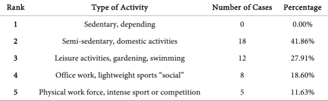
The level of activity of the patients has been appreciated according to the clas-sification in 5 grades of Devane (Table 3).
In preoperatory, 58.14% of patients were active, this is explained by the young age of the population studied.
Clinical Study
Table 3. Results of the patients activites in preoperatory according to the classification of Devane.
Rank Type of Activity Number of Cases Percentage
1 Sedentary, depending 0 0.00%
2 Semi-sedentary, domestic activities 18 41.86%
3 Leisure activities, gardening, swimming 12 27.91% 4 Office work, lightweight sports “social” 8 18.60% 5 Physical work force, intense sport or competition 5 11.63%
-Pain
The rating of the intensity of the pain was less than or equal to 3 points in 74.4% of cases, to 4 points in 23.4% and only a hip was rated to 5 points accord-ing to the rataccord-ing of LDCS, with an average of 2.8.
-The mobility
The rating of the mobility was less than or equal to 3 points in 68.09% and greater than or equal to 4 points in 31.91% with an average for the Mobility pre-operative of 3.
-Walking
The walk in preoperative was rated to 3 or less among 61.7% of our patients. It was rated to 4 among 31.9% of them with an average for the walk of 3.3.
In our series, the evaluation of the preoperative LDCS had found: a preopera-tive LDCS was bad among 28 hips (59.57%), poor in 15 hips (31.91%), and fi-nally fair among 4 hips (8.51%).
Radiological Study
-Standard Radiology
The radiological survey was conducted on x-rays of the face and profile of the Hip concerned and a radiography of basin of face. It was found constantly but in a variable manner: A pinch of the line spacing; Geodes and condensation; An osteophytose capital and/or cotyloidienne.
For the ONATF, the classification of Arlet Ficat and showed 52% of the hips in stage IV.
-Computed Tomography (CT)
Four of our patients have benefited of a CT scan of the basin. -Magnetic Resonance Imaging (MRI)
A patient has benefited from an MRI of the basin which has shown: a bilateral osteonecrosis of the femoral head.
-Study of operability -Clinical study
All our patients have benefited from a clinical examination of systematic and comprehensive in the search for an underlying pathology that can against-Indi- cate the surgical act.
Treatment
Surgical technique
-Preparation of the operative field
All our patients have benefited from a Preparation of the operative field on the eve of the intervention, which consisted of shaving the lower member and the pubis and a skin disinfection of the operative region by the dermal betadine before the operation.
The intervention took place in a room reserved exclusively to the aseptic sur-gery.
-Anesthesia
Among the 47 PTH, 30 have been put in place under general anesthesia (or 63.83%) and 17 put in place under rachianesthesie (or 36.17%).
-Installation of the patient
All our patients have been operated in lateral decubitus, with a sacred support pubic and to stabilize the patient during the surgical procedure.
-Tracks first:
All PTH of our series have been put in place by the track posterior external Moore.
-Additional Gestures
Of the bone gestures were required in 7 hips (14.89%) and all bone grafts were autologous.
A ring of supports Curboul type was placed in 1 case.
-Type of implanted prosthesis
According to the mode of fixation
The PTH implanted in our patients were cemented; in 7 cases, either 14.89%; not cemented in 40 cases, either 85.1% including: For 27 cases the acetabulum was screwed and for 13 cases the acetabulum was press-fit.
PTH dual mobility or conventional:
The PTH were of type “conventional” in 85% of cases and were of type double mobility in 15% of cases.
Metal/polyethylene (steel/polyethylene (PE) and cobalt-chrome/PE). The Friction Torque steel/polyethylene represented more than 68% in our se-ries.
Different implants used
-The sizes of implants -Parts cotyloidiennes
The size of the COINS cotyloidiennes used is reported in the Table 4.
The femoral parts
Femoral stems
The size of the femoral stems used is reported in the Table 5.
The neck was: Short in 23 cases (48.94%), medium in 14 cases (29.79%), long
in 9 cases (19.15%), extra-long in 1 cases (2.13%). Femoral heads
Table 4. Size of parts cotyloidiennes used.
External diameter
acetabulum 44 mm 46 mm 48 mm 50 mm 52 mm 54 mm 56 mm 58 mm 60 mm
Percentage 2.13% 8.51% 29.79% 25.53% 10.64% 12.77% 6.38% 2.13% 2.13%
Table 5. Size of stemmed femoral used.
Rod Size 1 2 3 4 5 6 7 7.5 8 9 10 11 12 13
Percent age 2.13% 2.13% 8.51% 4.26% 10.64% 8.51% 6.38% 2.13% 6.38% 12.77% 23.40% 6.38% 4.26% 2.13%
The postoperatory care:
The thrombophylaxy was started systematically in postoperatory in all the pa-tients on the basis of low molecular weight heparin.
-Analgesics:
The postoperatory analgesia has been ensured by administration of non-ste- roidal anti-inflammatory drugs (NSAIDS) and analgesics.
-Rehabilitation:
The rehabilitation has been started as soon as possible as well as the sitting position, the very day after the intervention. In the 2nd day lifted and wandering
has been accompanied using a strut. The rehabilitation was continuing in the exercises of articular mobilization and strengthening muscles by isometric con-tractions with an upgrade in charge protected by bequillage and under the su-pervision of a physiotherapist.
This rehabilitation has been to rule, among all our sick.
For patients who have had a bone graft, the charging was outlawed up to con-solidation.
Postoperatory Complications
-Early complications (the first 3 months)
General Complications
No thromboembolic complications, cardiac, or early death has been noted.
Local complications
No cases of superficial hematoma or femur fracture on prosthesis has been observed.
We have had 1 cases (2.13%) of Early sepsis on hardware, to J5 Post-opera- tive. An abundant washing and curettage of the infected tissues with bacterio-logical sampling has been carried out. The direct examination and culture was negative. Antibiotic therapy had been established. The evolution has been good. It was a patient aged 39 years operated on the left side for coxite in-flammatory. Followed for PR since the age of 10 years under prolonged corti-costeroid therapy in combination with methotrexate (Observation (obs) number (no) 16).
In our series, 3 cases of Early dislocation of PTH have been noted, the Evolu-tion was favorable year for all the CA:
-
In 2 cases: the dislocation occurred respectively to J5 (obs. n˚24) and J17 (obs. n˚30) Post-surgery. The dislocation occurred for non-compliance of move-ments proscribed in the 2 cases. The reduction was made in urgency: ortho-pediquement in 1 case (obs. n˚30) and in the other case, resumption of the PTH with a change in the head.-
In 1 cases: the dislocation had occurred at 1 months post-operative and was associated with a tilt of the cupule > 50˚ (obs. n˚42). The reduction has been made surgically after change of the head.Late complications (beyond the 3rd month post-operative)
-Descellements
We have noted 1 case of descellement bipolar aseptic occurred at 3 years post-op (obs. n˚17), having need a removal of the initial prosthesis and install a new prosthesis cemented together at the same time.
-Late Dislocation
We have listed a case of late dislocation of PTH to 7 years post-surgery, after a false movement during a bath in the Hammam (OBS. N˚1). The PTH has been resumed.
-Femur fracture
No cases of femur fracture has not been listed in our study. Results at the latest decline
Patients reviewed and average decline
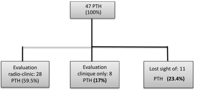
The latest decline of 32 patients (36 PTH) have been reviewed and 11 patients (11 PTH) have been lost sight of. The average decline for patients reviewed was 42 months with extremes ranging from 9 to 83 months. Among the patients re-viewed: 24 patients (28PTH) have been reviewed in consultation for an assess-ment of radio-clinic.
[image:8.595.211.526.573.716.2]8 patients (8 PTH) have not been able to move for reasons not related to the PTH and have been seen at home for a clinical evaluation. The radiological analysis at the latest decline has not been able to be carried out for these patients. (Last balance sheet dating back more than 2 years and not taken into account) (Figure 1).
Figure 1. Distribution of patients to the latest decline.
47 PTH (100%)
Evaluation radio-clinic: 28
PTH (59.5%)
Evaluation clinique only: 8
PTH (17%)
Functional Evaluation
For patients reviewed, we resumed the criteria of Postel and Merle of Aubigne (LDCS), in order to assess the clinical condition of the patient and the function of their hip after decline.
Results on the Pain
The average rating of the pain to the last decline was 5.63. The latest decline 69.44% of hips were painless.
Results on the mobility
The average rating of the mobility in the last decline was of 5.08.
Results on the walk
The rating of the walk to the latest decline was 5.4 on average. Score overall LDCS and appreciation to the latest decline
The score LDCS average overall to the decline was 16.1.
The functional outcome global according to the scoring LDCS, at the average rate of decline of 42 months, was excellent in 27.78% of cases, very good in 19.44% of cases and good in 44.44% of cases, fair in 2.78% of cases, poor in 2.78% of cases, wrong in 2.78% of cases (Figure 2).
We have grouped the functional results assessed according to the criteria of Postel-Merle of Aubigne (LDCS) by: Satisfactory for the excellent results, very good and good; Unsatisfactory for the results passable, poor and ill. The results were generally satisfactory (91%) to the average rate of decline of 42 months.
Comparison of preoperative LDCS and the latest decline
The average gain of the LDCS global at the latest decline was 7 points com-pared to the preoperative state. Among the items the score of the LDCS the gain the higher was the item “pain”, it went from 2.8 to 5.6.
[image:9.595.213.534.475.713.2]Radiological Evaluation
The radiological assessment has been based on x-ray images of the basin of strict face as well as radiographs of the hip surgery of the face and profile taking the totality of the prosthesis.
A careful study of the successive radiographs and their confrontation with the cliches post-operative, earlier had for the purpose of the search for signs of com-plications.
The radiological shots have been analyzed according to the following criteria: at the level of the workpiece cotyloidienne, we checked:
The angle of tilt of the Cup by report to the BI Line ischiatic. It is normally of
45˚.
The position of the acetabulum by report to the line innominee (Protrusion)
and the upper part of the valve stem hole. It is normally < 2 mm.
The liseres were analyzed in the 3 areas of Lee and Charnley. At the level of the femoral exhibit, we checked:
The position of the workpiece by femoral report to the axis of the femur: varus
valgus, or normo-focused.
Tail of the cement.
The femoral liseres were analyzed in the 14 zones of Gruen.
For the PERI ossification prosthetic-it has used the classification of Brooker.
In post-procedure
We have studied the radiographs of control of the post-operative of all pa-tients (47 PTH).
At the level of the Prosthetic acetabular
-
30 cases had a slope between 42˚ - 45˚.-
16 cases had a slope between 46˚ - 50˚.-
A case of vertical cup with an angle >50˚ has been noted.At the level of the femoral exhibit
-
Three PTH had a rod in a varus.-
Two PTH had a rod in valgus.-
The STEM was normo-focused in 42 PTH.No border line has been found in post-operative immediate.
The last decline:
It is recalled that the x-ray analysis focuses on 28 PTH, the results are the fol-lowing:
At the level of the Prosthetic acetabular:
No cases of verticalisation of the Cup was noted. All the wells had a good po-sitioning.
No wear has been observed at the level of the acetabulum to the last decline.
At the level of the femoral part:
-
1 case of varisation of the femoral stem.It was found 2 case of ossification peri-prosthetic appliances not having need for surgical recovery, according to the classification of Brooker:
-
1 case was of grade II: patient operated for neurogenic paraosteoarthropathie-
1 case was in Grade I: patient operated for coxarthrose post-traumatic (obs.n˚41).
In addition no sign pointing toward a descellement has been noted. At total after an average rate of decline of 42 months:
Five PTH on the 47 implanted (10.6%), have undergone a surgical recovery: 4 times with a change of the implant and a resumption without change of im-plant.
Late complications (beyond the 3rd month post-operative)
-Descellements
We have noted 1 case of descellement bipolar aseptic occurred at 3 years post-op (obs. n˚17), having need a removal of the initial prosthesis and install a new prosthesis cemented together at the same time.
-Late dislocation
We have listed a case of late dislocation of PTH to 7 years post-surgery, after a false movement during a bath in the Hammam (obs. n˚1). The PTH has been resumed.
-Femur fracture the post-operative
No cases of femur fracture has not been listed in our study. Results at the latest decline
-Patients reviewed and average decline
The latest decline 32 patients (36 PTH) have been reviewed and 11 patients (11 PTH) have been lost sight of. The average decline for patients reviewed was 42 months with extremes ranging from 9 to 83 months.
Functional evaluation:
For patients reviewed, we resumed the criteria of Postel and Merle of Aubigne (LDCS), in order to assess the clinical condition of the patient and the function of his hip after decline.
-Results on the pain:
The average rating of the pain to the last decline was 5.63. The latest decline 69, 44% of hips were painless.
Results on the mobility:
The average rating of the mobility in the last decline was of 5.08.
Results on the walk:
The rating of the walk to the latest decline was 5.4 on average.
-Score overall LDCS and appreciation to the latest decline:
The score LDCS average overall to the decline was 16.1.
The global functional outcome according to the scoring LDCS, at the average rate of decline of 42 months, was excellent in 27.78% of cases, very good in 19.44% of cases and good in 44.44% of cases, fair in 2.78% of cases, poor in 2.78% of cases, wrong in 2.78% of cases (Figure 2).
We have grouped the functional results assessed according to the criteria of Postel-Merle of Aubigne (LDCS) by:
The results were generally satisfactory (91%) to the average rate of decline of 42 months.
Comparison of preoperative LDCS and the latest decline
The average gain of the LDCS global at the latest decline was 7 points com-pared to the preoperative state. Among the items the score of the LDCS the gain the higher was the item “pain”, it went from 2.8 to 5.6.
Radiological evaluation
The radiological assessment has been based on x-ray images of the basin of strict face as well as radiographs of the hip surgery of the face and profile taking the totality of the prosthesis.
A careful study of the successive radiographs and their confrontation with the cliches post-operative, previous had for the purpose of the search for signs of complications.
The radiological shots have been analyzed according to the following criteria: at the level of the workpiece cotyloidienne, we checked:
The angle of tilt of the Cup by report to the BI Line ischiatic. It is normally of 45˚.
The position of the acetabulum by report to the line innominee (Protrusion) and the upper part of the valve stem hole. It is normally < 2 mm.
The liseres were analyzed in the 3 areas of Lee and Charnley. At the level of the femoral exhibit, we checked:
The position of the workpiece by femoral report to the axis of the femur: varus valgus, or normo-focused.
Tail of the cement.
The femoral liseres were analyzed in the 14 zones of Gruen.
For the PERI ossification prosthetic-it has used the classification of Brooker.
-In post-procedure
We have studied the radiographs of control the post-operative of all patients (47 PTH).
At the level of the prosthetic acetabular
-
30 cases had a slope between 42° - 45°.-
16 cases had a slope between 46° - 50°.-
A case of vertical cup with an angle > 50° has been noted.At the level of the femoral exhibit
-
Three PTH had a rod in a varus.-
Two PTH had a rod in valgus.-
The STEM was normo-focused in 42 PTH.No border line has been found in post-operative immediate.
-The last decline:
It is recalled that the x-ray analysis focuses on 28 PTH, the results are the fol-lowing:
At the level of the prosthetic acetabular:
No wear has been observed at the level of the acetabulum to the last decline.
At the level of the femoral part:
-
1 case of varisation of the femoral stem.It was found that 2 case of ossification peri-prosthetic appliances not having need for surgical recovery, according to the classification of Brooker:
-
1 case was of grade II: patient operated for neurogenic paraosteoarthropathie(obs. n˚ 32) associated with a score of LDCS to 11;
-
1 case was in Grade I: patient operated for coxarthrose post-traumatic (obs.n˚41).
In addition no sign pointing toward a descellement has been noted. At total after an average rate of decline of 42 months.
Five PTH on the 47 implanted (10.6%), have undergone a surgical recovery. 4 times with a change of the implant and a resumption without change of im-plant.
5. Discussion
Indications of the PTH
-General information on indications
The indications of the PTH remain dominated by the coxarthrose [1]-[6], but the main objective of the PTH is to combat the pain and to improve the function of the hip, thereby it finds its place in a lot of other traumatic pathology and de-generative diseases of the hip.
In France, the National Agency for the development of the Medical Assess-ment (ANDEM) [6] has made the following recommendations on the indica-tions of the PTH:
For the coxarthrose:
The PTH is indicated in the functionally coxarthroses causing a severe disabil-ity insufficiently daily improved by a medical treatment well led, after an obser-vation period of a few weeks to a few months.
For fractures of the neck of the femur:
No indication of the PTH in the fractures non-displaced from the neck of the femur.
In the cases of fractures in coxa Vara, beyond 70 years, a PTH can be used. However, before 50 years, the surgical treatment must give preference to the os-teosynthesis.
In the United States, NIH (National Institutes of Health) has recommended the installation of arthroplasties total hip (ATH) for patients with chronic pain of the hip with alteration of the Quality of Life. The Glory (Global Orthopaedic Registry), an international registry of patients who received an ATH in first in-tention, had found that the selection criteria for an ATH varied in functions of countries of patients and surgeons. However, there is no international consensus for the indications of the PTH [7].
Epidemiological data
More than 1 million of total hip replacement are performed each year in the world, and this figure is expected to double in the next two decades. The rate of ATH of first intention has increased by 50 per cent in the United States between 1990 and 2002. In the UK between 2005 and 2010, this rate has experienced an increase of 16% and similar increases have been noted in Finland and Norway
[7].
Kurtz, et al., have made a projection on the period from 2005 to 2030 in the United States. They have used the data from the Nationwide Inpatient Sample
(NIS) to formulate a model allowing for a projection of the number of cases per year and predict an increase in the number of ATH of first intention to 174% [8]. Kurtz, et al., in another study, using the same method, on the period 2010 to 2030, predict a 50% increase in the number of ATH in younger pa-tients [29].
In Morocco, an increase in acts of total hip replacement was also observed, especially since the introduction of the system of medical assistance (RAMED) ensuring an availability of the implants in the public hospitals.
The prosthetic surgery Hip in constant increase, also concerns the subjects of more and more young and active [7] [9] [10].
The current control of operational techniques, the improvement of drawings and quality of materials of prostheses have largely contributed to these increases of settlements [11].
-Age
The age as epidemiological element is important to take into consideration in the installation of a PTH. It is an important factor determining the functional outcome and the longevity of the prosthesis [32]. The results of the PTH before 50 years of age are variously assessed in the literature. It is traditional to say that they are overall less better on the long term than those of an older population
[12]-[22].
In fact, youth patients seek mechanically their prosthetic way more important. Thus, for Johnsson, each decade of less during the intervention increases the rel-ative risk of descellement of 1.8 [23] (Table 6).
The average age in our series is 36 years, she joined the Series P32 of the CHU Ibn Rochd [44] and is lower than those of the other series [32]-[42]. This lesser average age could be explained by the young age of the Moroccan population compared to the western population and by the frequency of pathologies affect-ing the young subject in our context: coxites inflammatory, coxarthrose post- traumatic.
-Sex
Classically, there is a predominance of women among patient candidates in a PTH [1] [7]. The female sex was predominant in our series, this joined the series of Aldinger [13], Chana [19] and Schmitz [20], Nich [25] and Archibeck
[26] (Table 7).
Table 6. Comparison of the average age with the series of less than 50 years.
Year Numbers of cases Average Age (years)
Delaunay et al. [32] 2008 73 40.7
Roof series 32 of the CHU
Ibn Rochd (P32 CHUIR) [44] 2009 31 36
Aldinger et al. [33] 2009 141 47
Migaud et al. [34] 2011 30 39.8
Hwang et al. [35] 2011 70 39.8
Teusink et al. [36] 2012 118 40.5
Takenaga et al. [37] 2012 100 40.1
Innmann et al. [38] 2013 91 42
Chana et al. [39] 2013 110 45
Schmitz et al. [40] 2013 126 37.6
Channel Babovic et al. [41] 2013 50 38.9
Ryan et al. [42] 2014 72 43.1
Our series 2015 43 36
Table 7. Distribution of PTH in young subjects on the basis of sex among various au-thors.
Authors Year Numbers of cases of men (H) (%) Percentage women (F) (%) Percentage of
Nich et al. [15] 2006 23 43.4 56.6
Archibeck et al. [16] 2006 91 49 51
Delaunay et al. [12] 2008 73 79.4 20.6
Series P32 CHUIR [14] 2009 31 51,61 48,38
Aldinger et al. [13] 2009 141 48.9 51.1
Migaud et al. [14] 2011 30 83.3 16.7
Hwang et al. [15] 2011 70 81.4 18.6
Teusink et al. [16] 2012 118 51.7 48.3
Takenaga et al. [17] 2012 100 63 37
Innmann et al. [18] 2013 91 67 33
Chana et al. [19] 2013 110 47.3 52.7
Schmitz et al. [20] 2013 126 48.4 51.6
Ryan et al. [12] 2014 88 51.4 48.6
Our series 2015 43 39.5 60.47
among women (3.8% F against 2.5% H), same as for Turner (5.5% F against 2.8% H) [28]. This difference can be attributed to a greater muscle weakness
[image:15.595.210.538.382.645.2]prac-tice of physical activities are more important than women of the same age, would entail a solicitation that are more important of the implant leading to terms to the descellement [30] [31].
C-indications:
When a HIP is reached by a pathological process (coxarthrose primitive or secondary, osteonecrosis of femoral head, coxites … etc.) the clinical expression is generally summarized to 3 functional signs associated with various degrees: pain, loss of mobility and lameness. These signs are reflected by a disability in the life of the sick. The indication of arthroplasties, is essentially based on the functional discomfort of patients [32].
The pathologies leading to the establishment of a total hip are multiple and well known. In young patients, the pathologies responsible of disability are specific. The legacies of diseases of the childhood are: primitive osteo-chondritis of the Hip (OPH), congenital dislocation of hip (LCH), Aseptic osteonecrosis of the femoral head in transplant recipients of marrow, sickle cell anemia, corticosteroids for various ailments, severe asthma or other. These are also of rheumatic diseases (juvenile idiopathic arthritis (JIA), an-kylosing spondylitis (SPA), rheumatoid arthritis (PR)), the villo synovi-tis-nodular and quickly coxarthroses scalable. The sequelae of trauma also represent an important part: fractures of the acetabulum and the neck of the femur. The sequelae of pathologies of the children constitute a population where the morphological alterations are frequent and may complicate the surgical management [11].
[image:16.595.56.537.475.737.2]In our series The ONATF was predominant which combined the results of Delau-nay [12], Migaud [14], Hwang [15], Schmitz [16], Ryan [22] and Kim [33] (Table 8).
Table 8. Comparison of etiologies having conducted to the indication of PTH.
Series Publication Year of hips # of Coxarthrose Primitive (%)
Coxarthrose post-traumatic
stress (%)
Coxite inflammatoire
(%)
Congenital Dysplasia
(%)
ONATF
(%) Epiphysiolyse (%) OPH (%) Septic arthritis
of the hip (%)
Delaunay et al. [12] 2008 83 23 4.8 - 27.7 42 - 2.4 -
Series P32CHUIR [24] 2009 35 11.42 22.85 34.27 - 25.7 - - 5.70
Aldinger et al. [23] 2009 154 31 5 1 33 19 - - -
Migaud et al. [24] 2011 39 2.56 2.56 - 38.4 51.3 - 5.1 -
Hwang et al. [25] 2011 78 7.7 - 7.7 7.7 72 - - 2.5
Kim et al. [33] 2011 228 14 - 4.5 1.95 66 - - 11
Teusink et al. [16] 2012 144 20.83 4.86 8,97 12.5 26.4 6.25 4.86 0.7
Takenaga et al. [17] 2012 115 34 7.8 2.61 12.2 30.5 1.73 7 0.87
Innmann et al. [18] 2013 100 38 6 4 27 18 - - -
Chana et al. [19] 2013 120 63.3 - 3.3 25 4.2 4.2 - -
Schmitz et al. [20] 2013 152 9.9 5.3 10.5 21.7 26.3 3.3 7.2 3.3
Ryan et al. [22] 2014 88 5.7 5.7 9.2 4.5 67 3.4 1.1 3.4
The impact of the different aetiologies in the chance of the PTH and how they may influence the results of surgical, however remains unclear in the literature
[22].
During the ONATF, the overall results of the PTH are classically less better in particular subjects under 50 years of age. In these young people patients with osteonecrosis aseptic, of anatomical differences compared to the primitive cox-arthrose have been observed, leading some surgeons to propose shoots on mea-surement in order to adapt the implant to the patient in the hope of a better long-term Outcome [34] [35].
Radl et al.[36], in a retrospective study, have assessed the results of a femoral stem which is not cemented in young patients, made for coxarthrose (49 pa-tients) and other for aseptic osteonecrosis (31) patients with Ficat stage III and IV), the average decline was 6 years. Using the technique EBRA (Einzel-Bild roentgen-analysis), a method of analysis assisted by computer to measure the displacement of implants, they noted a migration of the rod of 1.5 mm among operated for osteonecrosis versus 0.6 mm among those operated coxarthrose (p
< 0.001). The analysis of the survival of the stem at the latest decline, with as event revision of the rod, was 74% among the group osteonecrosis versus 98% among the group coxarthrose. They have suggested that this difference could be attributed to a decrease in the stock of bone peri-prosthetics occurred during the osteonecrosis.
However, Ince et al. [37], in a retrospective study using the same method of analysis of implants (EBRA), have evaluated the results of the 41 young patients who had a non-PTH cemented (porous coating), all made for osteonecrosis ad-vanced and they have found no revision of the rod or descellement (average rate of decline of 7 years). Similar results have been found by Min et al. [38], among young people under the age of 55 years with osteonecrosis of the femoral head, the rate of survival of the femoral stem was 95% to 10 years. However, the au-thors recommend a radiological monitoring particularly attentive in these pa-tients treated for osteonecrosis aseptic.
In the case of osteonecrosis secondary to a corticosteroid treated by a PTH which is not cemented, biological anchoring is reduced because the corticoste-roids directly inhibit osteoblasts and stimulate bone resorption. It has been shown that the results of the PTH for idiopathic ONA or post-traumatic stress are better than in the case of prolonged corticosteroid. It is therefore important to know the origin of the osteonecrosis to estimate the survival of the implant
[35].
In our sample, the inflammatory coxite was the 2nd etiology in terms of
disabil-ity and the surgery Prosthetics. These therapies are costly and are unfortunately not in the scope of more of the majority of our patients. The popularization of the joint arthroplasty surgery remains the last way to improve the prognosis of functioning and quality of life of these patients.
The inflammatory coxites during the ankylosing spondylitis and rheumatoid arthritis, occur in patients at a very young age and are most often bilateral. The surgery must be fairly early, before that the deletions joints are major (ankylosis, acetabular protrusion) and are not accompanied of surgical difficulties [29].
In the course of the PR, it has been reported that results in the long-term of the PTH cemented showed a high rate of sepsis and revision for descellement
[29]. Eskelinen et al., in their series based on the Finnish register, have studied 2557 PTH carried out in the framework of a PR, patients were all under the age of 55years. They noted that the risk of revision was more important for the fe-moral stems cemented that for those posed without cement [40]. The Hip arthri-tis is characterized by the frequency and timeliness of loss of bone substance on the two slopes of the articulation, this bone loss would be partly responsible for the aseptic descellements [41]. In addition, the PR is known as aggravating factor of infectious risks, through the auto character-IMMUN pathology and of thera-peutic used in this context. Nevertheless, it is recommended to stop or NSAIDS, neither the corticosteroids, nor the disease modifying antirheumatic drugs (DMARDS) including methotrexate, before surgery [41].
The SPA is willingly responsible for an enraidissement, or even of a true an-kylosis in a quarter of cases. The patients are usually operated young, between 35 and 40 years. Installing a PTH may require additional gestures: tenotomie ad-ductor, correction of a contracture of the Knee… etc. The suites can be compli-cated by the appearance ossifications periprothetiques which limit the mobility in 12% to 45% of cases in the literature; they are rarely voluminous (8 to 23 per cent of classes III and IV of Brooker) and lead exceptionally at the reankylose
[41].
In the course of arthroplasties total hip on coxarthrose post-dysplastic, the major problem encountered is the reconstruction of the acétabulum [42]. In ef-fect, the congenital dysplasia of the Hip can lead to severe anatomical modifica-tions of the articulation of the hip. These changes increase the surgical difficul-ties and can also threaten the long-term survival of the implant prosthetics [43]. Pidhorz Sedel et al., have analyzed 1347 PTH with an average rate of decline of 6.7 years. The coxarthrose post-dysplastic represented 27% of the indications of arthroplasties of this series. The PTH on cervical dysplasia posed problems of surgical technique which interfered more on the result that the quality of the materials and of the friction torque: 35% of dysplasias had need a transplant acetabular [44].
In our series, the coxarthrose post-dysplastic represented 10.64% (5 hips) In-dications. Among the 5 hips dysplastic, both hips (4.2%) had needed an aceta-bular reconstruction by transplantation Autologous bone.
risk of dislocation that the PTH on primitive coxarthrose, probably due to the relative lack of abducteurs muscles but also due to the diameter of the acetabu-lum often imposing cupules of small size and therefore of prosthetic heads of small diameter [45]. Thillemann et al., in their series of 56087 PTH, based on the Danish Registry of arthroplasties have compared the results of the PTH carried out for coxarthroses primitives (96%) to those of the aftermath of the patholo-gies of the children (cotyloidienne dysplasia (1.6%), LCH (1.0%), epiphysiolyse (0.5%), OPH (0.7%)). Patients with cotyloidienne dysplasia had a higher risk of recovery for a dislocated prosthesis during the 6 months post-operative com-pared to patients operated for coxarthrose primitive (relative risk = 2.8). In the Group of LCH this risk was not high in relation to the group primitive coxarth-rose [46]. By contrast, Furnes et al., have reported in their study a higher risk recovery for dislocation in this group (LCH) [47].
In our series of a hip dysplastic (2.13%), was resumed for a dislocated early prosthetic. The dislocation occurred in a context of non-compliance of luxant movements.
PTH on cervical dysplasia remains a challenge for the surgeon. A pre-ade- quate planning and an appropriate surgical technique, are essential to ensure a better outcome in the long terms in these patients [42] [43] [45].
In our series among the coxarthroses on congenital dysplasia (5 hips), 4 were sequellaires, a LCH of childhood.
The screening of the LCH at birth, although compulsory, is often neglected. In addition, it remains very little known or poorly practiced by physicians and the auxiliary health. This complicates the supported and the prognosis. The progress to be made in our context also concern well the time diagnosis that the time taken to load.
In our study, a patient (2.13%) had benefited from a PTH for para-neuro- genic osteoarthropathie (NENA).
The NENA was following in our patient to a head trauma, it has been seen at the stage of ankylosis. The para-neurogenic osteoarthropathie or neurogenic osteomes of hip are ossifications periarticulaires heterotypic intracapsulaires secondary to a nerve lesion central or peripheral. It is a complication often asymptomatic incidentally discovery during a radiological examination. In our context, our patients are often supported at a late stage [68]. They have a func-tional impact important due to the limitation which articular can evolve up to the Ankylosis [48] [49] [50]. Several surgical techniques are used to operate the NENA, the most used is the arthrolyse with excision. More rarely can real-ize a prosthetic replacement in the event of destruction articular. The choice of the time of the surgery is important. It should wait for the maturation of the ostéome according to some authors, whereas other advocates a surgery early more [48] [49].
Preoperatory Data
-Comparison of the average score LDCS with the literature
Table 9. Comparison of the average score of the Scoring LDCS in preoperative and the latest decline
Series Average score preoperative Average score at the latest decline
Nich et al. [25] 11.7 16.2
Delaunay et al. [12] 11.1 17.4
Series P32 [24] 9.65 13.9
Courpied et al. [51] 10.1 17.1
Vidalain et al. [52] 10.9 17.4
Our series 9.1 16.1
The average gain in the last decline was 7 points compared to the preoperative state. The average gain of our series was higher than that of the series P32, and was comparable to the other series of the literature. The average gain of the pain was remarkable, from 2.8 in pre-operative at 5.6 at the average rate of decline of 42 months, thus improving the quality of life of these young patients. The mo-bility and the walking were also improved in our patients.
Radiological study
The imaging plays a major role in the preoperative planning of prostheses, in their monitoring and in the search for possible complications at a distance.
Preoperative evaluation
The radiological cliches of the Hip provide essential information on the pa-thology and the model and the size of the prosthesis the more appropriate as well as the technical gestures such as the depth of the acetabular trimming, the level of resection of the femoral neck, positioning and orientation of the aceta-bulaires components and femoral [6] [53].
The preoperative planning, to determine the positioning of the cup and de-termine the femoral pivot adapted, requires a radiological balance sheet (Ra-diography: Pool facing, pathological hip face and profile) which the scale must be known in order to be able to use the layers of same magnification .The possible complementary Bone gestures are also studied through the Radiogra-phy: resection of osteophytes, osteotomy of the greater trochanter, bone graft-ing and acetabular strengthengraft-ing. The preoperative radiography also allows account to be taken of any anomaly that may be a fear of a bad mounting and orientation [53].
The quality is also an important parameter in the choice of the prosthesis. It is the X-ray, which crudely reflects the state of bone as well as its architecture [6].
Postoperative control
The radiological assessment is an essential complement of clinical follow-up of a hip replacement. It allows us to trace the main complications including the wear and the descellement which are inevitable, particularly in young subjects who have a higher life expectancy [41] [42].
In our series it was noted in the latest decline, a case of varisation of the stem but without sign of associated descellement.
Search for complications
The standard radiographic balance sheet also made the diagnosis of possible complications: descellement, osteolysis periprothetique, wear, instability, hete-rotypic ossification, and in a context that is often more acute periprothetique fracture or rupture of the implant.
Treatment Anesthesia
The prosthetic surgery is a heavy surgery and hemorrhagic, may commit the vital prognosis. This surgery has largely benefited from recent progress of Anes-thesia and resuscitation in particular in the field of transfusion economy (auto-logous transfusion programd (TAP), preoperative prescription of erythropoietin (EPO), devices for intraoperative recovery (RPO) [54] [55].
The two modalities of anesthesia used in the hip prosthetic surgery are: the general anesthesia (AG) and regional anesthesia (ALR) (rachianesthesie, epidur-al anesthesia).
The rachianesthesie is the technique of ALR most used for the installation of PTH. The choice between the different modalities anesthetic is still widely de-bated [55].
The meta-analysis of Rodgers et al., appealed for a reduction of thromboem-bolic risk, respiratory infections, transfusion needs in case ALR by report to the
AG [56]. The meta-analysis of Mauermann et al., on the anesthesia during the
total hip replacement, confirmed most of these results [57].
Other studies have shown that regional anesthesia is associated with analgesia the post-operative higher compared to the AG. In addition, the ALR shows a low incidence of nausea and vomiting [58] [59] [60]. In the Dutch Recommenda-tions of 2011 for the total hip replacement, the ALR was advocated as a tech-nique of Anesthesia [61].
However, the ALR often cause a urinary retention and a hypoblood pressure
[59] [60].
For the acts of long duration and complexes such as times of total hip pros-theses, general anesthesia is preferred [54] [55].
Outside of customary field problems in anesthesia (diabetes, heart disease, etc.), specific pathologies such as rheumatoid arthritis and ankylosing spondyli-tis can inquire a few problems: difficulty of intubation, poor tolerance to the Pain, risks associated with therapeutic with long course in these patients [55].
Antibioprophylaxis
The infection of a prosthesis hip by operative contamination remains the complication in the most formidable of this operation, the contamination sur-gical wounds at the end of the intervention is not exceptional, although it is of a clean surgery, with a complication rate sevenique often below 5% [62].
The impact of the post-operative infection in surgery articular prosthetic is 3% to 5%. The antibioprophylaxis allows to reduce the rate of infection to less than 0.5% [63].
In our series, the antibioprophylaxis was systematic for all patients. Tracks first and surgical technique
The choice of a track first from the hip to the implantation of a prosthesis in total intention is the more often dictated by the habits acquired in the course of the training received by the orthopedic surgeon. The access to the hip may be earlier, anterolateral, lateral, posterolateral or posterior. The quality of a track first of the hip is often considered superior or inferior to that of another track in function of the comparisons of functional results of the Prosthetic arthroplasty performed. The objective of the surgeon who wants a total prosthesis of hip will therefore be to obtain the best compromise between the quality of operating conditions and of tissues periprothetiques [64].
The PTH by navigation[64]
The navigation by computer in the domain of the hip prosthetic surgery is the use of the computer tool in the aim of helping the orthopaedic surgeon in the planning and/or the achievement of a surgical gesture.
The installation in the supine position is classic, patient flat on the operating table under general anesthesia or rachianesthesie. The principle is to produce a digital image that will be used as a benchmark to guide the procedure by incor-porating possibly by instruments.
It has been proven that the surgery of the PTH by navigation allows a better positioning of the acetabulum prosthetics.
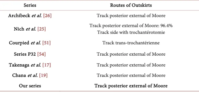
Comparison of tracks first with the literature (Table 10) The different types of total hip prosthesis
[image:22.595.210.534.586.738.2]The PTH are distinguished primarily by their mode of fixation and according to the friction torque.
Table 10. Comparison of tracks of outskirts.
Series Routes of Outskirts
Archibeck et al. [26] Track posterior external of Moore
Nich et al. [25] Track posterior external of Moore: 96.4% Track side with trochantérotomie
It prefers to cement a PTH as soon as the quality of the bone is presumed or proven wrong: In patients aged among osteoporotic patients or patients with a bone pathology likely to promote the occurrence of a fracture periprothetique per or postoperative [6] [25].
PTH not cemented
The prostheses not cemented were introduced at the end of the years 70 to find out a solution to a high failure rate of cemented implants in young and ac-tive patients. In effect, the survival of the PTH is particularly important in the young subject whose life expectancy is long and the request is strong and func-tional [6].
The PTH not cemented are most often used in patients young and active. In effect, this mode of fixation is preferable to young adults, because it esteems the bone capital and allows a real osteo-integration, union of the living bone to the inert implant. The deference of the bone capital is also important in these sub-jects young people whose recovery is inevitable [6].
Analysis of the means of securing in the literature
Archibek et al., have studied the results of 100 PTH using second generation implants not cemented in patients less than 50 years. Has 9 years of decline, they noted 13 times,1 Sepsis on hardware, 5 prosthetic luxations, 7 case of osteolysis (5 cotyloidiennes Limited and 2 associated with a aseptic descellement). No cases of femoral descellement has been reported. Overall survival at 10 years was 87.5%. If one takes the failure resumption of the femoral component as a crite-rion, the survival to 10 years was 100% and as a criterion of failure the resump-tion of the acetabular component, the survival was 97.1% [46].
Ryan et al., have recently reported (2014), the results of a series of 88 PTH not cemented in patients under the age of 55 years. The decline in 5 years, they noted 2 cases of recovery: 1 for prosthetic dislocation and 1 cases of sepsis on hardware. There had been no cases of descellement [22].
Kim et al., in a retrospective study, evaluated the results of 166 patients aged less than 50 years who have undergone a total hip replacement. There were 114 hybrid PTH with rods cemented and 114 PTH not cemented (all without ce-ment) with stems not cemented. The rate of revision of the femoral components and acetabulaires were not significant between the two groups. The overall sur-vival of the implants were similar in the 2 groups. As well, in the group "hybrid" overall survival at 10 years was 93.6% to 15 years 91.8% [33].
In the light of these results, the total prosthesis of hip implanted in patients young appears to be good, and this, whatever the mode of fixation used.
The various studies do not show the advantage of a mode of fixation by report to the other. A condition to respect the basic rules, the two modes of fixation are reliable.
been defined for the implantation of a PTH not cement. However, if the various studies carried out are state of an excellent survival for the prostheses having this mode of fixation, the problem of aseptic descellement is nevertheless not re-solved. Reactions of osteolysis related to wear particles are also found, even that of migration of stemmed femoral and pain of thighs. The ablation of a PTH not cemented can be very difficult at the level of the barrel femoral and sometimes requires a component of osteotomy diaphyseal [15].
In our series the dominance of prostheses not cemented was significant. In Effect 85% of PTH were non-cemented and 15% of PTH were cemented. This choice is explained by the young age of our patients with more often a good bone quality.
Friction Torque
Within the factors likely to affect the longevity of total prosthesis of hip, and this regardless of the mode of fixation, the friction torque plays a preponderant role.
The couples of friction of arthroplasties are in constant evolution. The choice is important for young subjects in order to reduce the Chess [65].
Up until the middle of the 1990s, the majority hip prostheses included a fric-tion torque with a metal head and a prosthesis acetabular polyethylene (met-al/polyethylene). Different studies have concluded that the wear of the polyethy-lene was the main cause of failure. To improve the results, that is to say the du-ration of these arthroplasties, two major channels have been proposed: improve the couples using polyethylene or change of torque using the so-called couples hard/drive: metal/metal or ceramic/ceramic [65].
Polyethylene torque-metal
The Friction Torque conventional polyethylene-metal is the reference torque which are compared to all other couples of friction. The prostheses to conven-tional friction torque polyethylene-metal have the longest clinical decline. Their survival is known up to 30 years for large series of patients. The survival rates reported are 85% to 20 years, 80% to 25 years and 78% to 30 years [15].
The relationship between the wear of the polyethylene and osteolysis respon-sible for the aseptic descellement of the prosthesis has been shown in the litera-ture.
The polyethylene remains the most widely used material because of its low cost and ease of manufacture [17].
Conventional polyethylene
This material of high molecular weight combines the crystals and long poly-meric chains, the mechanical properties are directly related to the crystallinity and spatial cohesion of macromolecules. The disadvantage lies in the existence of free radicals, source of areas where it will happen the oxidation and therefore of wear. The wear particles cause inflammatory reactions by the intermediary of leaching of cytokine. These reactions can lead to the prosthetic explaining the descellement [66].
0.1mm per year of the EP conventional. The survival of the implant is strongly conditioned by the presence of wear. There is a correlation between the rate of wear and tear and survival curve of the implants of PTH. Sochart et al., have demonstrated a direct relationship between the importance of the wear and the survival of the implants, if the wear is 0.1mm per year, the survival to 25 years is around 70%, if the wear is greater than 0.25mm per year, the survival passes to less than 20% at 20 years of retreat [67].
It has been shown that the significant parameter influencing the wear of the conventional PE was the activity of the patient and the age (more than three mil-lion cycles per year among an active subject) [66].
To compensate for the failure of the conventional PE a new type of cross- linked polyethylene, source of less than wear debris and osteolysis appeared [66].
Polyethylenes highly reticulated (cross-linked polyethylene “cross-linked”) The principle is to improve the reticulation chains between the crystals by ir-radiation and to avoid the free radical oxidation by a thermal treatment. This change of structure must significantly improve the resistance to wear and reduce the release of particles of wear of PE. The doses of irradiation vary from 5 to 10m rads and manufacturers announce a decrease of the wear between 42 and 100% compared to a conventional PE.
However, irradiation leads to alterations in the mechanical properties of po-lyethylene, in particular an increase in the rigidity [66].
Antonio, et al., in a retrospective and comparative study including 108 pros-theses, showed a significant difference between the two types of PE. They re-ported a linear wear annual at 59 months of decline in average of 0.055 mm/year for the cotyledons uncemented polyethylene in highly reticulate, and wear an-nual linear to 63 months of decline on average 0.138 mm/year for the cotyledons not cemented in conventional polyethylene [67].
Channel Babovic et al., reports the results to medium term of PTH with PE highly reticulate in 50 patients (54 PTH) under the age of 50 years. They re-ported a rate of annual wear of 0.020 ± 0.0047 mm/year. Having 10 years of de-cline, no cases of recovery for osteolysis nor descellements of implants has been noted. Overall survival at 10 years was 100% [21].
The wear measured in the short term of cotyledons in polyethylene highly re-ticulate is significantly lower than that of cotyledons in conventional polyethy-lene. The data for long-term survival of these implants are however not yet known [15].
Torque metal-metal
The implants to a torque metal-metal, have been reintroduced at the end of the 1980s to have been widely used in the years 1950 and 1960. They are com-posed of an alloy of chromium and cobalt.
The prostheses using a friction torque metal-metal have an excellent resis-tance to wear, bringing to favor their use among the topics most actively.
in-terface of friction are not yet known. The ions are eliminated by the urine, which against-indicates this torque in patients with renal impairment. A risk of carci-nogenesis is mentioned, but it has never been demonstrated, reactions allergic immune against the metal ions have also described [17].
Kim, et al., in 2004 reported a series of 70 prostheses in patients less than 50 years. The average decline was 7 years, the clinical results were excellent, and no cases of descellement nor stripe have been observed after use of the torque met-al-metal [68].
Ceramic torque/ceramic
The Friction Torque ceramics/ceramic has been developed and proposed as an alternative by the French Pierre Boutin since 1972. The purpose was to avoid the wear of prosthetic components and its consequences, in order to increase the longevity of implants and to enable their use in young and active patients more and more [69].
The torque ceramics/ceramic is composed of a head and an insert in ceramic massive. The insert is attached in an acetabular cup metal. Two ceramic type are used, the alumina and the zirconia.
The most used ceramic is the alumina. This torque is used since 30 years. The tolerance is excellent with a wear virtually zero: less than 0.0015 mm/year.
The behavior in friction of ceramics is very much higher than that of other couples used in arthroplasties, including the torque metal-metal. It reflects the characteristics of exceptional surface Ceramics (hardness, very low roughness, wettability, coefficient of friction very low) [69].
Chana, et al., have reported the results of 110 patients aged less than 55 years, having benefited of PTH to a torque Ceramic-ceramic. Has 10 years of decline, the wear was undetectable. The survival for the 2 components with as a criterion of failure recovery for whatever the cause was 96.5% [29].
Baek et al., in their series, analyzed the results of 71 PTH to a torque Ceram-ic-ceramic, without cement, implanted for osteonecrosis in patients less than 50 years. The average decline was 7.1 years. No cases of descellement, osteolysis or fracture of implant has been observed. In 13 patients (20%) of the phenomena of noise have been reported [70].
In 2007 report, the HAS recommends the friction torque Ceramic-ceramic, the fact of the low production of wear debris, among patients aged less than 50 years [15].
If the torque Ceramic-ceramic proves to be competitive for young patients in terms of longevity of prosthesis, it presents however of notable drawbacks [69]:
-
Risk of fracture of implants,-
Use impossible for sizes < 46 mm, which would make the insert too fragile,-
Phenomenon of noise (“squeaking”) of the torque,-
High cost.The dual mobility
the secondary fastening (by bone regrowth on the surface prosthetics) cupules metal-back is become reliable with the contribution of the hydroxyapatite. This type of cupule is indicated in patients with a high risk of dislocation: high age, neurological pathologies, alcoholism, low muscle trophicite, resumption of prosthesis, tumor pathology.
The problems of wear are occurring at the beginning of the experience be-cause the surface condition and the geometry of the Prosthetic collar also inter-vene in the phenomena of wear, bringing in favor of smooth collar devoid of notch of extraction.
Despite a rate of wear and survival comparable to the fixed polyethylene, the risk of dislocation intraprothetique (2%) own to this type of implant, must make their prudent indication in particular in young subjects and assets. On the other hand, it is possible to recommend their use because of their excellent stability as soon as it is a HIP to risk [17].
Phillipot et al., have reported the results of 46 PTH to double cupule im-planted mobility in subjects less than 50 years. 10 years of retreat 2 descellements acetabulaires isolated, 1 dislocated intra-prosthetics by wear of the retention and an advanced wear, have been observed. The rate of survival of overall actuarial to ten years of this cupule was 90.8%. There has been no episode of prosthetic dis-location in this series [71].
In our series 15% of PTH were of type double mobility, no cases of prosthetic dislocation or intra-prosthetics was observed in these patients.
Conclusions on the friction torque:
The metal torque-polyethylene is the most used and which has the longest de-cline. In younger patients, the failures of this torque, are in relation with the descellement due to the wear of the polyethylene resulting in an osteolysis. The wear of the polyethylene can be decreased by the use of a head of ceramic and improving the qualities of polyethylene by reticulation (cross-linked polyethy-lene).
The torque metal-metal has as benefits of limited wear and the possibility of use of implants of large diameter or very large diameter. Its main drawback is the ionic leaching. The consequences of the accumulation of these ions are cur-rently unknown.
The couple Ceramic-ceramic is the couple which has the rate of wear more low and without ionic leaching. The fracture of the ceramic, remains the main inconvenience of this couple.
In our series, the use of a torque PE/metal was used, because it is the least ex-pensive, most available, and because it is accustomed to work with this material.
6. Conclusions
makes them more likely to have undergone a resumption of their implants. In this retrospective study of 43 patients (47 hips) operated before the age of 50 years between January 2008 and March 2014, we find 5 times of PTH is hav-ing a failure rate of 10.6% for a duration of mean follow-up of 42 months. But despite this failure rate, the clinical results appear more important with an aver-age score LDCS clinic who spent 9.1 pre-operative at 16.1 in postoperative, ei-ther having average gain of 7 points.
The higher the gain has increased pain and 69.44% of hips were painless. In addition, the Final Clinical Evaluation of the patients shows an overall result judged satisfactory. These clinical results should encourage the surgeons to less restraint in the decision-making socket of the installation of a total hip among these young patients, including most of the time already multi-operated and suffering from a severe infringement of the articulation coxal-femoral. As to the choice of materials and means of mounting, the superiority (theoretical) of the friction is the torque metal-metal or ceramic.
Ceramics in terms of wear is an indisputable parameter in young subjects and assets. However, the friction torque metal-polyethylene gives good results and appears to be adapted to our context, given its availability and its cost price.
References
[1] Christofilopoulos, Lubbeke, A., Peter, R. and Hoffmeyer, P. (2010) The Point on the Total Hip. Revue Medicale Suisse, 6, 2454-2458.
[2] Delaunay, C., Aghayev, E. and Staub, L. (2009) Register of Total Prosthesis of Hip of the SOFCOT. French Society of Orthopaedic Surgery and Trauma. Report 2009. [3] Girard, J., May, O., Krantz, N. and Migaud, H. (2011) Surgical Treatment of the
Coxarthrose. EMC-Musculoskeletal Health, 2011, 1-14.
[4] Lequesne. (2009) Coxarthrose Coxopathies and of the Adult. Diagnosis and Treat-ment. EMC-Musculoskeletal Health, 2009, 1-21.
[5] French Society of Orthopaedic Surgery and Trauma (SOFCOT). Register of PTH of the SOFCOT, Annual Report 2013.
http://www.sofcot.fr/Pages/Registre-des-protheses-de-hanche
[6] National Agency for Accreditation and Evaluation in Health (2001) Total Prosthesis Primary Hip: Evaluation of the Choice of the Prosthesis and Operational Tech-niques. Paris: ANAES 2001.
[7] Pivec, R., et al. (2012) Hip Arthroplasty. Lancet, 380, 1768-1777.
https://doi.org/10.1016/S0140-6736(12)60607-2
[8] Kurtz, S.M., et al. (2009) Future Young Patient Demand for Primary and Revision Joint Replacement: National projections from 2010 to 2030. Clinical Orthopaedics and Related Research, 467, 2606-2612.https://doi.org/10.1007/s11999-009-0834-6
[9] McAuley, J.P., et al. (2004) Total Hip Arthroplasty in Patients 50 Years and Young-er. Clinical Orthopaedics and Related Research, 418, 119-125.
[10] Pourreyron, D., et al. (2008) Total Hip among the Young Patient. E-Memoirs of the National Academy of Surgery, 7, 42-46.