Ascorbate metabolism in vegetative and reproductive
organs of “cherry” tomato
G. Tsaniklidis
1, N. Nikoloudakis
2, C. Delis
3, G. Aivalakis
11
Laboratory of Plant Physiology and Morphology, Agricultural University of Athens,
Athens, Greece
2
Vegetative Propagation Material Control Station, Hellenic Ministry of Rural
Development and Food, Athens, Greece
3
Department of Agricultural Technology, Technological Institute of Kalamata,
Kalamata, Greece
Abstract
Tsaniklidis G., Nikoloudakis N., Delis C., Aivalakis G., 2014. Ascorbate metabolism in vegetative and re-productive organs of “cherry” tomato. Hort. Sci. (Prague), 41: 114–121.
Ascorbate metabolism is an essential procedure for all plant cells that plays important roles in several physiological processes such as plant development and reactive oxygen species detoxification. To shed more light on ascorbate metabolism in certain organs of tomato plants, we performed a detailed compartmentalized analysis of ascorbate con-centration, ascorbate peroxidase/dehydroascorbate reductase enzyme activities and transcript accumulation of genes related to ascorbate metabolism. Our results showed higher level of ascorbate concentration and ascorbate peroxidase and dehydroascorbate reductase activities in young leaves and shoot tips, while min. ascorbate concentration was recorded in root tips. The study of the expression of the genes involved in ascorbate metabolism revealed that several genesfollowed similar patterns. However, APX3 gene expression was considerably higher in reproductive organs, while plastidialAPX6 and DHAR2genes transcripts were barely detectable in root tips. Organ-specific expression of genes involved in ascorbate metabolism suggests that different isoenzymes have a specific role in regulation of the redox status of some of the organs in tomato plants.
Keywords: ascorbic acid; ascorbate peroxidase; dehydroascorbate reductase; monodehydroascorbate reductase; glutathione reductase
As a highly active, low-molecular-weight reduc-ing agent, ascorbate (AsA) is involved in various processes of cell metabolism (Smirnoff 2011). It is well established that AsA protects cells against oxidative damage caused by reactive oxygen spe-cies (ROS) including the extremely active hydrogen peroxide (H2O2), which is a by-product of aerobic metabolism (Gestet al. 2013). AsA redox reactions are often referred to in literature as ascorbate–glu-tathione circle, which plays an important role in plants in redox status regulation through the
sev-eral enzymes with active sites susceptible to oxida-tion (Prescott, John 1996). Addioxida-tional roles were also proposed for AsA in plants; thus several studies suggest that AsA is an essential element that par-ticipates in a number of growth and developmen-tal processes like cell division, cell expansion and root growth (Davey 2000; Cordoba-Pedregosa et al. 2003). In tomato plants, high accumulation of transcripts of the genes involved in AsA biosynthe-sis were correlated to fruit growth and maturation suggesting an important role of AsA in certain de-velopmental stages (Ioannidiet al. 2009). Consid-ering the differences in the physiological status and functionality of the plant organs we undertook the task to elucidate the distinguishing roles of certain isoenzymes involved in AsA metabolism among the vegetative and reproductive cherry tomato or-gans. Moreover, total AsA content was determined in parallel with the activity of key enzymes of AsA metabolism.
MATERIALS AND METHODS
Plant material and growth conditions. Plants
of cherry tomato (Solanum lycopersicum L. var.
cerasiforme cv. Conchita F1; de Ruiter seeds, Melbourne Australia) were cultivated in a glass-house of the Agricultural University of Ath-ens, Greece between December and May. Mean min. and max. temperatures in the greenhouse were 15.7 ± 2.0°C and 26.6 ± 4.3°C, respective-ly, (Spring: March–May) and 12.9 ± 1.9°C and 23.9 ± 4.4°C (Winter: Oct–Feb). Solar radiation var-ied between 700–1,400 μmol/m·s PAR. Mature and young leaves (from sixth and third knot, respec-tively), shoot tips (top 2 cm), shoot (with diameter 1,5 cm), root tips (2 cm), open flowers, pericarp and central region of red ripe fruits (pulp) (52 days after flowering) were collected and prepared simultane-ously. Each harvest was carried out at 11 a.m. during springtime with similar conditions and replicated three times creating three lots. Samples were imme-diately frozen in liquid nitrogen, homogenized using a pestle and mortar and then stored at –80oC.
qPCR analysis. Real-time polymerase chain
re-action (PCR) experiments were conducted with the gene-specific primers sequences as previously re-corded (Tsaniklidis et al. 2014).
Ascorbic acid determination. Total AsA
con-tents of tissues were determined as previously de-scribed (Tsaniklidis et al. 2014).
APX and DHAR enzyme assay. The activity of
APX and DHAR was assessed as previously de-scribed (Tsaniklidis et al. 2014).
Statistical analysis. Statistics were performed
using the Statgraphics Centurion (Statpoint Tech-nologies, Warrenton, USA). Significant differences between treatments were determined by two-way ANOVA and post-hoc comparisons by least signifi-cant difference (P < 5%).
RESULTS AND DISCUSSION
AsA metabolism in shoot tips and shoot
The fast growing shoot tips were among the most metabolically active plant tissues in terms of en-zyme activity (APX and DHAR) and transcript ac-cumulation levels (APX, MDHAR, DHAR and GR) (Figs 1–4). With the exceptions of APX3 and of both GR isoenzymes, all genes exhibited the highest ac-cumulation of transcripts in shoot tips. Moreover, AsA was accumulated in high levels in growing shoot tips. Zhang et al. (2011) reported that AsA is important for controlling the redox status dur-ing cell division and expansion. AsA possibly mod-erates plant growth by regulating numerous basic biological processes, such as (i) the biosynthesis of hydroxyproline-rich proteins which are essential for the advancement of the cell cycle (ii) the bonding of cell wall glycoproteins and other polymers, and (iii) redox reactions at the plasmalemma involved in elongation mechanisms. The AsA free radical also
b
a a
d d b
c cd
0 1 2 3 4 5 6 7 8 9 10
Re
la
tive gene
ex
pr
[image:2.595.314.520.528.675.2]ession
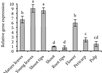
Fig. 1. Τotal AsA (tAsA) (reduced + oxidized AsA) content of the organs of cv. Conchita tomato plants
stimulates a high vacuolization accountable for cell elongation (Smirnoff 2011). The combination of cell division and expansion with photosynthesis is believed to have led to the observed up-regulation of the transcription of AsA metabolism-related genes. Concerning AsA metabolism in shoot, Antonova et al. (2009) showed a sound escalation of AsA con-centration in cambium, conductive phloem and the cell enlargement zone, with a following sharp de-crease during subsequent cell maturation and ligni-fication. AsA eliminates free radicals involved in xy-logen synthesis and controls the lignification of cell wall (Takahama 1993). The balance amongst AsA and hydrogen peroxide regulates the polymeriza-tion of xylogen monomers, modulating the cell wall lignification (Davey et al 2000). Another possible explanation of the comparably low levels of AsA in mature shoot is that the slow metabolism and actu-al photosynthesis of this organ probably do not re-quire high levels of AsA for antioxidant protection (Smirnoff 2000). The combination of cell division and expansion with photosynthesis is believed to lead to an up-regulation of the transcription of AsA metabolism-related genes. The findings of this study reinforce the hypothesis that AsA is crucial for cell expansion and morphogenesis.
Antonova et al. (2009) showed a sound escala-tion of AsA concentraescala-tion in cambium, conduc-tive phloem and the cell enlargement zone, with a following sharp decrease during subsequent cell maturation and lignification. AsA eliminates free radicals involved in xylogen synthesis, controls the polymerization of xylogen monomers and also ad-justs the lignification of cell wall by regulating the
free radicals into the apoplast (Takahama 1993; Davey et al. 2000). Another possible explanation of the comparably low levels of AsA in mature shoot is that the slow metabolism and actual photosyn-thesis of this organ probably do not require high levels of AsA for antioxidant protection (Smirnoff 2000).
AsA metabolism in young and mature leaves
Total AsA levels in young tomato leaves were higher comparing to mature leaves and ranged between 6.5 to 9 μM/g fresh weight (FW) (Fig. 1). Bartoli et al. (2000) also reported higher tAsA contents in young potato leaves with comparable concentrations. Moreover Li et al. (2010b) found that tASA concentration in apple leaves increased rapidly with their development and reached the highest level in 20-day-old leaves remaining nearly constant until senescence. The lower tASA concen-tration in mature leaves could be attributed to both lower AsA biosynthesis and reduction capacity (Bartoli et al. 2000). Zhang (2013) reported that low accumulation of AsA accelerates senescence while its high content delays the process.
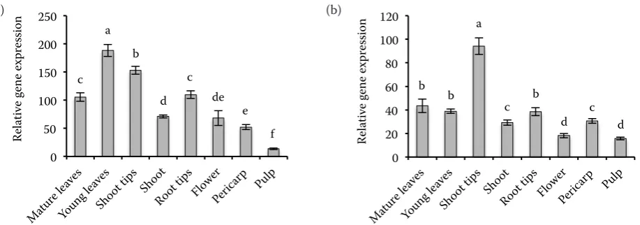
Moreover, our results showed higher APX activ-ity in young leaves than in mature ones. This find-ing indicates that a higher antioxidant requirement probably exists in developing young leaves (Fig. 2). In contrast, as in the case of apple leaves, DHAR activity was rather constant in both mature and young leaves, indicating a different metabolic func-tion (Li et al. 2010b).
c a
b
d c
de e
f 0
50 100 150 200 250
Re
la
tive gene
ex
pr
ession
b b
a
c b
d c d
0 20 40 60 80 100 120
Re
la
tive gene
ex
pr
[image:3.595.71.530.82.243.2]ession
Fig. 2. APX (a) and DHAR (b) enzyme activity in the organs of cv. Conchita tomato plants
bars represent means (+ standard error) of three biological replications; indicator letters in common denote a lack of a signifi-cant difference
e d d
c b
f g
0 1.0 2.0 3.0 4.0 5.0
Re
la
tive gene
ex
pr
ession
h g d e f c
b
0 0.1 0.2 0.3 0.4 0.5 0.6
Re
la
tive gene
ex
pr
ession
g d
a
b
f c
e f 0
0.3 0.6 0.9 1.2 1.5
Re
la
tive gene
ex
pr
ession
c d a
e c b
f f 0
0.3 0.6 0.9 1.2
Re
la
tive gene
ex
pr
ession
cd d a
d b
cd c e
0 0.5 1.0 1.5 2.0
Re
la
tive gene
ex
pr
ession
c cd a
d e
cd b
cd
0 0.1 0.2 0.3 0.4 0.5 0.6 0.7
Re
la
tive gene
ex
pr
ession
bc bc a
b
d c d d 0
0.005 0.010 0.015 0.020 0.025 0.030
Re
la
tive gene
ex
pr
[image:4.595.58.532.75.760.2]ession
Fig. 3. Accumulation of APX isoenzyme gene transcripts in organs of cv. Conchita tomato plants
(a) APX1, (b) APX2, (c) APX3, (d) APX4, (e) APX5, (f) APX6, (g) APX7
bars represent means (+ standard error) of three biological replications; indicator letters in common denote a lack of a significant difference
a
a
(a) (b)
(d)
(f) (c)
(e)
d cd a
c cd b
c e
0 0.1 0.2 0.3 0.4
Re
la
tive gene
ex
pr
ession
c a
cd b
bc
de e 0
0.05 0.10 0.15 0.20 0.25 0.30 0.35 0.40
Re
la
tive gene
ex
pr
ession
bc b a
c
ef d e f
0 0.5 1.0 1.5 2.0
Re
la
tive gene
ex
pr
ession
c b
a a
d a
e f
0 0.3 0.6 0.9 1.2 1.5 1.8
Re
la
tive gene
ex
pr
ession
e ef a
d c b
f f 0
0.05 0.10 0.15 0.20 0.25
Re
la
tive gene
ex
pr
ession
a
bcd
d c bcd cd b bc 0
0.01 0.02 0.03 0.04 0.05 0.06 0.07 0.08 0.09
Re
la
tive gene
ex
pr
ession
Interestingly, differences of transcript accumu-lation for most genes among young and mature leaves were minor. Similar results for APX tran-scription were obtained by Panchuk et al. (2005). In Arabidopsis, Li et al. (2010a) also found minor
differences in the transcripts levels of MDAHRand DAHR between young and mature leaves, as re-corded in the current study as well. Thus, it could be said that different molecular mechanisms par-ticipate in controlling the transcriptional
regula-Fig. 4. Accumulation of transcripts of AsA recycle enzymes in organs of cv. Conchita tomato plants (a) MDHAR1, (b) DHAR1, (c) GR1, (d) MDHAR2, (e) DHAR2, (f) GR2
bars represent means (+ standard error) of three biological replications; indicator letters in common denote a lack of a signifi-cant difference
a
(a)
(c)
(e)
(b)
(d)
[image:5.595.63.529.82.589.2]tion of ASA metabolism genes in young and mature leaves. However, both cytosolic and plastidial GR isoenzymes followed completely different patterns of transcription among mature and young leaves and also in other tissues (Fig. 4). It is established that glutathione and by extension GR, are involved in numerous physiological processes comparing to other enzymes of AsA metabolism with more specific roles and probably have different specific-ity/function (Nagalakshmi, Prasad 2001). Young and mature leaves as well as shoots exhibited similar DHAR activity, while in shoot tips the activity was considerable higher and in flowers it was fairly lower (Fig. 2). On the other hand, APX activity was high-er in young leaves and shoot tips following similar pattern to tAsA levels. These results suggest that in cherry tomatoes, although APX activity is related to tAPX levels, the DHAR activity follows a different pattern possibly related to the redox status of APX.
AsA metabolism in root tips
AsA is considered to be actively involved with photoprotection of photosynthesis and in reactions to biotic and abiotic stress conditions (Smirnoff 2011). The fact that in root tips photosynthesis is absent makes the study of AsA metabolism appeal-ing.
Root tips showed the lowest tASA levels among all other plant tissues examined. (Fig. 1). Lin et al. (2004) reported that tAsA levels in tomato roots were around 0.45 μM/g FW, which is com-parable to the tASA concentration of the present study (0.7 μM/g FW), while in onion roots up to 0.19 μM/g FW were found (Cordoba-Pedregosa et al. 2003). Interestingly, despite low tASA levels, APX and DHAR activities in root tips were relative-ly high (Fig. 2), which could point to a higher turn-over between oxidized and reduced forms of AsA. Comparable results were obtained by Cordoba-Pedregosa et al. (2003) and Lin et al. (2004) for tomato and onion roots respectively. AsA level is also related to growth of root architecture and root reaction to gravity. Transcript accumulation in root tips revealed a complicated picture; transcripts lev-els of APX1, APX2, MDHAR1, MDHAR2 and GR1 were comparable to the other vegetative organs, while APX4, APX5, APX6 and DHAR2 were con-siderably lower (Figs 3 and 4). The corresponding thylacoid bound APX isoenzyme of rice was also not detected in roots. However, a plastidial APX
isoenzyme of rice showed high levels of transcript accumulation as was the case of plastidial APX7 (Teixeira et al. 2006).
AsA metabolism in flower and fruit compartments
respira-tion rate between mature fruits and fast growing organs as flowers and root tips. Indeed, both fac-tors are reported to have an effect on AsA metabo-lism (Bartoli et al. 2006).
CONCLUSION
Our analysis confirmed the presence of AsA me-tabolism in all organs studied. Moreover, it was shown that both AsA metabolite concentration and transcripts accumulation depend on the func-tionality and the physiological role of the organ examined. The specificity of the accumulation of transcripts of some genes could suggest that sever-al isoenzymes have differentisever-al importance in AsA redox status in each organ.
References
Antonova G.F., Stasova V.V., Varaksina T.N., 2009. Ascorbic acid and development of xylem and phloem cells in the pine trunk. Russian Journal of Plant Physiology, 56: 190–199.
Attolico A.D., De Tullio M.C., 2006. Increased ascorbate content delays flowering in long-day grown Arabidopsis
thaliana (Heyn.). Plant Physiology and Biochemistry, 44:
462–466.
Bartoli C.G., Pastori G.M., Foyer C.H., 2000. Ascorbate biosynthesis in mitochondria is linked to the electron transport chain between complexes III and IV1. Plant Physiology, 123: 335–343.
Bartoli C.G., Yu J., Gomez F., Fernandez L., McIntosh L., Foyer C.H., 2006. Inter-relationships between light and respiration in the control of ascorbic acid synthesis and accumulation in Arabidopsis thaliana leaves. Journal of Experimental Botany, 57: 1621–1631.
Conklin P.L., Barth C., 2004. Ascorbic acid, a familiar small molecule intertwined in the response of plants to ozone, pathogens, and the onset of senescence. Plant Cell and Environment, 27: 959–970.
Cordoba-Pedregosa C.M., Cordoba F., Villalba J.M., Gonzalez-Reyes J.A., 2003. Zonal changes in ascor-bate and hydrogen peroxide contents, peroxidase, and ascorbate-related enzyme activities in onion roots. Plant Physiology, 131: 697–706.
Davey M.W., Van Montagu M., Inze D., Sanmartin M., Kanellis A., Smirnoff N., Benzie I.J.J., Strain J., Favell D., Fletcher J., 2000. Plant L-ascorbic acid: chemistry, function, metabolism, bioavailability and effects of pro-cessing. Journal of the Science of Food and Agriculture,
80: 825–860.
Fotopoulos V., De Tullio M.C., Barnes J., Kanellis A.K., 2008. Altered stomatal dynamics in ascorbate oxidase over-expressing tobacco plants suggest a role for dehydroascor-bate signalling. Journal of Experimental Botany, 59: 729–737. Gest N., Gautier H., Steevens R. 2013. Ascorbate as seen
through plant evolution: the rise of a successful molecule? Journal of Experimental Botany, 64: 33–53.
Ioannidi E., Kalamaki M.S., Engineer C., Pateraki I., Alexandrou D., Mellidou I., Giovannonni J., Kanel-lis A.K., 2009. Expression profiling of ascorbic acid-related genes during tomato fruit development and ripening and in response to stress conditions. Journal of Experimental Botany, 60: 663–678.
Li M., Ma F., Shang P., Zhang M., Hou C., Liang D., 2009. Influence of light on ascorbate formation and metabolism in apple fruits. Planta, 230: 39–51.
Li F., Wu Q.Y., Sun Y.L., Wang L.Y., Yang X.H., Meng Q.W., 2010a. Overexpression of chloroplastic monodehy-droascorbate reductase enhanced tolerance to temperature and methyl viologen-mediated oxidative stresses. Physi-ologia Plantarum, 139: 421–434.
Li M., Ma F., Guo C., Liu J., 2010b. Ascorbic acid forma-tion and profiling of genes expressed in its synthesis and recycling in apple leaves of different ages. Plant Physiology and Biochemistry, 48: 216–224.
Lin K.H., Weng C.C., Lo H.F., Chen J.T., 2004. Study of the root antioxidative system of tomatoes and eggplant under waterlogged conditions. Plant Science, 167: 355–365. Massot C., Stevens R., Genard M., Longuenesse J.J.,
Gautier H., 2012. Light affects ascorbate content and ascorbate-related gene expression in tomato leaves more than in fruits. Planta, 235: 153–163.
Mittler R., 2002. Oxidative stress, antioxidants and stress tolerance. Trends in Plant Science, 7: 405–410.
Muller G.L., Drincovich M.F., Andreo C.S., Lara M.V., 2010. Role of photosynthesis and analysis of key enzymes involved in primary metabolism throughout the lifespan of the tobacco flower. Journal of Experimental Botany, 61: 3675–3688.
Nagalakshmi N., Prasad M.N.V., 2001. Responses of glutathione cycle enzymes and glutathione metabolism to copper stress in Scenedesmus bijugatus. Plant Science,
160: 291–299.
Panchuk I.I., Zentgraf U., Volkov R.A., 2005. Expression of the Apx gene family during leaf senescence of
Arabidop-sis thaliana. Planta, 222: 926–932.
Prescott A.G., John P., 1996. Dioxygenases: molecular structure and role in plant metabolism. Annual Review of Plant Physiology and Plant Molecular Biology, 47: 245–271. Smirnoff N., 2000. Ascorbate biosynthesis and function in
Smirnoff N., 2011. Vitamin C: The metabolism and func-tions of ascorbic acid in plants. Advances in Botanical Research, 9: 109–177.
Takahama U., 1993. Regulation of peroxidase-dependent oxidation of phenolics by ascorbic acid: Different effects of ascorbic-acid on the oxidation of coniferyl alcohol by the apoplastic soluble and cell wall-bound peroxidases from epicotyls of Vigna angularis. Plant Cell Physiology,
34: 809–817.
Talla S., Riazunnisa K., Padmavathi L., Sunil B., Ra-jsheel P., Raghavendra A.S., 2011. Ascorbic acid is a key participant during the interactions between chloro-plasts and mitochondria to optimize photosynthesis and protect against photoinhibition. Journal of Biosciences,
36: 163–173.
Teixeira F.K., Menezes-Benavente L., Galvao V.C., Margis R., Margis-Pinheiro M., 2006. Rice ascorbate peroxidase gene family encodes functionally diverse iso-forms localized in different subcellular compartments. Planta, 224: 300–314.
Tsaniklidis G., Delis C., Nikoloudakis N., Katinakis P., Passam H.C., Aivalakis G., 2014. L-ascorbic acid me-tabolism in parthenocarpic and seeded cherry tomatoes. Plant Growth Regulation, 72: 141–153.
Zhang C., Liu J., Zhang Y., Cai X., Gong P., Zhang J., Wang T., Li H., Ye Z., 2011. Overexpression of SlGMEs leads to ascorbate accumulation with enhanced oxidative stress, cold, and salt tolerance in tomato. Plant Cell Reports,
30: 389–398.
Zhang Y., 2013. Ascorbic Acid in Plants. New York, Springer: 7–33.
Zushi K., Matsuzoe N., 2012. Comparative analysis of oxidative parameters, antioxidant content, and antioxidant enzyme activity during fruit ripening in tomato pericarp and pulp. Journal of the Japanese Society for Horticultural Science, 81: 109–116.
Received for publication October 20, 2013 Accepted after corrections April 29, 2014
Corresponding author:
Dr. Georgios Tsaniklidis, Agricultural University of Athens, Laboratory of Plant Physiology and Morphology, Iera Odos 75, 11855 Botanikos, Athens, Greece