(3
S
,7
R
)-7,14,16-Trihydroxy-3-methyl-3,4,5,6,7,8,9,10,11,12-decahydro-1
H
-2-benzoxacyclotetradecin-1-one.
Sarah Drzymala,* Werner Kraus, Franziska Emmerling and Matthias Koch
BAM Federal Institute for Materials Research and Testing, Department of Analytical Chemistry, Reference Materials, Richard-Willsta¨tter-Strasse 11, D-12489 Berlin, Germany
Correspondence e-mail: sarah.drzymala@bam.de Received 3 September 2012; accepted 1 October 2012
Key indicators: single-crystal X-ray study;T= 296 K; mean(C–C) = 0.004 A˚;
Rfactor = 0.059;wRfactor = 0.147; data-to-parameter ratio = 9.9.
The asymmetric unit of the title compound, C18H26O5, which is
known as -zearalanol, contains two molecules having the same conformation, with a r.m.s. deviation of less than 0.03 A˚ for all non-H atoms. In each independent molecule, an intramolecular O—H O hydrogen bond stabilizes the molecular conformation. In the crystal, O—H O hydrogen bonds link the molecules, forming infinite chains along [110] and [110].
Related literature
For the chemical preparation of-zearalanol, see: Urryet al.
(1966). For its natural occurrence as a metabolite, see: Baldwin
et al.(1983) and for its use as an animal growth promoter, see: Wang & Wang (2007). For the crystal structures of related derivatives, see: Panneerselvamet al.(1996); Gelo-Pujic´et al.
(1994); Zhaoet al.(2008); Ko¨ppenet al.(2012); Drzymalaet al.
(2012).
Experimental
Crystal data
C18H26O5 Mr= 322.39
Triclinic,P1
a= 5.0734 (11) A˚
b= 11.618 (2) A˚
c= 14.718 (3) A˚
= 87.388 (13) = 86.595 (15)
= 89.780 (15)
V= 865.0 (3) A˚3 Z= 2
MoKradiation
= 0.09 mm1 T= 296 K
0.430.220.10 mm
Data collection
Bruker APEXII CCD diffractometer
Absorption correction: multi-scan (SADABS; Bruker, 2001)
Tmin= 0.186,Tmax= 0.350
19642 measured reflections 4264 independent reflections 3421 reflections withI> 2(I)
Rint= 0.095
Refinement
R[F2> 2(F2)] = 0.059 wR(F2) = 0.147 S= 0.95 4264 reflections 431 parameters 7 restraints
H atoms treated by a mixture of independent and constrained refinement
max= 0.24 e A˚
3
min=0.16 e A˚
[image:1.610.312.567.304.374.2] [image:1.610.46.238.568.719.2]3
Table 1
Hydrogen-bond geometry (A˚ ,).
D—H A D—H H A D A D—H A
O5—H5A O2 0.82 1.83 2.549 (3) 146
O50—H50A O20 0.82 1.82 2.540 (3) 146
O4—H4A O3i
0.83 (2) 1.93 (3) 2.745 (3) 171 (3)
O40 —H40A
O30ii
0.82 (3) 1.94 (3) 2.740 (3) 163 (4)
O3—H3A O4iii
0.82 (3) 2.29 (3) 3.080 (3) 162 (3)
O30—H30A O40iii
0.82 (3) 2.27 (3) 3.067 (3) 165 (4)
Symmetry codes: (i)xþ1;yþ1;z; (ii)x1;yþ1;z; (iii)x;y1;z.
Data collection:APEX2(Bruker, 2001); cell refinement:SAINT
(Bruker, 2001); data reduction:SAINT; program(s) used to solve structure:SHELXS97(Sheldrick, 2008); program(s) used to refine structure: SHELXL97 (Sheldrick, 2008); molecular graphics:
SHELXTL(Sheldrick, 2008); software used to prepare material for publication:SHELXTL.
Supplementary data and figures for this paper are available from the IUCr electronic archives (Reference: FJ2595).
References
Baldwin, R. S., Williams, R. D. & Terry, M. K. (1983). Regul. Toxicol. Pharmacol.3, 9–25.
Bruker (2001).APEX2, SAINTandSADABS. Bruker AXS Inc., Madison, Wisconsin, USA.
Drzymala, S., Kraus, W., Emmerling, F. & Koch, M. (2012).Acta Cryst.E68, o1577.
Gelo-Pujic´, M., Antolic´, S., Kojic´-Prodic´, B. & Sˇunjic´, V. (1994).Tetrahedron,
50, 13753–13764.
Ko¨ppen, R., Riedel, J., Emmerling, F. & Koch, M. (2012).Acta Cryst.E68, o832.
Panneerselvam, K., Rudin˜o-Pin˜era, E. & Soriano-Garcı´a, M. (1996).Acta Cryst.C52, 3095–3097.
Sheldrick, G. M. (2008).Acta Cryst.A64, 112–122.
Urry, W. H., Wehrmeister, H. L., Hodge, E. B. & Hidy, P. H. (1966).
Tetrahedron Lett.7, 3109–3114.
Wang, S. & Wang, X. H. (2007).Food Addit. Contam.24, 573–582. Zhao, L.-L., Gai, Y., Kobayashi, H., Hu, C.-Q. & Zhang, H.-P. (2008).Acta
Cryst.E64, o999. Acta Crystallographica Section E
Structure Reports Online
supporting information
Acta Cryst. (2012). E68, o3071 [doi:10.1107/S1600536812041141]
(3
S
,7
R
)-7,14,16-Trihydroxy-3-methyl-3,4,5,6,7,8,9,10,11,12-decahydro-1
H
-2-benzoxacyclotetradecin-1-one.
Sarah Drzymala, Werner Kraus, Franziska Emmerling and Matthias Koch
S1. Comment
α-Zearalanol (α-ZAL, generic name Zeranol) is a resorcylic acid lactone (RAL) with estrogenic and anabolic activity. α -ZAL can be obtained chemically by reduction of zearalenone (ZEN) (Urry et al. 1966), a mycotoxin produced by a variety of Fusarium fungi and well known crop contaminant. α-ZAL also occurs naturally as a metabolite of zearalanone (ZAN), another ZEN derivative (Baldwin et al., 1983). Crystal structures of ZEN and ZEN derivatives have been elucidated by Panneerselvam et al. (1996), Gelo-Pujić et al. (1994), Zhao et al. (2008), Köppen et al. (2012) and Drzymala et al. (2012).
ZEN-related structures have a more or less pronounced hormonal activity. Particularly α-ZAL proved to be an effective anabolic hormone. Marketed under the trade name Ralgro, it is widely used as a growth promoter in cattle. In contrast to the U.S.A., Canada and several other countries, α-Zearalanol was banned by the EU in 1985 (Wang & Wang, 2007) resulting in a series of legal issues between the US and the EU. Due to its growth promoting effects α-ZAL also belongs to the list of substances prohibited in sports as classified by the World Anti-Doping Agency.
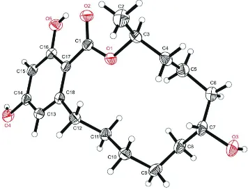
The compound has a macrocyclic structure and crystallizes in the triclinic space group P1. The molecular structure of the compound and the atom-labeling scheme are shown in Fig 1. The absolute configuration could not be defined confidently based on the single-crystal diffraction data. The isomeric purity of the title compound was confirmed by 1
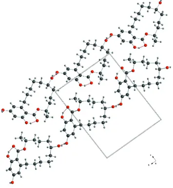
H-NMR, HPLC-DAD and –MS/MS data. Fig. 4 shows the difference in conformation between the known β-Zearalanol (Gelo-Pujić et al., 1994) and the title compound. Every molecule in the asymmetric unit builds an infinite chain with the help of hydrogen bonds of the hydroxyl groups. The two chains in relation to the unit cell are depicted in Fig. 2. The analysis of polymeric structures shows two infinite one dimensional chains with the base vectors of [1 1 0] and [1 - 1 0], Fig 3.
S2. Experimental
α-Zearalanol was obtained from Sigma-Aldrich Chemie GmbH (Germany, purity 97.0%). 5 mg (15.5 µmol) were weighed in a 1.5 ml HPLC glass vial and solved in 0.6 ml diethyl ether. Subsequently, 0.2 ml of n-hexane were added. Colorless crystals of the title compound were formed after 14 days of slow solvent evaporation at room temperature.
S3. Refinement
All H-atoms were positioned geometrically and refined using a riding model with d(C—H) = 0.93 Å, Uiso=1.2Ueq (C) for
aromatic 0.98 Å, Uiso = 1.2Ueq (C) for CH, 0.97 Å, Uiso = 1.2Ueq (C) for CH2, 0.96 Å, Uiso = 1.5Ueq (C) for CH3 atoms, and
0.82 Å, Uiso = 1.5Ueq (C) for hydroxyl group of O5. The hydrogen atoms from the other hydroxyl groups were treated
enantiomer has been assigned by reference to an unchanging chiral centre in the synthetic procedure.
Figure 1
Figure 2
Figure 3
View of the unit cell of the title compound, showing the two chains with planes of the basevectors. Turquoise for [1 1 0] and lime for [1 - 1 0]. Hydrogen bonds are drawn as dashed red lines.
Figure 4
The difference in conformation between the known β-Zearalanol (yellow, Gelo-Pujić et al., 1994) and the title compound.
(3S,7R)-7,14,16-Trihydroxy-3-methyl-3,4,5,6,7,8,9,10,11,12- decahydro-1H-2-benzoxacyclotetradecin-1-one.
Crystal data
C18H26O5
Mr = 322.39
Triclinic, P1
a = 5.0734 (11) Å
b = 11.618 (2) Å
c = 14.718 (3) Å
α = 87.388 (13)°
[image:5.610.132.481.376.582.2]γ = 89.780 (15)°
V = 865.0 (3) Å3
Z = 2
F(000) = 348
Dx = 1.238 Mg m−3
Mo Kα radiation, λ = 0.71073 Å
Cell parameters from 6563 reflections
θ = 2.3–26.4°
µ = 0.09 mm−1
T = 296 K Block, colourless 0.43 × 0.22 × 0.10 mm
Data collection
Bruker APEXII CCD diffractometer
Radiation source: fine-focus sealed tube Graphite monochromator
φ and ω scans
Absorption correction: multi-scan (SADABS; Bruker, 2001)
Tmin = 0.186, Tmax = 0.350
19642 measured reflections 4264 independent reflections 3421 reflections with I > 2σ(I)
Rint = 0.095
θmax = 28.3°, θmin = 1.4°
h = −6→6
k = −15→15
l = −19→19
Refinement
Refinement on F2
Least-squares matrix: full
R[F2 > 2σ(F2)] = 0.059
wR(F2) = 0.147
S = 0.95 4264 reflections 431 parameters 7 restraints
Primary atom site location: structure-invariant direct methods
Secondary atom site location: difference Fourier map
Hydrogen site location: inferred from neighbouring sites
H atoms treated by a mixture of independent and constrained refinement
w = 1/[σ2(F
o2) + (0.0803P)2]
where P = (Fo2 + 2Fc2)/3
(Δ/σ)max < 0.001
Δρmax = 0.24 e Å−3
Δρmin = −0.16 e Å−3
Special details
Geometry. All e.s.d.'s (except the e.s.d. in the dihedral angle between two l.s. planes) are estimated using the full covariance matrix. The cell e.s.d.'s are taken into account individually in the estimation of e.s.d.'s in distances, angles and torsion angles; correlations between e.s.d.'s in cell parameters are only used when they are defined by crystal symmetry. An approximate (isotropic) treatment of cell e.s.d.'s is used for estimating e.s.d.'s involving l.s. planes.
Refinement. Refinement of F2 against ALL reflections. The weighted R-factor wR and goodness of fit S are based on F2,
conventional R-factors R are based on F, with F set to zero for negative F2. The threshold expression of F2 > σ(F2) is used
only for calculating R-factors(gt) etc. and is not relevant to the choice of reflections for refinement. R-factors based on F2
are statistically about twice as large as those based on F, and R- factors based on ALL data will be even larger.
Fractional atomic coordinates and isotropic or equivalent isotropic displacement parameters (Å2)
x y z Uiso*/Ueq
C3′ 0.6061 (5) 0.2899 (2) 0.87071 (17) 0.0436 (6) H3′B 0.4548 0.2832 0.8329 0.052* C4′ 0.6511 (6) 0.1755 (2) 0.92215 (19) 0.0484 (6) H4′B 0.7733 0.1878 0.9690 0.058* H4′C 0.7338 0.1224 0.8801 0.058* C5′ 0.4010 (6) 0.1196 (2) 0.96673 (18) 0.0497 (6) H5′B 0.2853 0.1009 0.9194 0.060* H5′C 0.3099 0.1750 1.0047 0.060* C6′ 0.4513 (6) 0.0096 (2) 1.02532 (19) 0.0548 (7) H6′A 0.2827 −0.0263 1.0432 0.066* H6′B 0.5524 −0.0437 0.9882 0.066* C7′ 0.5984 (6) 0.0283 (2) 1.11146 (18) 0.0472 (6) H7′A 0.7575 0.0733 1.0933 0.057* C8′ 0.4374 (6) 0.0953 (2) 1.18242 (19) 0.0511 (6) H8′A 0.3308 0.0415 1.2210 0.061* H8′B 0.3183 0.1471 1.1514 0.061* C9′ 0.6079 (7) 0.1660 (2) 1.24287 (19) 0.0594 (8) H9′A 0.4982 0.1905 1.2947 0.071* H9′B 0.7455 0.1166 1.2660 0.071* C10′ 0.7366 (6) 0.2724 (2) 1.1937 (2) 0.0537 (7) H10C 0.8532 0.3073 1.2345 0.064* H10D 0.8440 0.2479 1.1414 0.064* C11′ 0.5409 (6) 0.3635 (2) 1.16147 (18) 0.0491 (6) H11C 0.4388 0.3909 1.2140 0.059* H11D 0.4194 0.3281 1.1227 0.059* C12′ 0.6742 (5) 0.4672 (2) 1.10867 (18) 0.0440 (6) H12C 0.7903 0.5050 1.1480 0.053* H12D 0.7810 0.4401 1.0571 0.053* C13′ 0.3928 (6) 0.6373 (2) 1.13423 (18) 0.0456 (6) H13B 0.4704 0.6401 1.1898 0.055* C14′ 0.1995 (5) 0.7179 (2) 1.11290 (17) 0.0444 (6) C15′ 0.0797 (6) 0.7162 (2) 1.03129 (18) 0.0450 (6) H15B −0.0523 0.7690 1.0180 0.054* C16′ 0.1590 (5) 0.6347 (2) 0.96934 (16) 0.0417 (6) C17′ 0.3551 (5) 0.55111 (19) 0.98913 (16) 0.0376 (5) C18′ 0.4720 (5) 0.55347 (19) 1.07492 (16) 0.0389 (5) H4A 0.388 (4) 1.371 (3) 0.431 (2) 0.073 (11)* H3′A 0.543 (5) −0.113 (4) 1.169 (3) 0.108 (17)* H3A −0.175 (5) 0.418 (3) 0.436 (3) 0.087 (14)* H4′A −0.005 (5) 0.823 (4) 1.163 (4) 0.13 (2)*
Atomic displacement parameters (Å2)
U11 U22 U33 U12 U13 U23
O5 0.0702 (13) 0.0535 (11) 0.0519 (11) −0.0147 (10) −0.0194 (10) −0.0050 (9) C1 0.0458 (14) 0.0372 (12) 0.0393 (13) 0.0009 (10) 0.0025 (10) −0.0048 (9) C2 0.066 (2) 0.0638 (19) 0.072 (2) −0.0032 (16) 0.0199 (16) −0.0031 (15) C3 0.0473 (14) 0.0422 (13) 0.0400 (12) −0.0025 (11) −0.0010 (10) 0.0090 (10) C4 0.0490 (15) 0.0413 (14) 0.0494 (14) −0.0106 (11) −0.0020 (11) 0.0078 (11) C5 0.0513 (16) 0.0489 (15) 0.0584 (16) −0.0009 (13) −0.0098 (13) −0.0034 (12) C6 0.072 (2) 0.0400 (14) 0.0628 (17) −0.0007 (13) −0.0070 (15) −0.0040 (12) C7 0.0560 (16) 0.0319 (12) 0.0593 (16) −0.0116 (11) 0.0059 (13) −0.0059 (11) C8 0.0623 (18) 0.0402 (14) 0.0568 (16) −0.0132 (13) 0.0130 (14) −0.0098 (12) C9 0.078 (2) 0.0482 (15) 0.0433 (13) −0.0210 (14) −0.0054 (13) −0.0057 (11) C10 0.0565 (16) 0.0425 (13) 0.0466 (13) −0.0088 (12) −0.0128 (12) 0.0004 (10) C11 0.0509 (15) 0.0368 (12) 0.0456 (13) −0.0098 (11) −0.0031 (11) −0.0029 (10) C12 0.0397 (13) 0.0377 (12) 0.0462 (13) −0.0041 (10) −0.0068 (10) 0.0001 (10) C13 0.0559 (16) 0.0344 (12) 0.0420 (13) −0.0015 (11) −0.0082 (11) −0.0033 (10) C14 0.0554 (15) 0.0291 (11) 0.0461 (13) −0.0034 (11) 0.0019 (11) −0.0035 (10) C15 0.0515 (14) 0.0356 (12) 0.0506 (14) −0.0083 (11) −0.0005 (11) −0.0105 (10) C16 0.0462 (14) 0.0379 (13) 0.0413 (13) −0.0012 (11) −0.0031 (11) −0.0091 (10) C17 0.0438 (13) 0.0297 (11) 0.0384 (11) −0.0005 (10) −0.0001 (10) −0.0067 (9) C18 0.0377 (12) 0.0324 (11) 0.0432 (12) 0.0003 (10) −0.0024 (10) −0.0050 (9) O1′ 0.0606 (11) 0.0368 (9) 0.0418 (9) 0.0061 (8) −0.0066 (8) −0.0057 (7) O2′ 0.0787 (14) 0.0458 (10) 0.0406 (10) 0.0043 (9) −0.0097 (9) 0.0003 (8) O3′ 0.0700 (15) 0.0376 (10) 0.0724 (13) 0.0094 (10) −0.0038 (11) 0.0034 (9) O4′ 0.0712 (14) 0.0415 (11) 0.0567 (11) 0.0081 (10) 0.0065 (10) −0.0081 (8) O5′ 0.0785 (14) 0.0532 (11) 0.0521 (11) 0.0131 (10) −0.0191 (10) 0.0004 (9) C1′ 0.0474 (14) 0.0343 (12) 0.0420 (13) −0.0040 (10) −0.0006 (11) 0.0016 (9) C2′ 0.070 (2) 0.0622 (19) 0.082 (2) −0.0024 (17) 0.0238 (18) −0.0024 (16) C3′ 0.0473 (14) 0.0415 (13) 0.0417 (12) −0.0006 (11) 0.0031 (10) −0.0074 (10) C4′ 0.0502 (15) 0.0445 (14) 0.0504 (14) 0.0071 (12) 0.0012 (11) −0.0080 (11) C5′ 0.0538 (16) 0.0470 (15) 0.0486 (14) −0.0032 (12) −0.0060 (12) −0.0012 (11) C6′ 0.0695 (19) 0.0388 (14) 0.0564 (16) −0.0056 (13) −0.0047 (14) −0.0040 (12) C7′ 0.0563 (16) 0.0320 (12) 0.0528 (14) 0.0023 (11) 0.0000 (12) −0.0001 (10) C8′ 0.0619 (17) 0.0403 (13) 0.0499 (14) 0.0036 (12) 0.0029 (12) 0.0013 (11) C9′ 0.093 (2) 0.0404 (14) 0.0452 (14) 0.0140 (15) −0.0131 (15) 0.0002 (11) C10′ 0.0653 (18) 0.0387 (13) 0.0596 (17) 0.0054 (12) −0.0225 (14) −0.0040 (12) C11′ 0.0591 (16) 0.0382 (13) 0.0500 (14) 0.0053 (12) −0.0073 (12) 0.0019 (11) C12′ 0.0495 (15) 0.0388 (13) 0.0452 (13) 0.0012 (11) −0.0107 (11) −0.0059 (10) C13′ 0.0550 (16) 0.0367 (13) 0.0452 (13) −0.0047 (11) −0.0045 (11) −0.0019 (10) C14′ 0.0549 (15) 0.0313 (12) 0.0452 (13) −0.0010 (11) 0.0090 (11) 0.0015 (10) C15′ 0.0498 (15) 0.0321 (12) 0.0521 (14) 0.0041 (11) 0.0010 (11) 0.0045 (10) C16′ 0.0492 (15) 0.0344 (12) 0.0406 (12) −0.0055 (11) −0.0030 (10) 0.0074 (10) C17′ 0.0413 (13) 0.0309 (11) 0.0402 (12) −0.0052 (10) −0.0010 (10) 0.0017 (9) C18′ 0.0409 (13) 0.0298 (12) 0.0456 (13) −0.0046 (10) −0.0033 (10) 0.0024 (10)
Geometric parameters (Å, º)
O3—C7 1.460 (3) O3′—C7′ 1.456 (3) O3—H3A 0.818 (10) O3′—H3′A 0.821 (10) O4—C14 1.372 (3) O4′—C14′ 1.370 (3) O4—H4A 0.826 (10) O4′—H4′A 0.821 (10) O5—C16 1.359 (3) O5′—C16′ 1.365 (3)
O5—H5A 0.8200 O5′—H5′A 0.8200
C1—C17 1.489 (3) C1′—C17′ 1.476 (3) C2—C3 1.503 (4) C2′—C3′ 1.504 (4)
C2—H2A 0.9600 C2′—H2′A 0.9600
C2—H2B 0.9600 C2′—H2′B 0.9600
C2—H2C 0.9600 C2′—H2′C 0.9600
C3—C4 1.517 (4) C3′—C4′ 1.523 (4)
C3—H3B 0.9800 C3′—H3′B 0.9800
C4—C5 1.507 (4) C4′—C5′ 1.527 (4)
C4—H4B 0.9700 C4′—H4′B 0.9700
C4—H4C 0.9700 C4′—H4′C 0.9700
C5—C6 1.540 (4) C5′—C6′ 1.538 (4)
C5—H5B 0.9700 C5′—H5′B 0.9700
C5—H5C 0.9700 C5′—H5′C 0.9700
C6—C7 1.527 (4) C6′—C7′ 1.533 (4)
C6—H6A 0.9700 C6′—H6′A 0.9700
C6—H6B 0.9700 C6′—H6′B 0.9700
C7—C8 1.532 (4) C7′—C8′ 1.527 (4)
C7—H7A 0.9800 C7′—H7′A 0.9800
C8—C9 1.523 (5) C8′—C9′ 1.542 (4)
C8—H8A 0.9700 C8′—H8′A 0.9700
C8—H8B 0.9700 C8′—H8′B 0.9700
C9—C10 1.520 (4) C9′—C10′ 1.532 (4)
C9—H9A 0.9700 C9′—H9′A 0.9700
C9—H9B 0.9700 C9′—H9′B 0.9700
H8A—C8—H8B 107.6 H8′A—C8′—H8′B 107.7 C10—C9—C8 114.9 (2) C10′—C9′—C8′ 114.1 (2) C10—C9—H9A 108.5 C10′—C9′—H9′A 108.7 C8—C9—H9A 108.5 C8′—C9′—H9′A 108.7 C10—C9—H9B 108.5 C10′—C9′—H9′B 108.7 C8—C9—H9B 108.5 C8′—C9′—H9′B 108.7 H9A—C9—H9B 107.5 H9′A—C9′—H9′B 107.6 C9—C10—C11 114.1 (2) C11′—C10′—C9′ 114.4 (3) C9—C10—H10A 108.7 C11′—C10′—H10C 108.7 C11—C10—H10A 108.7 C9′—C10′—H10C 108.7 C9—C10—H10B 108.7 C11′—C10′—H10D 108.7 C11—C10—H10B 108.7 C9′—C10′—H10D 108.7 H10A—C10—H10B 107.6 H10C—C10′—H10D 107.6 C12—C11—C10 112.7 (2) C10′—C11′—C12′ 113.4 (2) C12—C11—H11A 109.1 C10′—C11′—H11C 108.9 C10—C11—H11A 109.1 C12′—C11′—H11C 108.9 C12—C11—H11B 109.1 C10′—C11′—H11D 108.9 C10—C11—H11B 109.1 C12′—C11′—H11D 108.9 H11A—C11—H11B 107.8 H11C—C11′—H11D 107.7 C11—C12—C18 112.0 (2) C18′—C12′—C11′ 111.6 (2) C11—C12—H12A 109.2 C18′—C12′—H12C 109.3 C18—C12—H12A 109.2 C11′—C12′—H12C 109.3 C11—C12—H12B 109.2 C18′—C12′—H12D 109.3 C18—C12—H12B 109.2 C11′—C12′—H12D 109.3 H12A—C12—H12B 107.9 H12C—C12′—H12D 108.0 C18—C13—C14 122.0 (2) C18′—C13′—C14′ 121.6 (2) C18—C13—H13A 119.0 C18′—C13′—H13B 119.2 C14—C13—H13A 119.0 C14′—C13′—H13B 119.2 O4—C14—C15 122.8 (2) O4′—C14′—C15′ 122.5 (2) O4—C14—C13 116.8 (2) O4′—C14′—C13′ 116.7 (2) C15—C14—C13 120.4 (2) C15′—C14′—C13′ 120.8 (2) C14—C15—C16 119.3 (2) C14′—C15′—C16′ 119.0 (2) C14—C15—H15A 120.4 C14′—C15′—H15B 120.5 C16—C15—H15A 120.4 C16′—C15′—H15B 120.5 O5—C16—C15 115.8 (2) O5′—C16′—C15′ 115.6 (2) O5—C16—C17 123.2 (2) O5′—C16′—C17′ 122.9 (2) C15—C16—C17 121.0 (2) C15′—C16′—C17′ 121.5 (2) C18—C17—C16 118.8 (2) C16′—C17′—C18′ 118.2 (2) C18—C17—C1 125.7 (2) C16′—C17′—C1′ 116.0 (2) C16—C17—C1 115.6 (2) C18′—C17′—C1′ 125.8 (2) C13—C18—C17 118.6 (2) C13′—C18′—C17′ 118.8 (2) C13—C18—C12 116.4 (2) C13′—C18′—C12′ 116.2 (2) C17—C18—C12 125.0 (2) C17′—C18′—C12′ 124.9 (2)
Hydrogen-bond geometry (Å, º)
D—H···A D—H H···A D···A D—H···A
O5′—H5′A···O2′ 0.82 1.82 2.540 (3) 146 O4—H4A···O3i 0.83 (2) 1.93 (3) 2.745 (3) 171 (3)
O4′—H4′A···O3′ii 0.82 (3) 1.94 (3) 2.740 (3) 163 (4)
O3—H3A···O4iii 0.82 (3) 2.29 (3) 3.080 (3) 162 (3)
O3′—H3′A···O4′iii 0.82 (3) 2.27 (3) 3.067 (3) 165 (4)