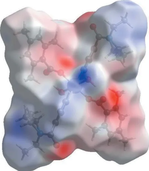
Crystal structure and Hirshfeld surface analysis of diaquabis(N,N diethylnicotinamide κN1)bis(2,4,6 trimethylbenzoato κO)manganese(II)
Full text
Figure


Related documents
With a larger temperature variation and therefore a higher Rayleigh num- ber, we observe in Figure 5 that the amplitudes of velocities increased in the vi- cinity of the heated
In the other patient, no increase was observed even after 6 months compared with the value at the ini- tial consultation ( Figure 1 ). In Group 2, the resting salivary flow rate
Term infants with abnormal MR imaging findings had a slightly higher prevalence of complete CoW and subsequently a lower prevalence of anatomic variations compared with those
Using high-frequency data on exchange rates across 30 countries, I measure exchange rate adjustment around the window of the unanticipated oil shock and link the heterogeneity
The overshoot column for the total sample includes participants who enrolled in training programs with a valid occupation code for the training occupation reported in TAPR
Figure 1. Assessment and evaluation information flow plan.. ences; 4) information technology or mathematics; and 5) the social or behavioural sciences. The final consideration is to
Figure 6: Conceptual overview. Routine oral dosing of microbes inhibits muscle-wasting syndromes including cachexia and sarcopenia. Microbiota stimulate thymopoiesis and
Present study observed a significant reduction in the mean AUA symptom score, PVR volume and significant reduction in urinary hesitancy, intermittent flow, straining during uri-
