FORMULATION DEVELOPMENT AND CHARECTERIZATION OF
NOVEL PREDNISONE MUCOADHESIVE LIPOSOMAL
FORMULATION
Parthiban S., Priyanka Raj G.* and Senthil Kumar G. P.
Dept. of Pharmaceutics, Bharathi College of Pharmacy, Bharathinagara, Mandya,
Karnataka-571422, India.
ABSTRACT
The aim of the present investigation was to design a mucoadhesive
liposomes containing Prednisone. Liposomes were prepared by the thin
film hydration method by using chloroform as a solvent.
Mucoadhesive liposomes containing prednisone formulations were
prepared by different concentration of lecithin and cholesterol by thin
film hydration technique. These formulations were evaluated for
entrapment efficiency, particle size, zeta potential, surface morphology
and in-vitro drug release. Particle size and zeta potential of the F4
formulation was found to be 212 nm, -164.9 mV respectively. Coating
of liposomes resulted increase in particle size and also increases the
zeta potential. Highest entrapment efficiency was observed in F1 and
F3 92% and 94%. The percent drug release from F4and CF1 was observed as follows
F1-88.57%, F2-73.31%, F3-76.29%, F4-90.97%, and F1 coated as a CF1-69.85%. Release
kinetics follows the Higuchi plot and non-Fickian diffusion mechanism.
KEYWARDS: Predisone, mucoadhesive liposome, thin film hydration method, in-vitro
release.
INTRODUCTION
Mucoadhesive Drug Delivery System
Mucoadhesive drug delivery systems are delivery systems which utilize the property of
bioadhesion of certain polymers which become adhesive on hydration and hence can be used
for targeting a drug to a particular region of the body for extended periods of time.[1] In case
of bioadhesive drug delivery, the term bioadhesion is used to describe the adhesion between
Volume 8, Issue 7, 1309-1317. Research Article ISSN 2277– 7105
Article Received on 28 March 2019,
Revised on 18 April 2019, Accepted on 09 May 2019 DOI: 10.20959/wjpr20197-15008
*Corresponding Author
Priyanka Raj G.
Dept. of Pharmaceutics,
Bharathi College of
Pharmacy, Bharathinagara,
Mandya, Karnataka-571422,
polymers, either synthetic or natural and soft tissues or the gastrointestinal mucosa. In cases
where the bond is formed with the mucus the term mucoadhesion may be used synonymously
with bioadhesion. Mucoadhesion can be defined as a state in which two components, of
which one is of biological origin, are held together for extended periods of time by the help of
interfacial forces.[2]
MATERIALS AND METHOD
Prednisone was gifted from micro labs LTD. Karnataka, Soya lecithin was purchased from
Pharma Sonic Biochem Extractions Ltd. Indore, Cholesterol, and other solvent like
Chloroform and Methanol purchased from S d fine chem Ltd. Mumbai. Phosphate buffer PH
6.8 were prepared as described in the indian pharmacopoeia (1996).
PREPARATION OF PREDNISONE LIPOSOME
A. Preparation of liposomes[3]
Cationic multilamellar liposomes can be prepared by hydration of lipid film. The lipid
mixture is dissolved in a small amount of chloroform and placed in a rotary evaporator at
40°C until a thin film is obtained, and allowed to stand overnight in a vacuum chamber to
ensure complete solvent removal. Phosphate buffer pH 6.8 is used to hydrate the thin film.
The hydrated thin film is melted in water bath at 70°C for 1 min and blended to obtain
multilamellar liposomes. Then prepared liposome will be sonicated to reduce particle size.
B. Coating of liposomes[4]
Chitosan was previously dissolved in glacial acetic acid overnight at room temperature. In
order to coat the liposomes with chitosan, 1 mL of chitosan with a concentration of 1 mg/mL
was added dropwise to the same amount liposome volume under continuous magnetic stirring
for 1 h. Finally, the liposomes were sonicated for 30 min with an ultrasonic batho produce
uniform chitosan-coated liposomes.
Table 1: Formulation design for the preparation uncoated liposomes containing
prednisone.
Formulation code Drug Soya lecithin Cholesterol
F1 100 200 100
F2 100 300 100
F3 100 400 100
Table 2: Coating of liposome containing Prednisone.
Formulation code Drug Soya lecithin cholesterol Chitosan % w/v
CF1 100 400 100 0.2%
CHARACTERIZATION OF LIPOSOMES
Drug entrapment efficiency
The drug entrapment efficiency was calculated using the total drug content of liposome
dispersion and unentrapped drug content of the dispersion. The total dug content of the
dispersion is determined estimating total drug entrapped and unentrapped. 5 ml of liposome
dispersion was taken in a volumetric flask. The dispersion was subjected to sonication in bath
sonicator for 30 minutes. Then the mixture was filtered and estimated after suitable dilution
at by using UV Visible Spectrophotometer (Shimadzu, UV1800). For the free unentrapped
drug, 5 ml of the liposome dispersion subjected to centrifugation at 18000 rpm using Remi
centrifuge for 40 min at 50° C. The supernatant clear solution was collected separately and
the free drug present in the supernatant was estimated after suitable dilution at 243.60nm
wavelength by using UV Visible Spectrophotometer.
In vitro diffusion studies
In vitro diffusion studies were carried by using cellophane membrane. The membrane was
soaked overnight in phosphate buffer pH 6.8. 10 ml of prepared liposomal dispersion which
contains 10 mg of drug was taken and placed in theone end of of the open tube. Dialysis tube
was was placed in 500 ml beaker, containing 250 ml phosphate buffer pH 6.8. The
temperature of the receptor phase was maintained at 37 ± 0.5° C and the it was stirred with
magnetic stirrer to maintain homogeneous condition. The samples were withdrawn at
different time intervals. Fresh medium was used to replace with equal volume of the sample
withdrawn. The samples were analyzed at 243.60 nm in a UV-Visible spectrophotometer and
amount of drug released at different time intervals was calculated.[5]
In vitro wash-off test for mucoadhesive testing
The mucoadhesive property of the polymer-coated liposomes was evaluated by an in vitro
adhesion test. The method used was the modified in-vitro wash-off test. The mucoadhesion of
the polymer-coated liposomes was compared with that of a non mucoadhesive material,
uncoated liposomes containing Prednisone. Freshly excised pieces of sheep intestinal mucosa
(2 × 2 cm) were tightened onto glass slides (3 × 1 inches) with thread. A volume of 0.5 ml of
each wet-rinsed tissue specimen and immediately incubated at 37 ℃. The tissue specimens
were taken out at 1 and 3 hrs. The samples were washed with 10.0 ml of PBS at each time
interval.
Determination of mucoadhesive strength
From the 10.0 ml of the eluted buffer containing nonadhered drug, 500 μl aliquots were taken
and liposomal lipids were dissolved by methanol. It was measured by a UV
spectrophotometer. The concentration of prednisone eluted in the phosphate buffer pH 6.8
was measured and the remaining drug was assumed to be present in liposomes adhered to the
intestinal mucosa. Hence, the percentage of mucoadhesive strength can be calculated by
Eq.[6,7]
RESULT AND DISCUSSION
FTIR spectra of pure Prednisone showed sharp characteristic peaks 1622, 1707.66, 1666.2
and 3289 Physical mixture showed all the characteristic peaks of pure drug, confirmed no
interaction between the drug and excipients. Comparative studies of FTIR graphs are showed
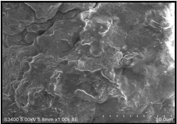
in Figure.1-2. The surface morphology was studied by Scanning electron microscopy (SEM).
The SEM photographs of liposomes formulation F4 as shown in Figure. 3. Vesicle size
distribution of F1-F4-CF1 graphs are showed in Fig.4. The % entrapment efficiency was
found to decrease with increasing the cholesterol concentration. It is shown in Figure.5. Zeta
potential of formulation F4 shown in figure.6. And it was found to -164.9 mV, respectively
which indicate that they are sufficient to be stable. In vitro drug release of liposomes in
phosphate buffer pH 6.8 was performed using dialysis tube diffusion technique. The in vitro
drug release profile of liposomes formulations obtained from dialysis experiment was shown
in Figure.7. The release of liposome Chitosan containing coated liposomes was varied
according to concentration of soya lecithin and cholesterol. The progressive decrease in the
amount of drug diffused through cellophane membrane from formulations F1 – F4 and CF1
attributed to gradual increase in soya lecithin and cholesterol content. It has been concluded
that, if we increase the concentration of soya lecithin and cholesterol, the diffusion of drug
also decreases. The amount of drug diffused from formulation CF1 was showed 69.85 %, was
lower among the formulations F1 to F4 and CF1. Release kinetics follows the Higuchi plot
Mucoadhesive strength was measured by a UV spectrophotometer. The Mucoadhesive
strength was measured by a UV spectrophotometer. Mucoadhesive strength formulation CF1.
The amount of drug released in the formulation CF1 63% which showed sufficient
mucoadhesive property.
Figure.1: FT-IR Spectroscopy of Prednisone.
Figure.3: SEM of liposomes formulation F4.
Figure. 4: vesicle size analysis of liposomal formulation.
[image:6.595.141.454.566.744.2]Figure.7: % in-vitro drug release of formulation F1- F4-CF1.
RESULT OF MODELLING FITTING
Table 3: Data for different kinetic model.
Formulation
code Zero order
First order
Higuchi plot Peppas plot
r2 ‘n’
F1 0.963 0.622 0.973 0.714 1.306
F2 0.959 0.478 0.970 0.578 1.166
F3 0.938 0.567 0.987 0.668 1.234
F4 0.966 0.611 0.969 0.705 1.315
[image:8.595.143.455.73.240.2]CF1 0.920 0.558 0.975 0.659 1.225
Figure 8: Higuchi drug release kinetics for F1-F4-CF1.
CONCLUSION
In this study, a mucoadhesive liposomal formulation of Prednisone was developed with
desirable drug delivery properties. The chitosan-coated liposomes had good in vitro stability,
strong mucoadhesiveness, and enhanced cellular uptake. Therefore, the chitosan-coated
carbopol gel formulation appears to have the potential to improve the bioavailability of
[image:8.595.104.490.320.619.2]REFERENCES
1. Mahajan P, Kaur A, Aggarwal G, Harikumar SL. Mucoadhesive drug delivery system: A
review. Int J Drug Dev Res., 2013; 5(1): 11-20.
2. Pranshu T, Satheesh NV. Oral mucoadhesive drug delivery systems: As a review. Int J
Pharma, 2011; 2(1): 436-46.
3. Patel DB, Patel JK. Liposomal drug delivery of Metronidazole for the local treatment of
vaginitis. Int J Pharm Sci Nanotechnol, 2009; 2(1): 248-57.
4. Mengoni T, Adrian M, Pereira S, Santos CB, Kaiser M, Goycoolea F. A Chitosan—
Based Liposome Formulation Enhances the In Vitro Wound Healing Efficacy of
Substance P Neuropeptide. Pharmaceutics, 6, 2017; 9(4): 1-17.
5. Srinivas LK, Vinaikumar T, Rajasri A. Preparation and evalution of liposomes
formulation for poorly soluble drug Itraconazole by complexation. Der Pharmacia Lettre.,
2015; 7(8): 1-7.
6. Chowdary KP, Rao YS. Design and in vitro and in vivo evaluation of mucoadhesive
microcapsules of glipizide for oral controlled release: a technical note. AAPS Pharm Sci
Tech., Sep 1, 2003; 4(3): 1-6.
7. Rao S, Varalakshmi ST, Chandana RV. Design and in vitro evaluation of mucoadhesive
microcapsules of aceclofenac for oral controlled release. Int J Pharm Pharm Sci, 4(5):