organic papers
o328
Bond and Davies C7H9N DOI: 10.1107/S1600536802003379 Acta Cryst.(2002). E58, o328±o330Acta Crystallographica Section E
Structure Reports
Online ISSN 1600-5368
3,4-Lutidine
Andrew D. Bond* and John E. Davies
Department of Chemistry, University of Cambridge, Lensfield Road, Cambridge CB2 1EW, England
Correspondence e-mail: [email protected]
Key indicators Single-crystal X-ray study T= 150 K
Mean(C±C) = 0.002 AÊ Rfactor = 0.053 wRfactor = 0.135
Data-to-parameter ratio = 19.1
For details of how these key indicators were automatically derived from the article, see http://journals.iucr.org/e.
#2002 International Union of Crystallography Printed in Great Britain ± all rights reserved
The crystal structure of 3,4-lutidine (3,4-dimethylpyridine, C7H9N), has been determined at 150 (2) K followingin situ
crystal growth from the liquid. In space groupC2/c, there are four independent molecules in the asymmetric unit, linked into dimersviaCÐH N interactions.
Comment
As part of a study devoted to improving the techniques for determining the crystal structures of substances that are liquids at room temperature, we have reported previously the crystal structures of 2,6-lutidine (Bond et al., 2001), 3,5-lut-idine (Bond & Davies, 2002a) and 2,5-lutidine (Bond & Davies, 2002b). We report here the crystal structure of 3,4-lutidine, (I), determined at 150 (2) K followingin situ crystal growth from the liquid.
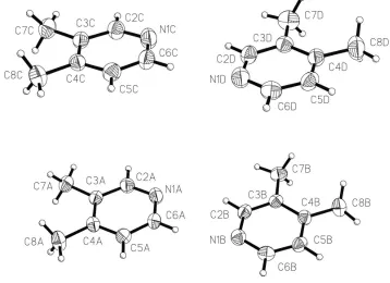
Compound (I) crystallizes in the monoclinic space group
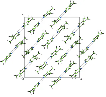
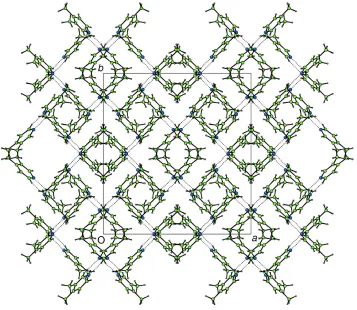
C2/cwith four independent molecules in the asymmetric unit (Fig. 1). Within the asymmetric unit, molecules are linked into dimersviaCÐH N interactions (Table 1). In the structures of the other lutidines reported to date, molecules are linked into linear chainsviaCÐH N interactions involving the H atom at the 4-position; this interaction is clearly prohibited in (I). Two orientations may be envisaged for dimerization in which a centrosymmetric motif would result: either both molecules interact through the H atoms at their 2-positions, or both interact through the H atoms at their 6-positions. The observed dimer involves one molecule of (I) interacting through H at the 2-position and one interacting through H at the 6-position, giving rise to an asymmetric motif (point symmetry 1). In both independent dimers within the asym-metric unit, the interaction C6ÐH6 N1 is signi®cantly shorter and closer to linear than the C2ÐH2 N1 interaction (Table 1). This is not obviously an intra-dimer effect and may be a result of inter-dimer interactions between methyl substituents. The dimers may be considered to form layers parallel to (001), with the planes through the dimer units lying alternately parallel to (110) and (110) in adjacent layers (Figs. 2 and 3).
Experimental
The sample (99%) was obtained from the Aldrich company and used without further puri®cation. The crystal was grown in a 0.3 mm glass capillary tube atca235 K (a temperature only slightly less than the melting point of the solid in the capillary) using a technique described earlier (Davies & Bond, 2001). Once grown, the crystal was cooled to 150 (2) K for data collection. The length of the cylindrical crystal was not estimated, but it exceeded the diameter of the collimator (0.35 mm).
Crystal data
C7H9N Mr= 107.15
Monoclinic,C2=c a= 20.9006 (8) AÊ
b= 20.6112 (9) AÊ
c= 12.9921 (3) AÊ
= 115.135 (2)
V= 5066.9 (3) AÊ3 Z= 32
Dx= 1.124 Mg mÿ3
MoKradiation Cell parameters from 9883
re¯ections
= 1.0±27.5 = 0.07 mmÿ1 T= 150 (2) K Cylinder, colourless 0.15 mm (radius)
Data collection
Nonius KappaCCD diffractometer Thin-slice!and'scans Absorption correction: none 9752 measured re¯ections 5740 independent re¯ections 4041 re¯ections withI> 2(I)
Rint= 0.026 max= 27.5 h= 0!26
k= 0!26
l=ÿ16!15
Re®nement
Re®nement onF2 R[F2> 2(F2)] = 0.053 wR(F2) = 0.135 S= 1.03 5740 re¯ections 301 parameters
H-atom parameters constrained
w= 1/[2(F
o2) + (0.0488P)2
+ 2.9233P]
whereP= (Fo2+ 2Fc2)/3
(/)max< 0.001
max= 0.20 e AÊÿ3
min=ÿ0.16 e AÊÿ3
Table 1
Hydrogen-bonding geometry (AÊ,).
DÐH A DÐH H A D A DÐH A
C6AÐH6A N1B 0.95 2.48 3.368 (2) 156
C2BÐH2B N1A 0.95 2.83 3.630 (2) 143
C6CÐH6C N1D 0.95 2.45 3.339 (2) 156
C2DÐH2D N1C 0.95 2.88 3.661 (2) 140
H atoms were placed geometrically and re®ned with isotropic
Figure 2
Projection on to (001) of a single layer in (I), showing dimers linked by
Figure 3
Projection on to (001) of the entire structure of (I), showing layers of dimers oriented alternately parallel to (110) and (110) (CAMERON; Watkinet al., 1996).
Figure 1
The asymmetric unit and atom-labelling scheme in (I), showing displacement ellipsoids (C/N atoms) at the 50% probability level (XP; Sheldrick, 1993). Independent molecules are denoted by the suf®xesA,B,
organic papers
o330
Bond and Davies C7H9N Acta Cryst.(2002). E58, o328±o330chemically equivalent H atoms (one parameter for all methyl H atoms, four parameters in total). Each methyl group was allowed to rotate about its local threefold axis.
Data collection:COLLECT(Nonius, 1998); cell re®nement:HKL SCALEPACK(Otwinowski & Minor, 1997); data reduction:HKL DENZO and SCALEPACK (Otwinowski & Minor, 1997); program(s) used to solve structure:SIR-92 (Altomareet al., 1994); program(s) used to re®ne structure:SHELXL97 (Sheldrick, 1997); molecular graphics:XP(Sheldrick, 1993) andCAMERON(Watkinet al., 1996); software used to prepare material for publication:
SHELXL97.
We thank the EPSRC for ®nancial assistance towards the purchase of the Nonius CCD diffractometer.
References
Altomare, A., Cascarano, G., Giacovazzo, C., Guagliardi, A., Burla, M. C., Polidori, G. & Camalli, M. (1994).J. Appl. Cryst.27, 435.
Bond, A. D., Davies, J. E. & Kirby, A. J. (2001).Acta Cryst.E57, o1242±o1244. Bond, A. D. & Davies, J. E. (2002a).Acta Cryst.E58, o5±o7.
Bond, A. D. & Davies, J. E. (2002b).Acta Cryst.E58, o326±o327. Davies, J. E. & Bond, A. D. (2001).Acta Cryst.E57, o947±o949. Nonius (1998).COLLECT. Nonius BV, Delft, The Netherlands.
Otwinowski, Z. & Minor, W. (1997). Methods in Enzymology, Vol. 276,
Macromolecular Crystallography, Part A, edited by C. W. Carter and R. M. Sweet, pp. 307±326. London: Academic Press.
Sheldrick, G. M. (1993).XP. University of GoÈttingen, Germany. Sheldrick, G. M. (1997).SHELXL97. University of GoÈttingen, Germany. Watkin, D. J., Prout, C. K. & Pearce, L. J. (1996).CAMERON. Chemical
supporting information
Acta Cryst. (2002). E58, o328–o330 [doi:10.1107/S1600536802003379]
3,4-Lutidine
Andrew D. Bond and John E. Davies
S1. Comment
As part of a study devoted to improving the techniques for determining the crystal structures of substances that are
liquids at room temperature, we have reported previously the crystal structures of 2,6-lutidine (Bond et al., 2001),
3,5-lutidine (Bond & Davies, 2002a) and 2,5-lutidine (Bond & Davies, 2002b). We report here the crystal structure of
3,4-lutidine, (I), determined at 150 (2) K following in situ crystal growth from the liquid.
Compound (I) crystallizes in the monoclinic space group C2/c with four independent molecules in the asymmetric unit
(Fig. 1). Within the asymmetric unit, molecules are linked into dimers via C—H···N interactions (Table 1). In the
structures of the other lutidines reported to date, molecules are linked into linear chains via C—H···N interactions
involving the H atom at the 4-position; this interaction is clearly prohibited in (I). Two orientations may be envisaged for
dimerization in which a centrosymmetric motif would result: either both molecules interact through the H atoms at their
2-positions, or both interact through the H atoms at their 6-positions. The observed dimer involves one molecule of (I)
interacting through H at the 2-position and one interacting through H at the 6-position, giving rise to an asymmetric motif
(point symmetry 1). In both independent dimers within the asymmetric unit, the interaction C6—H6···N1 is significantly
shorter and closer to linear than the C2—H2···N1 interaction (Table 1). This is not obviously an intra-dimer effect and
may be a result of inter-dimer interactions between methyl substituents. The dimers may be considered to form layers
parallel to (001), with the planes through the dimer units lying alternately parallel to (110) and (110) in adjacent layers
(Figs. 2 and 3).
S2. Experimental
The sample (99%) was obtained from the Aldrich company and used without further purification. The crystal was grown
in a 0.3 mm glass capillary tube at ca 235 K (a temperature only slightly less than the melting point of the solid in the
capillary) using a technique described earlier (Davies & Bond, 2001). Once grown, the crystal was cooled to 150 (2) K
for data collection. The length of the cylindrical crystal was not estimated, but it exceeded the diameter of the collimator
(0.35 mm).
S3. Refinement
H atoms were placed geometrically and refined with isotropic displacement parameters, with common parameters
assigned to chemically equivalent H atoms (one parameter for all methyl H atoms, four parameters in total). Each methyl
supporting information
[image:5.610.125.483.78.338.2]sup-2
Acta Cryst. (2002). E58, o328–o330Figure 1
The asymmetric unit and atom-labelling scheme in (I), showing displacement ellipsoids (C/N atoms) at the 50%
Figure 2
Projection on to (001) of a single layer in (I), showing dimers linked by C—H···N interactions orientated parallel to (110)
supporting information
[image:7.610.128.485.70.380.2]sup-4
Acta Cryst. (2002). E58, o328–o330Figure 3
Projection on to (001) of the entire structure of (I), showing layers of dimers oriented alternately parallel to (110) and
(110) (CAMERON; Watkin et al., 1996).
3,4-dimethylpyridine
Crystal data
C7H9N Mr = 107.15
Monoclinic, C2/c a = 20.9006 (8) Å
b = 20.6112 (9) Å
c = 12.9921 (3) Å
β = 115.135 (2)°
V = 5066.9 (3) Å3 Z = 32
F(000) = 1856
Dx = 1.124 Mg m−3 Melting point: 261 K
Mo Kα radiation, λ = 0.7107 Å Cell parameters from 9883 reflections
θ = 1.0–27.5°
µ = 0.07 mm−1 T = 150 K
Cylinder, colourless 0.15 mm (radius)
Data collection
Nonius KappaCCD diffractometer
Radiation source: fine-focus sealed tube Thin–slice ω and φ scans
9752 measured reflections 5740 independent reflections
4041 reflections with I > 2σ(I)
Rint = 0.026
θmax = 27.5°, θmin = 3.7° h = 0→26
k = 0→26
Refinement
Refinement on F2 Least-squares matrix: full
R[F2 > 2σ(F2)] = 0.053 wR(F2) = 0.135 S = 1.03 5740 reflections 301 parameters 0 restraints
Primary atom site location: structure-invariant direct methods
Secondary atom site location: difference Fourier map
Hydrogen site location: inferred from neighbouring sites
H-atom parameters constrained
w = 1/[σ2(F
o2) + (0.0488P)2 + 2.9233P] where P = (Fo2 + 2Fc2)/3
(Δ/σ)max < 0.001 Δρmax = 0.20 e Å−3 Δρmin = −0.16 e Å−3
Special details
Experimental. Crystal grown in situ in a 0.30 mm Lindemann tube.
Geometry. All e.s.d.'s (except the e.s.d. in the dihedral angle between two l.s. planes) are estimated using the full covariance matrix. The cell e.s.d.'s are taken into account individually in the estimation of e.s.d.'s in distances, angles and torsion angles; correlations between e.s.d.'s in cell parameters are only used when they are defined by crystal symmetry. An approximate (isotropic) treatment of cell e.s.d.'s is used for estimating e.s.d.'s involving l.s. planes.
Refinement. Refinement of F2 against ALL reflections. The weighted R-factor wR and goodness of fit S are based on F2, conventional R-factors R are based on F, with F set to zero for negative F2. The threshold expression of F2 > σ(F2) is used only for calculating R-factors(gt) etc. and is not relevant to the choice of reflections for refinement. R-factors based on F2 are statistically about twice as large as those based on F, and R- factors based on ALL data will be even larger.
Fractional atomic coordinates and isotropic or equivalent isotropic displacement parameters (Å2)
x y z Uiso*/Ueq
N1A 0.23300 (7) 0.60065 (6) 0.01432 (10) 0.0421 (3)
C2A 0.26063 (8) 0.64157 (7) −0.03603 (12) 0.0394 (3)
H2A 0.2424 0.6398 −0.1166 0.048 (2)*
C3A 0.31361 (7) 0.68629 (7) 0.01988 (11) 0.0323 (3)
C4A 0.33999 (7) 0.68930 (7) 0.13817 (11) 0.0329 (3)
C5A 0.31180 (8) 0.64716 (7) 0.19112 (12) 0.0363 (3)
H5A 0.3287 0.6478 0.2715 0.050 (2)*
C6A 0.25910 (8) 0.60414 (7) 0.12734 (12) 0.0375 (3)
H6A 0.2406 0.5758 0.1657 0.047 (2)*
C7A 0.34097 (8) 0.72962 (8) −0.04567 (13) 0.0432 (4)
H7AA 0.3154 0.7205 −0.1271 0.0730 (13)*
H7AB 0.3915 0.7215 −0.0214 0.0730 (13)*
H7AC 0.3338 0.7751 −0.0312 0.0730 (13)*
C8A 0.39661 (9) 0.73682 (8) 0.20617 (14) 0.0498 (4)
H8AA 0.4119 0.7284 0.2874 0.0730 (13)*
H8AB 0.3780 0.7811 0.1884 0.0730 (13)*
H8AC 0.4370 0.7320 0.1871 0.0730 (13)*
N1B 0.15531 (6) 0.49769 (6) 0.18226 (10) 0.0391 (3)
C2B 0.12057 (7) 0.48533 (7) 0.07082 (12) 0.0355 (3)
H2B 0.1335 0.5098 0.0206 0.048 (2)*
C3B 0.06718 (7) 0.43973 (7) 0.02281 (11) 0.0319 (3)
C4B 0.04803 (7) 0.40381 (6) 0.09667 (12) 0.0332 (3)
C5B 0.08360 (8) 0.41670 (7) 0.21207 (12) 0.0366 (3)
supporting information
sup-6
Acta Cryst. (2002). E58, o328–o330C6B 0.13596 (8) 0.46290 (7) 0.25080 (12) 0.0367 (3)
H6B 0.1595 0.4704 0.3304 0.047 (2)*
C7B 0.03213 (8) 0.43004 (8) −0.10363 (12) 0.0437 (4)
H7BA 0.0531 0.4595 −0.1399 0.0730 (13)*
H7BB −0.0185 0.4391 −0.1320 0.0730 (13)*
H7BC 0.0389 0.3851 −0.1216 0.0730 (13)*
C8B −0.00871 (9) 0.35304 (8) 0.05216 (15) 0.0505 (4)
H8BA −0.0152 0.3333 0.1157 0.0730 (13)*
H8BB 0.0051 0.3195 0.0121 0.0730 (13)*
H8BC −0.0531 0.3731 −0.0004 0.0730 (13)*
N1C 0.47325 (7) 0.34998 (7) −0.02172 (12) 0.0504 (4) C2C 0.50067 (8) 0.39217 (8) −0.07027 (13) 0.0449 (4)
H2C 0.4799 0.3939 −0.1510 0.048 (2)*
C3C 0.55689 (8) 0.43360 (7) −0.01279 (12) 0.0369 (3)
C4C 0.58764 (7) 0.43149 (7) 0.10593 (12) 0.0363 (3)
C5C 0.56011 (8) 0.38758 (8) 0.15718 (13) 0.0421 (4)
H5C 0.5801 0.3844 0.2377 0.050 (2)*
C6C 0.50382 (8) 0.34839 (8) 0.09192 (14) 0.0458 (4)
H6C 0.4860 0.3189 0.1296 0.047 (2)*
C7C 0.58322 (9) 0.47896 (8) −0.07719 (14) 0.0493 (4)
H7CA 0.5552 0.4729 −0.1590 0.0730 (13)*
H7CB 0.6330 0.4696 −0.0578 0.0730 (13)*
H7CC 0.5786 0.5239 −0.0566 0.0730 (13)*
C8C 0.64820 (9) 0.47483 (9) 0.17554 (14) 0.0517 (4)
H8CA 0.6614 0.4672 0.2565 0.0730 (13)*
H8CB 0.6341 0.5203 0.1572 0.0730 (13)*
H8CC 0.6886 0.4654 0.1585 0.0730 (13)*
N1D 0.39991 (7) 0.24523 (7) 0.14888 (11) 0.0498 (4)
C2D 0.36153 (8) 0.23333 (8) 0.03857 (13) 0.0429 (4)
H2D 0.3701 0.2598 −0.0142 0.048 (2)*
C3D 0.31041 (8) 0.18578 (7) −0.00550 (12) 0.0378 (3)
C4D 0.29759 (8) 0.14682 (7) 0.07185 (13) 0.0401 (3)
C5D 0.33786 (9) 0.15837 (8) 0.18685 (13) 0.0452 (4)
H5D 0.3310 0.1326 0.2420 0.050 (2)*
C6D 0.38758 (9) 0.20700 (8) 0.22098 (13) 0.0479 (4)
H6D 0.4145 0.2136 0.3002 0.047 (2)*
C7D 0.27101 (9) 0.17719 (9) −0.13189 (13) 0.0547 (4)
H7DA 0.2880 0.2089 −0.1707 0.0730 (13)*
H7DB 0.2204 0.1838 −0.1546 0.0730 (13)*
H7DC 0.2789 0.1332 −0.1528 0.0730 (13)*
C8D 0.24239 (10) 0.09446 (9) 0.03272 (18) 0.0620 (5)
H8DA 0.2429 0.0712 0.0989 0.0730 (13)*
H8DB 0.2525 0.0640 −0.0165 0.0730 (13)*
Atomic displacement parameters (Å2)
U11 U22 U33 U12 U13 U23
N1A 0.0428 (7) 0.0406 (7) 0.0423 (7) −0.0108 (6) 0.0175 (6) −0.0042 (5) C2A 0.0423 (8) 0.0411 (8) 0.0346 (7) −0.0047 (7) 0.0162 (6) −0.0016 (6) C3A 0.0312 (7) 0.0299 (7) 0.0379 (7) 0.0023 (6) 0.0165 (6) 0.0034 (6) C4A 0.0290 (7) 0.0285 (7) 0.0380 (7) 0.0010 (6) 0.0112 (6) 0.0015 (5) C5A 0.0392 (8) 0.0351 (8) 0.0336 (7) 0.0000 (6) 0.0145 (6) 0.0020 (6) C6A 0.0404 (8) 0.0340 (8) 0.0414 (8) −0.0057 (6) 0.0206 (6) 0.0022 (6) C7A 0.0441 (9) 0.0427 (9) 0.0477 (9) −0.0017 (7) 0.0243 (7) 0.0071 (7) C8A 0.0455 (9) 0.0462 (10) 0.0473 (9) −0.0138 (8) 0.0097 (7) 0.0005 (7) N1B 0.0366 (7) 0.0401 (7) 0.0396 (7) −0.0025 (6) 0.0152 (5) −0.0012 (5) C2B 0.0374 (8) 0.0360 (8) 0.0378 (7) −0.0001 (6) 0.0204 (6) 0.0050 (6) C3B 0.0314 (7) 0.0312 (7) 0.0349 (7) 0.0054 (6) 0.0159 (6) 0.0030 (5) C4B 0.0325 (7) 0.0265 (7) 0.0423 (8) 0.0022 (6) 0.0174 (6) 0.0018 (6) C5B 0.0432 (8) 0.0337 (7) 0.0381 (8) 0.0061 (6) 0.0223 (6) 0.0086 (6) C6B 0.0376 (8) 0.0384 (8) 0.0319 (7) 0.0054 (6) 0.0126 (6) 0.0002 (6) C7B 0.0432 (8) 0.0507 (10) 0.0361 (8) 0.0043 (7) 0.0156 (6) −0.0003 (7) C8B 0.0507 (10) 0.0434 (9) 0.0594 (10) −0.0119 (8) 0.0254 (8) −0.0024 (7) N1C 0.0469 (8) 0.0502 (8) 0.0525 (8) −0.0143 (7) 0.0197 (6) −0.0051 (6) C2C 0.0431 (9) 0.0489 (9) 0.0392 (8) −0.0055 (7) 0.0142 (7) −0.0005 (7) C3C 0.0342 (7) 0.0330 (8) 0.0446 (8) 0.0016 (6) 0.0179 (6) 0.0020 (6) C4C 0.0325 (7) 0.0330 (7) 0.0429 (8) 0.0006 (6) 0.0155 (6) −0.0030 (6) C5C 0.0423 (8) 0.0445 (9) 0.0404 (8) −0.0006 (7) 0.0184 (7) 0.0012 (6) C6C 0.0463 (9) 0.0430 (9) 0.0528 (9) −0.0068 (7) 0.0257 (7) 0.0025 (7) C7C 0.0494 (10) 0.0475 (9) 0.0535 (9) −0.0023 (8) 0.0244 (8) 0.0088 (8) C8C 0.0463 (9) 0.0494 (10) 0.0522 (10) −0.0108 (8) 0.0140 (8) −0.0054 (8) N1D 0.0521 (8) 0.0485 (8) 0.0500 (8) −0.0098 (7) 0.0228 (6) −0.0029 (6) C2D 0.0478 (9) 0.0405 (8) 0.0461 (9) −0.0027 (7) 0.0253 (7) 0.0053 (7) C3D 0.0351 (8) 0.0371 (8) 0.0415 (8) 0.0051 (7) 0.0165 (6) 0.0020 (6) C4D 0.0368 (8) 0.0339 (8) 0.0540 (9) 0.0016 (7) 0.0236 (7) 0.0019 (6) C5D 0.0545 (10) 0.0434 (9) 0.0481 (9) 0.0044 (8) 0.0317 (8) 0.0100 (7) C6D 0.0519 (10) 0.0516 (10) 0.0396 (8) 0.0004 (8) 0.0188 (7) −0.0042 (7) C7D 0.0517 (10) 0.0616 (11) 0.0432 (9) 0.0057 (9) 0.0129 (7) 0.0006 (8) C8D 0.0549 (11) 0.0496 (11) 0.0827 (13) −0.0127 (9) 0.0304 (10) 0.0006 (9)
Geometric parameters (Å, º)
N1A—C6A 1.3335 (18) N1C—C6C 1.337 (2)
N1A—C2A 1.3393 (19) N1C—C2C 1.338 (2)
C2A—C3A 1.386 (2) C2C—C3C 1.386 (2)
C2A—H2A 0.950 C2C—H2C 0.950
C3A—C4A 1.3963 (19) C3C—C4C 1.397 (2)
C3A—C7A 1.5047 (19) C3C—C7C 1.506 (2)
C4A—C5A 1.3855 (19) C4C—C5C 1.385 (2)
C4A—C8A 1.501 (2) C4C—C8C 1.498 (2)
C5A—C6A 1.382 (2) C5C—C6C 1.381 (2)
supporting information
sup-8
Acta Cryst. (2002). E58, o328–o330C6A—H6A 0.950 C6C—H6C 0.950
C7A—H7AA 0.980 C7C—H7CA 0.980
C7A—H7AB 0.980 C7C—H7CB 0.980
C7A—H7AC 0.980 C7C—H7CC 0.980
C8A—H8AA 0.980 C8C—H8CA 0.980
C8A—H8AB 0.980 C8C—H8CB 0.980
C8A—H8AC 0.980 C8C—H8CC 0.980
N1B—C6B 1.3328 (18) N1D—C6D 1.330 (2)
N1B—C2B 1.3399 (18) N1D—C2D 1.335 (2)
C2B—C3B 1.388 (2) C2D—C3D 1.382 (2)
C2B—H2B 0.950 C2D—H2D 0.950
C3B—C4B 1.3985 (19) C3D—C4D 1.398 (2)
C3B—C7B 1.5010 (19) C3D—C7D 1.502 (2)
C4B—C5B 1.387 (2) C4D—C5D 1.389 (2)
C4B—C8B 1.501 (2) C4D—C8D 1.502 (2)
C5B—C6B 1.375 (2) C5D—C6D 1.375 (2)
C5B—H5B 0.950 C5D—H5D 0.950
C6B—H6B 0.950 C6D—H6D 0.950
C7B—H7BA 0.980 C7D—H7DA 0.980
C7B—H7BB 0.980 C7D—H7DB 0.980
C7B—H7BC 0.980 C7D—H7DC 0.980
C8B—H8BA 0.980 C8D—H8DA 0.980
C8B—H8BB 0.980 C8D—H8DB 0.980
C8B—H8BC 0.980 C8D—H8DC 0.980
C6A—N1A—C2A 116.38 (12) C6C—N1C—C2C 115.92 (13)
N1A—C2A—C3A 125.25 (13) N1C—C2C—C3C 125.53 (14)
N1A—C2A—H2A 117.4 N1C—C2C—H2C 117.2
C3A—C2A—H2A 117.4 C3C—C2C—H2C 117.2
C2A—C3A—C4A 117.43 (12) C2C—C3C—C4C 117.56 (13)
C2A—C3A—C7A 120.62 (12) C2C—C3C—C7C 120.61 (14)
C4A—C3A—C7A 121.95 (13) C4C—C3C—C7C 121.83 (14)
C5A—C4A—C3A 117.79 (13) C5C—C4C—C3C 117.43 (13)
C5A—C4A—C8A 120.81 (13) C5C—C4C—C8C 121.08 (14)
C3A—C4A—C8A 121.40 (13) C3C—C4C—C8C 121.49 (14)
C6A—C5A—C4A 120.21 (13) C6C—C5C—C4C 120.42 (14)
C6A—C5A—H5A 119.9 C6C—C5C—H5C 119.8
C4A—C5A—H5A 119.9 C4C—C5C—H5C 119.8
N1A—C6A—C5A 122.94 (13) N1C—C6C—C5C 123.13 (15)
N1A—C6A—H6A 118.5 N1C—C6C—H6C 118.4
C5A—C6A—H6A 118.5 C5C—C6C—H6C 118.4
C3A—C7A—H7AA 109.5 C3C—C7C—H7CA 109.5
C3A—C7A—H7AB 109.5 C3C—C7C—H7CB 109.5
H7AA—C7A—H7AB 109.5 H7CA—C7C—H7CB 109.5
C3A—C7A—H7AC 109.5 C3C—C7C—H7CC 109.5
H7AA—C7A—H7AC 109.5 H7CA—C7C—H7CC 109.5
H7AB—C7A—H7AC 109.5 H7CB—C7C—H7CC 109.5
C4A—C8A—H8AB 109.5 C4C—C8C—H8CB 109.5
H8AA—C8A—H8AB 109.5 H8CA—C8C—H8CB 109.5
C4A—C8A—H8AC 109.5 C4C—C8C—H8CC 109.5
H8AA—C8A—H8AC 109.5 H8CA—C8C—H8CC 109.5
H8AB—C8A—H8AC 109.5 H8CB—C8C—H8CC 109.5
C6B—N1B—C2B 116.15 (13) C6D—N1D—C2D 116.07 (14)
N1B—C2B—C3B 125.28 (13) N1D—C2D—C3D 125.55 (14)
N1B—C2B—H2B 117.4 N1D—C2D—H2D 117.2
C3B—C2B—H2B 117.4 C3D—C2D—H2D 117.2
C2B—C3B—C4B 117.39 (12) C2D—C3D—C4D 117.38 (13)
C2B—C3B—C7B 120.51 (12) C2D—C3D—C7D 120.31 (14)
C4B—C3B—C7B 122.11 (13) C4D—C3D—C7D 122.31 (14)
C5B—C4B—C3B 117.51 (13) C5D—C4D—C3D 117.46 (14)
C5B—C4B—C8B 121.53 (13) C5D—C4D—C8D 120.97 (14)
C3B—C4B—C8B 120.95 (13) C3D—C4D—C8D 121.57 (15)
C6B—C5B—C4B 120.41 (13) C6D—C5D—C4D 120.10 (14)
C6B—C5B—H5B 119.8 C6D—C5D—H5D 119.9
C4B—C5B—H5B 119.8 C4D—C5D—H5D 119.9
N1B—C6B—C5B 123.26 (13) N1D—C6D—C5D 123.42 (15)
N1B—C6B—H6B 118.4 N1D—C6D—H6D 118.3
C5B—C6B—H6B 118.4 C5D—C6D—H6D 118.3
C3B—C7B—H7BA 109.5 C3D—C7D—H7DA 109.5
C3B—C7B—H7BB 109.5 C3D—C7D—H7DB 109.5
H7BA—C7B—H7BB 109.5 H7DA—C7D—H7DB 109.5
C3B—C7B—H7BC 109.5 C3D—C7D—H7DC 109.5
H7BA—C7B—H7BC 109.5 H7DA—C7D—H7DC 109.5
H7BB—C7B—H7BC 109.5 H7DB—C7D—H7DC 109.5
C4B—C8B—H8BA 109.5 C4D—C8D—H8DA 109.5
C4B—C8B—H8BB 109.5 C4D—C8D—H8DB 109.5
H8BA—C8B—H8BB 109.5 H8DA—C8D—H8DB 109.5
C4B—C8B—H8BC 109.5 C4D—C8D—H8DC 109.5
H8BA—C8B—H8BC 109.5 H8DA—C8D—H8DC 109.5
H8BB—C8B—H8BC 109.5 H8DB—C8D—H8DC 109.5
Hydrogen-bond geometry (Å, º)
D—H···A D—H H···A D···A D—H···A
C6A—H6A···N1B 0.95 2.48 3.368 (2) 156
C2B—H2B···N1A 0.95 2.83 3.630 (2) 143
C6C—H6C···N1D 0.95 2.45 3.339 (2) 156