Proximal humerus fracture rehabilitation.
HODGSON, Steve A.
Available from Sheffield Hallam University Research Archive (SHURA) at:
http://shura.shu.ac.uk/20723/
This document is the author deposited version. You are advised to consult the publisher's version if you wish to cite from it.
Published version
HODGSON, Steve A. (2006). Proximal humerus fracture rehabilitation. Doctoral, Sheffield Hallam University (United Kingdom)..
Copyright and re-use policy
Learning and IT Services Collegiate Learning Centre I Collegiate Crescent Cam pus i I S heffield S1G2BP j
1 0 1 853 2 7 0 6
ProQuest Number: 10702821
All rights reserved
INFORMATION TO ALL USERS
The quality of this reproduction is dependent upon the quality of the copy submitted.
In the unlikely event that the author did not send a com plete manuscript and there are missing pages, these will be noted. Also, if material had to be removed,
a note will indicate the deletion.
uest
ProQuest 10702821
Published by ProQuest LLC(2017). Copyright of the Dissertation is held by the Author.
All rights reserved.
This work is protected against unauthorized copying under Title 17, United States C ode Microform Edition © ProQuest LLC.
ProQuest LLC.
789 East Eisenhower Parkway P.O. Box 1346
Proximal Humerus Fracture Rehabilitation
Steve Hodgson
A thesis submitted in partial fulfilment
of the requirements of Sheffield Hallam
University for the degree of Doctor of
Philosophy
WaiS'i:^§y
I. Declaration
I hereby declare that, to my best knowledge and belief, it contains no material
previously published or written by another person nor material which to a substantial
extent has been accepted for the award of any other degree or diploma of the
university or other institution of higher learning, except where due acknowledgement
has been made in the text.
Steve Hodgson
II. Acknowledgements
The idea for the study originated from a discussion with David Stanley who willingly
gave up his time to consider the proposal and to give advice. His support, allowing
his patients to enter the study, reading papers and passing comment, has been
invaluable in the whole process.
Inputting data and ensuring that the follow-up assessments were completed were both
expertly managed by Julie Harris; her skill and dedication were crucial to the study
and made the whole process easier.
I would like to thank all the physiotherapists at the Northern General hospital who
treated the patients and willingly contributed their time to help with the smooth
running of the study.
For their statistical advice I would like to thank Stephen Walters and Mike Grimley
who never tired of my constant questioning and were always helpful with their
comments.
I would like to thank both Dr Sue Mawson and Dr John Saxton who supervised my
dissertation for their constant support and encouragement.
Finally, I would like to thank all the patients who willingly gave up their time to
contribute to the study and whose only motivation was to help other people with a
similar injury.
III. Dedication
This is dedicated to my children, Jenna and Helena, for asking why I still had to do
homework at my age and to my parents for their constant support and encouragement.
IV. Figures & Tables
Figures
Page
Figure 1: Proximal humerus fracture 20
Figure 2: Fracture incidence by type of fracture 39
Figure 3: Exponential growth in PH fracture incidence 42
Figure 4: Incidence of PH fractures by gender 43
Figure 5: Age-specific annual incidence of PH fractures 45
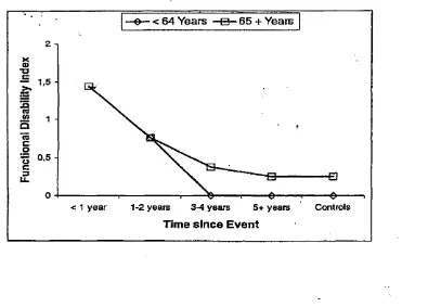
Figure 6: Level of disability after a fracture grouped by age 73
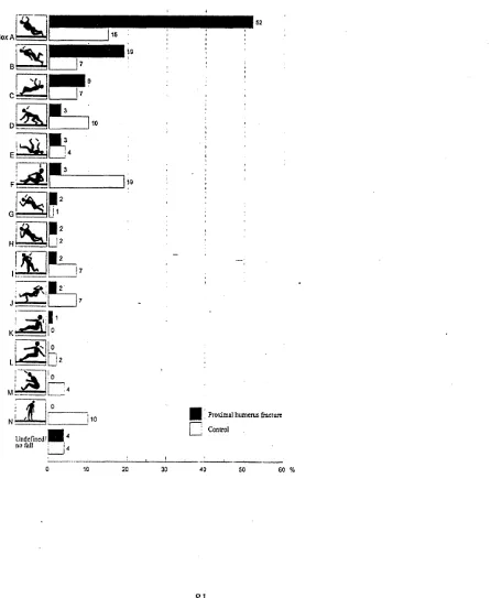
Figure 7: Fall Direction with a Proximal Humerus fracture 81
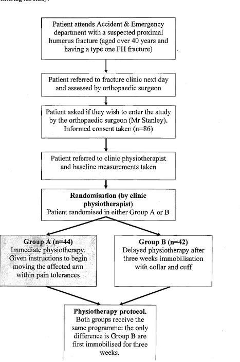
Figure 8: Flow-chart (admission to entering study) 143
Figure 9: Patient Flow-chart at follow-up 179
Figure 10: Treatment sessions by group allocation 181
Figure 11: Constant shoulder score at 8 to 52 weeks 183
Figure 12: Regression Modelling Analysis CSS 185
Figure 13: Regression Analysis Modelling SF-36 (RLP) 191
Figure 14: Regression Analysis Modelling SF-36 (PF) 192
Figure 15: Regression Analysis Modelling SF-36 (P) 192
Figure 16: CSDQ scores for each question at two year follow-up 196
Figure 17: SF-36 Score (Pain) by gender 200
Figure 18: SF-36 Score (Physical Function) by gender 200
Figure 19: SF-36 Score (Pain) by level of deprivation. 201
Tables
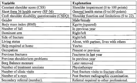
Table 1: Outcome variables 155
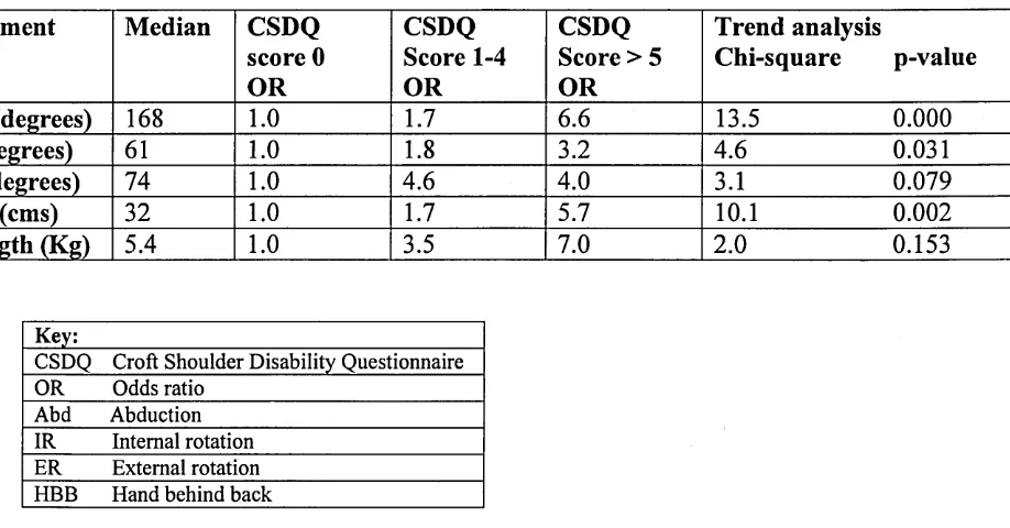
Table 2: CSDQ validation data 166
Table 3: Response rate 174
Table 4: Subjects’ baseline characteristics 176
Table 5: Complications reported over three years of the study 182
Table 6: Constant Shoulder Score difference (52 weeks) 184
Table 7: Constant shoulder score results up to one year 186
Table 8: Short form (SF-36) health survey 189
Table 9: Follow-up at one & two years 193
Table 10: Patient characteristics;at two years 194
Table 11: Shoulder disability (CSDQ) by group allocation 195
Table 12: Model output (binary logistic modelling) 197
V. Abbreviations
ADL Activities of Daily Living
AO Arbeitsgemeinschaft fur Osteosynthesefragen BMC Bone Mineral Content
BMI Body Mass Index
BMD Bone Mineral Density
P Bodily pain (SF-36)
CSDQ Croft Shoulder Disability Questionnaire CSS Constant Shoulder Score
CT Computerised Tomography
CVA Cardiovascular Accident DoH Department of Health
EQ EuroQoL
GP General Practitioner/ General Practice ICC Intraclass correlation coefficient LLI Long term limiting illness
M Million
MS Musculoskeletal
N Newtons (force)
NHS National Health System
OA Osteoarthritis
PF Physical function (SF-36)
PH Proximal Humerus
POP Plaster of Paris
RCT Randomised Controlled Trial RLP Role limitation-physical (SF-36)
ROM Range of Movement
RR Relative Risk
SES Socioeconomic status
SD Standard Deviation
SDQ-NL Dutch Shoulder Disability Questionnaire SF-36 Short Form 36 Health Survey
SPADI Shoulder Pain and Disability Index
STHFT Sheffield Teaching Hospitals Foundation Trust SRQ Shoulder Rating Questionnaire
UK United Kingdom
VI. Abstract
Background
The western world faces an explosion in the number of patients who will fracture their
proximal humerus (PH) as a result of the rapidly changing demographics and the
increase in osteoporosis. In 1998 there were 110 000 PH fractures in the United
Kingdom (UK) and epidemiological studies indicate that the PH fracture incidence is
increasing. Scant evidence exists to the optimum management and rehabilitation of
these fractures and the aims of the study were to investigate the effect of an
accelerated rehabilitation programme on patients’ recovery.
Method
A Randomised Controlled Trial (RCT) comparing two rehabilitation programmes
(n=86) with patients who sustained two-part fractures of the proximal humerus was
performed. Patients were randomised to receive immediate physiotherapy within one
week (Group A) or delayed physiotherapy (Group B) after 3 weeks immobilisation.
Assessment was at 8, 16 and 52 weeks with the Constant Shoulder Score (CSS), Short
form generic health survey (SF-36) and Croft Shoulder Disability Questionnaire
(CSDQ). Additional reassessment was undertaken at two years. Regression analysis
modelling was conducted to identify the risk factors for developed long-term shoulder
disability.
Results
At the primary outcome point (16 weeks) Group A experienced less pain (p<0.01) and
had greater shoulder function (pO.OOl) compared to Group B. At 52 weeks the
shoulder function (mean difference in AUC 6.4 [95% Cl: 2.5 to 10.5], p< 0.002). At
one year, shoulder disability (CSDQ) was 42.8% in Group A and 72.5% in Group B
(p<0.01). By two years, shoulder disability in Group A remained unchanged (43.2%),
but had reduced in Group B (59.5%).
Discussion
Immediate physiotherapy following a proximal humerus fracture results in faster
recovery with maximal functional benefit being achieved at one year and requires
fewer treatment sessions (9 versus 14 treatments, Group-A and B respectively).
Delayed rehabilitation by three weeks shoulder immobilisation produces a slower
recovery. The belief that patients make an excellent recovery after one year is
questionable as 25 patients (33.5%) still reported considerable shoulder disability after
two years of their injury. Gender (female), age and high levels of social deprivation
were identified as risk factors for continued shoulder problems at two years after the
fracture.
Conclusions and recommendations
This work suggests that patients who fracture their PH should not be immobilised
before referral for physiotherapy as immediate referral to physiotherapy (within 1
week) results in faster recovery and less reported pain. Physiotherapy should be
targeted towards those patients who are identified as having a greater risk of
developing long-standing problems. Currently, a wide variation in PH fracture
management exists in UK hospitals and implementing clinical care pathways will help
Contents
Declaration I
Acknowledgements II
Dedication III
List of Figures and tables IV
Abbreviations V
Abstract VI
Chapter 1: Introduction 17
1.1 An ageing population: implications for the proposed research 17
1.2 Justification for this Research Study 19
1.3 Originality in the Proposed Research 20
1.4 Immobilisation in Current Health Care 22
1.5 Historical Perspective 23
Chapter 2: Literature Review 25
2.1 Introduction to Literature Review 25
2.1.1 S earch Strategy 3 5
2.2 Epidemiology: Proximal Humerus Fractures 37
2.2.1 Introduction 37
2.2.3 Osteoporotic Fractures 37
2.2.4 Proximal Humerus Fractures 41
2.2.5 Prevalence of Shoulder Disability in the Population 46
2.2.6 Summary 48
2.3.1 Introduction 49
2.3.2 Gender & Age 50
2.3.3 Osteoporosis 52
2.3.4 Other Factors 54
2.3.5 Summary 56
2.4 Epidemiology: Factors Affecting Shoulder Function 58
2.4.1 Introduction 58
2.4.2 Shoulder Pain 61
2.4.3 Gender 64
2.4.4 Co-morbidity 65
2.4.5 Socioeconomic Factors 68
2.4.6 Summary 70
2.5. Aetiology 72
2.5.1 Introduction 72
2.5.2 Falls and Trauma 74
2.5.3 Risk Factors for Falling 76
2.5.4 Upper Limb Trauma 79
2.5.5 Summary 83
2.6 Proximal Humerus Fractures: Medical Management 85
2.6.1 Introduction 85
2.6.2 Proximal Humerus Fracture Classification 86
2.6.3 Fracture Management 91
2.6.4 Immobilisation and Bone Repair 94
2.7 Proximal Humerus Fracture Rehabilitation 104
2.7.1 Introduction 104
2.7.2 Rehabilitation 105
2.7.3 Shoulder Rehabilitation 111
2.7.4 Systematic Reviews 111
2.7.5 Electrotherapy 113
2.7.6 Exercise 114
2.7.7 Joint Mobilisation 117
2.7.8 Immobilisation 118
2.7.9 Complications and Accelerated Rehabilitation 121
2.7.10 Summary 126
2.8 Aims and Hypotheses 128
2.8.1 Study Aims 128
2.8.2 Hypotheses 128
Chapter 3: Methods
1313.1 Introduction 131
3.2 National UK Survey 131
3.3 Randomised Controlled Trial 135
3.4 Pilot Study 136
3.5 Study Design 138
3.5.1 Randomised Controlled Trial 138
3.5.2 Recruitment Procedure 139
3.5.3 Randomisation Process 144
3.6.1 Study Population 144
3.6.2 Inclusion & Exclusion Criteria 145
3.7 Assessment and Follow Up 146
3.7.1 Assessment 146
3.7.2 Follow-up 147
3.7.3 Summary 149
3.8 Physiotherapy Protocol 149
3.8.1 Early Phase 150
3.8.2 Intermediate Phase 151
3.8.3 Late Phase 152
3.8.4 Treatment Providers 153
3.9 Outcome Measures 154
3.9.1 Constant Shoulder Score 156
3.9.2 Short Form 36 General Health Questionnaire (SF-36) 160
3.9.3 Croft Shoulder Disability Questionnaire 165
3.9.4 Summary 168
3.10 Statistical Analysis 168
3.10.1 Power Calculation 168
3.10.2 Analysis 169
3.10.3 Regression Analysis 170
3.11 Reliability 171
Chapter 4: Results 174
4.1 Introduction 174
4.2 National UK Survey 174
4.3 Experimental 175
4.3.1. RCT Results (one year) 175
4.3.2 Attrition 177
4.3.3 Treatment 180
4.3.4 Complications Reported (over 36 months) 181
4.4 Constant Shoulder Score Results 183
4.4.1 Regression Analysis (Constant Shoulder Score) 184
4.5 SF-36 Results 187
4.5.1 Skewness 187
4.5.2 Regression Analysis Modelling (SF-36) 188
4.6 RCT Long-Term Results (Two Years) 193
4.6.1 Response rate ' 193
4.6.2 Shoulder Disability 194
4.7 Binary Logistic Modelling 196
4.8. Repeat Regression Modelling 198
Chapter 5: Discussion 203
5.2 Parti: UK Survey 205
5.2.1 Summary 208
5.3 Part 2: Randomised Controlled Trial (HI & H2) 209
5.3.1 Introduction 209
5.3.2 Baseline Characteristics 209
5.3.3 Shoulder Function after immediate and delayed physiotherapy (HI) 213
5.3.4 Physiotherapy 216
5.3.5 Immobilisation 218
5.3.6 General Health Questionnaire at up to Year One (SF-36) 220
5.3.7 Summary 223
5.4 Part 3: Long-term Disability 224
5.4.1 Introduction 224
5.4.2 Shoulder Disability 225
5.4.3 Summary 232
5.5 PART 4: Risk Factors for developing shoulder disability 233
5.5.1 Introduction 233
5.5.2 Regression Modelling 233
5.5.3 Gender 234
5.5.4 Socioeconomic Factors 236
5.5.5 Conclusion 238
5.6 Limitations of Study 239
5.6.1 Study Design 239
5.6.2 Outcome Measures 239
Chapter 6: Recommendations & Conclusion 243
6.1 Recommendations 243
6.1.1 Introduction 243
6.1.2 Medical Management 244
6.1.3 Rehabilitation 248
6.1.4 Summary 251
6.2 Conclusion 252
References 258
CHAPTER
1: INTRODUCTION
Please note: Within the text any reference to the ‘author’s research’, ‘author’s work’ or ‘proposed research’, refers to the research undertaken as part of the PhD thesis by myself, Stephen Hodgson.
1.1 An ageing population: implications for the proposed research
Society will experience an explosion in the elderly population in the future and this
will have considerable implications for many aspects of health and social care. The
increases are not evenly distributed across each age group, but are concentrated in the
older categories and are further inflated by the decline in younger age groups. In 2002
there were 19.8 million people aged 50 years and over in the UK, an increase of 24 %
from 1961 (16 million), but by 2031 the population projection is 27 million (Statistics
2005). This represents a 37% increase in people aged 50 and over in only three
decades and this trend shows no sign of diminishing.
There are many implications of an ageing population, but the rise in osteoporosis and
the related increase in fractures caused as a direct result of this bone loss, causes the
greatest concern. The so called ‘osteoporotic fractures’ (e.g. hip, proximal humerus,
wrist) are placing an ever increasing burden on health and social services’ budgets as
they struggle to balance both the requirements of managing an acute injury and the
long-term impact of disability to the person and their carers. Proximal humerus (PH)
fractures are the third most common osteoporotic fracture after the wrist and hip
(Melton III 1988). Furthermore, as well as the obvious implications to the person who
fractures their PH, the rise in number of PH fractures places increasing pressures on
carers and this does not always diminish with age. As a recent population census
than 50 hours per week as unpaid carers. The size of the problem in the UK (and
other western countries) is incalculable. This thesis addresses the long-term problems
of PH fractures by investigating how they are managed and rehabilitated. The
findings from the proposed research will help to reduce the burden faced by the
person who fractures their PH, their carer and a society that must deal with the
consequences of this increasingly common injury by improving the effectiveness of
management and treatment.
The research covers PH fractures, but recent research suggests that ‘osteoporotic
fractures’ share many similar characteristics and should be viewed as part of a
continuum. For example, sustaining a wrist fracture increases the risk of having a PH
fracture (Lauritzen et al. 1993) in the future and this subsequently increases a person’s
chance of having a hip fracture. ‘Osteoporotic fractures’ must not be viewed in
isolation as a single episode, but as part of a larger picture that requires early
intervention to prevent further injury. However, this approach is not adopted by the
current trauma services that deal with the acute injury and rarely orders further
investigations or starts preventative programmes. The author examines the links
between these fractures as the findings from this research have implications for many
other types of fractures. If patients are developing long-term disability from a PH
fracture, this will increase their future risk of other ‘osteoporotic fractures’ as they are
more likely to fall if their neuromuscular function (ability to balance and compensate
in response to changes in centre of gravity) is compromised (Kelsey et al. 1992). This
research has established an optimum rehabilitation programme which will potentially
Conventional rehabilitation to date has been based along traditional lines and practice
varies between and within hospitals, compounded by the poor evidence base from
which to inform clinicians (Gibson et al. 2001). The new approach to fracture
management presented in this thesis represents another original aspect to the thesis.
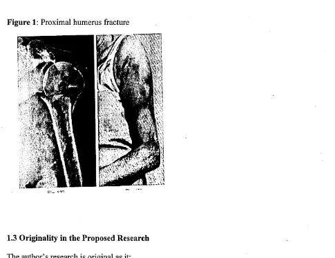
PH fractures (fig. 1), as seen in the literature review, are increasing exponentially and
this trend shows no sign of declining (Buhr and Cooke 1959; Kannus et al. 1996;
Kannus et al. 2000). Additionally, the number of falls that result in an injury are
increasing and the greatest changes are in the older adult category (Kannus et al.
1999) with fractures forming the largest group of fall-induced injuries. However,
research in the field of PH fractures is generally of a poor standard and in several key
areas is almost absent. In any thesis it is important to highlight the ‘gaps’ in the
evidence base and this research addresses fundamental issues around PH fractures. To
allow discussion of the problems facing the clinical management of PH fractures, this
thesis incorporates evidence from other areas of fracture management and shoulder
rehabilitation to develop ideas and to recommend future research.
1.2 Justification for this Research Study
The desire to conduct research within this field stems from personal observations
when working as a physiotherapist in a fracture clinic. The traditional approach to
modem PH fracture management and rehabilitation, combined with the lack of good
quality evidence, limited the efficacy of clinical practice and this resulted in patients
receiving unacceptable treatment. The patriarchal stmcture of orthopaedics in which
innovation is stifled and barriers are constantly erected that prevents change has
This research challenges some entrenched views about PH management and proposes
a radical new approach to rehabilitation. Thus, potentially limiting some of the long
term problems facing the person who fractures their PH and the burden faced by their
[image:23.615.55.540.161.535.2]carers.
Figure 1: Proximal humerus fracture
1.3 Originality in the Proposed Research
The author’s research is original as it:
• Investigates the effects of immediate rehabilitation before a period of
immobilisation (current practice immobilises before rehabilitation).
• Includes an active rehabilitation programme based on best available evidence
• Incorporates long-term, prospective evaluation (two year) of patients’ status
following a PH fracture to investigate, for the first time, the long-term impact
of a PH fracture on a vulnerable population.
• Evaluates shoulder disability and general health status in combination with
impairment measures (e.g. ROM, strength, pain) to fully assess the wider
health implications of a PH fracture. Previous research in this area has relied
mainly on impairment measures to judge outcome, thus missing crucial
information from which to base conclusions and recommendations.
• Recognises the importance of patient risk factors in predicting long-term
recovery and challenging current practice (characterised by over-reliance on
radiographic appearance).
• Compares socioeconomic status to patient outcome to establish if a link exists
between general health status and PH fracture recovery. Research in other
‘osteoporotic fractures’ confirms the influence on general health status after
fracture, but these wider issues have not been addressed in previous research.
• Challenges perceived wisdom that states that patients make excellent recovery
• Includes data from the first national survey in the UK establishing current PH
fractures management from which to base future recommendations and
guidelines.
Each point is covered in greater detail within the relevant section, but the research is
innovative in that it is the first to challenge the belief that fractures require a period of
immobilisation before rehabilitation starts. The proposed rehabilitation programme is
a radical departure from current thinking and will challenge many to consider their
practice. Rehabilitation in many upper limb conditions, and especially PH fractures,
has virtually no reliable evidence on which to base clinical decisions.
Many surgeons believe that PH fractures require immobilisation for healing to occur
and early movement results in non-union (fractures failing to heal) or at the very least,
exacerbation of pain. The research evidence from the few studies that exist does not
support this view and the findings from the proposed study will help clarify whether
immobilisation is necessary for bone healing and if early movement leads to non
union (failure of the normal fracture repair process).
1.4 Immobilisation in Current Health Care
Following an extensive search, no previous research has been found that has
investigated the effects of immediate rehabilitation on patient function following a PH
fracture. Two key issues arise from the extensive literature review. First, little
evidence exists to support current clinical practice and second, bone (as with all forms
(Carter 1984; Buckwalter 1996). This concept is reinforced in other areas of
healthcare, for example, after cardiac surgery the myocardium continues to heal
without immobilisation or following a hernia repair the patient often walks out of the
operating theatre, stressing the incision, but with little ill effect and minimising post-
surgical complications. Increasingly, more operations are being undertaken as day
cases with people walking out of operating theatres and encouraged to resume their
normal function. This tendency to avoid prolonged hospitalisation could be explained
by financial restraints and the increase in hospital infections, but it is also a response
to the benefits of early return of function. Damaged structures require controlled
stress from movement to maximise recovery and the seminal work linking ‘form and
function’ was made by Woolf as far back as 1892 (Woolf 1892). The quantity of
evidence supporting this concept is so overwhelming that it is inconceivable why
current fracture management remains so intransigent to change, although most of this
research is on animals or lower limb fractures.
1.5 Historical Perspective
The dichotomy between the ‘movers’ and ‘resters’ in the history of orthopaedic
surgery remains an ongoing controversy (Salter 1982). The ‘father’ of British
orthopaedics, Hugh Owen Thomas, was a strong advocate of rest and stated that:
"... rest or immobilisation must be complete, prolonged and uninterrupted."
This assertion from the latter part of the nineteenth century continues to resonate
today and immobilisation remains largely unchallenged in fracture management. For
example, modem PH fracture management immobilises the patient for three weeks
before starting rehabilitation, although as can be seen from the only UK survey
conducted as part of this research (see results section for further detail) it is not
uncommon to immobilise patients for up to eight weeks. The wide variation in the
period of immobilisation reinforces the belief that surgeons are not basing their
decisions on firm evidence, but continue along a time-honoured approach.
The aim of the author’s research was to question the basis for this practice and test the
efficacy of immobilisation versus immediate rehabilitation; allowing surgeons and
physiotherapists to make informed decisions and about PH fracture management and
rehabilitation. This new approach was found to be effective and it will reduce long
term disability and improve the patient’s quality of life after a PH fracture.
The next part reviews the literature base underpinning the author’s research and
considers the size of the problem facing society with an increasing older population.
The associated rise in PH fractures is discussed and the current evidence base to both
management and rehabilitation is critically evaluated. From this the justification to
CHAPTER 2: LITERATURE REVIEW
2.1 Introduction to Literature Review
Before reviewing the literature relevant to this study, it is important to define two key
terms in the study: the orthopaedic management of the PH fractures (proximal
humerus) and the subsequent rehabilitation of the patient following the fracture. PH
fracture management is undertaken by an orthopaedic surgeon and includes decisions
on the type of approach, for example ‘conservative’ or surgical. Additionally, the
surgeon will monitor progress and recognise possible complications resulting from the
fracture (mal-union, non-union, nerve/vascular damage). This study does not include
patients undergoing surgery and only includes those managed by conservative means.
Rehabilitation is defined as:
“ ...restoration (to the maximum degree possible) either o f function (physical or
mental) or o f role (within the family, social network or workforce)."
Nocon & Baldwin 1998 (Nocon and Baldwin 1998)
Rehabilitation is usually undertaken by the physiotherapist, but the decision when this
process starts is made by the referring surgeon. The physiotherapist’s role is to return
the patient to their previous functional state, or as close as reasonably possible using
education, passive movement and a graded home exercise programme. Both the
This study investigates both the management and rehabilitation of the PH fracture and
the results have far ranging implications for both these aspects of care, but the
findings also have relevance to all fracture treatment.
A PH fracture is not always the result of a chance event, but is often the final
consequence of a series of risk factors (risk factors are specific patient characteristics
that individually or collectively increase a person’s chance of having a fracture e.g.
gender, age, activity level). The probability of long-term functional loss is the
interaction between risk and mediating factors. Mediating factors prevent the person
with specific risk factors developing long-term shoulder disability e.g. two people
with the same risk factors but with different levels of social deprivation; the one with
the higher level of deprivation is more likely to develop shoulder disability. The
reporting of musculoskeletal problems and illness is higher in areas with high levels
of socioeconomic deprivation (Davies et al. 1994; Saul 1995; Paul et al. 1998). No
previous research has investigated the effects of socioeconomic status (SES) on
shoulder function, but evidence for joint pain, low back pain (Walsh et al. 1992;
Urwin et al. 1998), neck pain (Webb et al. 2003)knee pain (Webb et al. 2004) and
disability levels (McEntegart et al. 1997; Melzer et al. 2000; Bajekal 2005; Rautio et
al. 2005) has consistently demonstrated that people living in areas of high social
deprivation have more problems compared to those living in areas of low deprivation.
The factors linking pain to social deprivation remain to be elucidated (Aggarwal et al’
2003), but possible explanations are divided between behavioural and materialistic
deprivation is associated with activities that compromise health. For example,
smoking is associated with higher rates of low back and shoulder pain (Adamson et al.
2006) and the authors suggest that habitual flexed postures and repetitive arm
movements could explain this observation. Additionally, less social contact and low
levels of physical activity (Stuck et al. 1999) are risk factors in predicting functional
decline and both these factors are seen in areas of high deprivation (Guralnik and
Kaplan 1989; Verbrugge 1989; Vita et al. 1998).
There is increasing evidence that smoking has an deleterious effect on bone healing in
various fractures including the scaphoid (Little et al. 2006), calcaneus (Folk et al.
1999) and tibia (Schmitz et al. 1999; Harvey et al. 2002; Dahl and Toksvig-Larsen
2004; Castillo et al. 2005). These studies measured the time for clinical bone union to
occur between groups of smokers and non-smokers and the increased risk of delayed
union for smokers varied between a relative risk (RR) of 1.2 (Folk et al. 1999) and
2.5 (Dahl and Toksvig-Larsen 2004). Furthermore, a study by Glassman et al.
(Glassman et al. 2000) who reviewed patients following a spinal fusion (n=357),
found that non-union rates were 14.2% and 26.5% for non-smokers and smokers
respectively. The researchers also identified a group of patients who quit smoking
following the surgery and their risk of developing non-union reduced to a value
similar to that of the non-smokers (17.1%). This led the author to suggest that
smoking cessation helps reverse the impact of smoking and implies that its effects are
transitory. However, the effects of cigarettes are hard to isolate in clinical studies as
possible co-founders exist that might also influence bone healing. Nevertheless,
with rabbits who were given intermittent smoking for six weeks compared with
controls (Lee et al. 2005). Bone lengthening in rabbits (Ueng et al. 1997) grouped into
smoking (n=19) or non-smoking (n=19) demonstrated lower levels of maximal torque
(22%) across the fracture site at eight weeks in the smoke inhalation group.
Although, animal studies must be viewed with some caution, short periods of smoking
do appear to delay bone healing. Many smokers who sustain a fracture have probably
had the habit for many years and this would probably further delay fracture healing.
Factors that possibly explain the differences between levels of social deprivation in
the materialistic category include education (Leveille et al. 1992) and income (Jette
and Branch 1985; Leveille et al. 1992; Maddox and Clark 1992). Lower educational
attainment is associated with higher rates of functional decline (Maddox and Clark
1992; Maddox et al. 1994) and there is convincing evidence that people living in areas
of high deprivation do less well in healthcare systems (Feinstein 1993). An example
of this is the work by Criswell and Katz (Criswell and Katz 1994) who reported that
patients with Rheumatoid arthritis and who had high educational attainment received
better healthcare than patients with lower education. The reason given for this
difference was that higher education allowed the person to negotiate for treatment.
Furthermore, low income (associated with low educational attainment) predicts the
decline in physical function (Leveille et al. 1992; Koster et al. 2005) and physical
disability (Jette and Branch 1985). Low income might limit access to healthcare and
The association between certain psychological profiles (e.g. high deprivation causes
increased stress on the individual) and high levels of social deprivation have also been
given as reasons for increased levels of morbidity (Urwin et al. 1998). However,
when this hypothesis is tested, the results are unclear with some authors reporting that
psychological factors did predict functional decline (Kempton et al. 1999;
Martikainen et al. 1999), and others finding only weak links between psychological
factors and functional decline.
Overall, an explanation for the links between social deprivation and morbidity remain
equivocal and further research is needed to test the complex relationship between the
many different factors. The work has started in the area of functional decline and
disability, but little work exists in the field of specific musculoskeletal conditions.
For this reason PH fracture risk factors, functional loss and mediating factors are
included in the literature review to allow the author to explore those characteristics
that clinicians managing PH fractures must consider. Additionally, this allows
exploration of potential problems for recovery and ongoing disability.
With any review of the literature, it is important to highlight the ‘gaps’ in current
knowledge and how this research adds to the body of evidence. However, in the field
of management of proximal humerus fractures what little evidence that exists is weak
and making informed decisions based on current research is impossible. The
Cochrane review of PH fracture management (Gibson et al. 2001) came to similar
conclusions and stated that only tentative recommendations could be made with the
less well researched as most programmes are based on empirical evidence alone. So,
for both management of PH fractures and rehabilitation, the evidence is not available
or often of a low standard.
This research aims to investigate both management and rehabilitation approaches and
proposes a new approach to fracture management that is previously untested and
could potentially revolutionise modem fracture management. Furthermore, current
rehabilitation is reviewed to formulate the best programme for PH fractures, but with
the lack of evidence base within this field, other shoulder conditions are included to
justify the programme. The research is original in two key aspects: first, initially
investigating the effects of immobilisation on recovery by comparing three weeks
immobilisation with immediate restoration of function is something that previous
research has ignored. Secondly, by recognising the limitations of basing fracture
management solely on radiographic appearance and incorporating wider patient
characteristics and socioeconomic variables in formulating a truer representation of an
individual’s risk of long-term shoulder disability. Both these approaches are new and,
as such, the literature review is far ranging in its scope to reflect these aims.
The literature review covers four main areas: epidemiology, aetiology, medical
management and finally, rehabilitation. Overall, epidemiology has the strongest
evidence base and this clearly delineates the current problems in fracture management
with an exponential growth in PH fractures. The epidemiology highlights the
problems facing health providers as they attempt to manage this common injury with
changes only serve to highlight its importance as management and rehabilitation of
these fractures must be optimised to cope with the predicted future demands. Without
the development of ‘clinical pathways’ (as routinely seen in stroke and falls
management) to take the patient from injury to functional independence many patients
may receive inadequate treatment and develop long-term problems, thus impacting on
their quality of life and placing an additional, burden on health and social services.
This study helps provide the evidence base on which to form these clinical decisions.
The inclusion of risk factors in developing shoulder pain and dysfunction within the
epidemiology section is paramount to the later exploration of the results that highlight
the factors that mediate long-term shoulder disability. These seemingly disparate
factors are increasingly acknowledged among practitioners as key characteristics in
the development of many major health problems, ranging from cancer and
cardiovascular disease to arthritis. Likewise, recovery from a shoulder fracture is
influenced by these factors and must be considered when planning treatment and is
therefore included in the review. Current practice makes little or no consideration of
these factors and including them will challenge the conventional management of both
PH fractures and other fractures in an older population.
Most PH fractures in an older population are as a consequence of a fall, but only in
approximately 5% of falls does a fracture occur (Oakley et al. 1996). Falling is
relatively common, but the relationship between the force of the fall and injury
mechanism is complex and recent research suggests that the type of the upper limb
(Palvenen et al. 2000). For example, falls resulting in a PH fracture are usually
sideways and the person fails to extend their arm, whereas the fall resulting in a wrist
fracture is usually forward and the arm is broken as the patient’s arm is extended.
Identifying fall patterns that result in a PH fracture will allow incorporation of
preventative measures in rehabilitation programmes for older adults.
Mechanism of injury resulting in a PH fracture is discussed in Aetiology (page 72)
and is important to consider for a number of reasons: a component of fracture
rehabilitation is prevention of further injuries and knowledge of injury mechanics
allows the tailoring of exercise programmes to diminish the harmful effects of the fall,
thus limiting future fractures.
Medical management of PH fractures is based mainly on radiographic appearance
with little, or no, importance given to the patient or their personal circumstances. For
example, their level of physical activity or previous shoulder pain before the fracture
could influence their rate of recovery and management should consider these factors.
Many PH fractures are complex with considerable fragmentation of bone and
displacement. In an attempt to help standardise radiographic interpretation the Neer
classification scheme (Neer 1970; Neer 1970) was developed to classify each fracture
into certain groups (the largest group, and the one that is included in this research, is
the ‘minimally displaced’ or type one fracture) and it measures displacement and
angulation of each fracture segment. Interpretation of a three-dimensional fracture
from a two-dimensional image is fraught with difficulties and research has repeatedly
that is unreliable, and even if it were reliable, would fail to consider other important
factors in deciding on management. The system is reviewed as it allows comparisons
to be made in the discussion between current practice and the author’s results,
therefore allowing recommendations to be made regarding future management.
As previously stated, management is based on a radiographic system with
questionable reliability, but current clinical practice remains unswerving in its
commitment to this system. In the case of ‘minimally displaced fractures’,
rehabilitation commences after a variable period of immobilisation and challenging
the necessity for immobilisation is a central theme in this thesis. The review
considers evidence that casts doubt on the necessity to immobilise fractures with both
laboratory and clinical research indicating that tissue responds to movement and
immobilisation only produces secondary damage.
This research aims to establish if PH fracture healing requires a period of
immobilisation for optimum repair. The first task is to review evidence for
immobilisation in fracture healing. This is widened to include lower as well as upper
limb fractures and the results are extrapolated to PH fractures. The case for
immobilisation is discussed and balanced against current evidence and, from this key
research, questions are based.
Following the review, the main problems surrounding the medical management of PH
fractures are identified and this links to the study’s proposed aims. The main goal of
results in better shoulder function and enhanced general health gains. The literature
review first considers existing management before comparing it with this new
approach.
There is a growing evidence base for the physiotherapy treatment efficacy in several
shoulder conditions, for example shoulder instability (laxity of capsule and supporting
ligaments/muscles) and impingement (catching of tendons against the coracoacromial
arch). Current evidence would support an active approach to rehabilitation using
education, exercise programmes and an accelerated return to function, therefore,
avoiding the use of electrotherapy modalities and other passive interventions that
demonstrate poor clinical value.
These differing approaches to rehabilitation are reviewed to justify the programme
used in the author’s research and to highlight trends in rehabilitation that this work
builds on. The research is generally of a low standard with weaknesses in sample
size, flawed designs and unreliable outcome measures and is compounded by the
retrospective nature of most fracture research (variable review dates and unreliable
outcome measures). This contributes to the uncertainty around fracture rehabilitation,
but does allow the author to highlight the necessity for this and future research. This
research also challenges modem fracture management and rehabilitation, something
that has remained unchanged for decades, and proposes a novel approach to PH
rehabilitation. Any new system that questions conventional practice is certain to face
opposition from certain quarters. This research is no different and it raises ethical
fracture to heal or to develop a mal alignment). The importance that some surgeons
attribute to rest in fracture repair is strong and concerns that early movement across a
fracture site results in more pain and an increase in fracture complications is not
uncommon. The review discusses the evidence base of these concerns and questions
their credibility by comparing the results in other areas of fracture repair that have
avoided immobilisation. Furthermore, as part of the ethical considerations for the
study it was important to justify early movement and give a reasoned argument for its
inclusion to present to the ethics committee.
In summary, robust epidemiological evidence points to an increasing problem facing
health care providers coping with an explosion in PH fracture rates. The evidence
base to management and rehabilitation of these fractures is weak and treatment is
mainly based along traditional lines. Evaluation is incomplete with no measure of
disability or consideration of generic health status. This review considers these key
aspects to justify the proposed research and to highlight why this research is original
in several fundamental areas. Immediate rehabilitation, avoidance of immobilisation,
active patient-centred rehabilitation and realistic outcome measures all challenge
current practice and represent a new approach in the field of fracture treatment.
2.1.1 Search Strategy
Electronic searches were performed in the following databases:
a. MEDLINE (Silverplatter) 1980-2004
c. CINAHL 1980-2004
d. The Cochrane Library (http://www.update-soflware.com) 2004 Issue 1
e. National Register of Research Trials 2003
f. PeDRO database Physiotherapy database based at Sydney University
http://ptwww.cchs.usyd.edu.au/pedro/ (October 2004)
g. Other searches: References from key papers were checked for new research
areas and key authors were identified. Additionally, colleagues working in
specialist areas were contacted for their views on current research areas and
authors were suggested.
Search terms included the following:
HUMERUS: injuries/ fractures/ epidemiology/ physiotherapy/ physical therapy/
rehabilitation/ orthopaedic/ aetiology
PROXIMAL HUMERUS: Injuries/ fractures/ epidemiology/ physiotherapy/ physical
therapy/ rehabilitation/ orthopaedic/ aetiology
SHOULDER FRACTURES: Injuries/ fractures/ epidemiology/ physiotherapy/
rehabilitation/ orthopaedic/ aetiology/ upper limb/ immobilisation.
HIP FRACTURE: epidemiology/ immobilisation/ co-morbidity
WRIST FRACTURE: epidemiology/ immobilisation/ co-morbidity
2.2: Epidemiology: Proximal Humerus Fractures
2.2.1 Introduction
In order to establish the extent of the problem facing society, the author first needs to
consider the epidemiology of PH fractures and their close association with the rise of
osteoporosis. Quantifying the number of patients who fracture their proximal
humerus each year within the United Kingdom is difficult for several reasons: the
problems of fracture classification, case ascertainment, (providing a specific reference
code that accurately identifies each condition) and incomplete records ensure that any
data must be viewed with some caution. This is further complicated when trying to
predict future levels of shoulder fractures when secular trends and an increasingly
ageing population are put into the equation. Initially, the Epidemiology section will
cover those trials conducted within the last 30 years in the UK in which a specified
population was sampled. The inclusion of International studies helps to highlight
some of the deficiencies of the UK data and allows a full exploration of the
demographic variables.
Increasingly, it is recognised that people who fracture, or have an increased risk of
fracturing, their proximal humerus (PH) share certain characteristics that identify
them from other types of osteoporotic fractures, for example, wrist fracture. Patients
who fracture their wrist or PH are often osteoporotic, but the fall mechanism that
results in each fracture is different. The fall resulting in a wrist fracture is usually
forwards (Palvenen et al. 2000) and the patient attempts to break the fall by using the
sideways and the force of the fall is directly onto the shoulder; thus resulting in a
fracture. The patient is unable to ‘break’ the fall with their arm and this inability to
save themselves is an indication of their poor neuromuscular control mechanism
(Kelsey et al. 1992). Identification of these specific risk factors is fundamental in
planning prevention strategies and is important to the author’s work as these factors
are investigated in the later analyses. Furthermore, identifying the risk factors that
predict those patients that will continue to have long-term problems is a departure
from present fracture management that fails to consider why some patients make
excellent recovery whilst others continue to experience ongoing problems.
As Baron stated:
" ...individual fractures have distinct epidemiological patterns, there may he discreet
etiologic factors that require separate preventative efforts."
Baron etal. 1996 (Baron etal. 1996)
Finally, the prevalence of shoulder dysfunction in a population not actively seeking
medical interventions is considered as this has implications for the proposed research.
The natural history of many shoulder problems is unknown and this is important
because rehabilitation would be unnecessary if all patients recovered spontaneously
2.2.3 Osteoporotic Fractures
In 1988, 1.3 million people sustained ‘osteoporotic fractures’ (Melton III 1988) in the
USA with estimated costs running to $18 billion. The cost to a UK hospital (1993/4)
for any closed upper limb fracture is estimated at £1200 (CHKS 1995) and for women
reaching the age of 50 years, 40 out of 100 will have one or more fractures (Lips
1997). Worryingly, these figures continue to increase exponentially. Osteoporotic
fractures (e.g. hip, PH, wrist, rib, spine) are characterised by increasing incidence with
[image:42.616.72.479.334.696.2]increasing age compared with non-osteoporotic fractures (fig. 2).
Figure 2: Fracture incidence by type of fracture in UK (Donaldson et al. 1990).
300n
(a) Femoral neck B ) Upper end o f humerus
a
200-0 - 200-0 Males » f t Females
100
-2 0- | 70-|
Shaft o f tibia and fibula Lower end o f radius and ulna
co
60-<Q 3
o.
8
o
8o
50-
40-a>
19
30-20
-8
10
--9
The database for hip fractures is the most reliable and figures are closely monitored as
small changes in fracture incidence have significant economic implications for health
providers. In an attempt to quantify worldwide projections for hip fractures, Gullberg
et al. (Gullberg et al. 1997) estimated that 1.26M hip fractures occurred in 1990, with
this rising to 2.6M by the year 2025 and an estimated 4.5M in 2050. Caution must be
taken with any estimate of future fracture rates as the unknown factors such as
demographic changes and secular trends can not be included in the final calculations.
Older people sustaining ‘osteoporotic fractures’ will continue to take an even greater
proportion of the health and social funds in the future, thus the proposed study has
clear fiscal implications for health providers faced with a finite budget.
In the last 40 years, five major epidemiological studies have investigated fracture
incidence in the United Kingdom. Consistently, their results show that the incidences
of fractures increase with age and more women sustain fractures than men. In the
largest, and probably the most reliable study (n=5M), Van Staa et al. (Van Staa et al.
2001) demonstrated that women aged 55 years and over were more than twice as
likely to sustain any fracture than men. Similar results were seen with Donaldson et
al. (Donaldson et al. 1990), however great variation exists between other studies and
absolute figures are inconsistent. Studies based in large cities (Singer et al. 1998)
(Johansen et al. 1997) give higher fracture incidence figures and this could be
explained by demographic variations and coding errors within the sample. Both these
studies used populations of just over 15 000 people, a relatively small population
show that osteoporotic fracture rates are rising and exponentially. Future projections
are high and the trend does not appear to be reversing, or even slowing down.
2.2.4 Proximal Humerus Fractures
No study has exclusively examined the rate of PH fractures in the UK and most
published studies have grouped all fractures together. The lack of clear and specific
data within this field reduces the accuracy of future projects and reflects the low
priority given to PH fracture research. Only one epidemiological study has presented
results that have included national data (Kannus et al. 2000) (based in Finland) and
many only provide extrapolations from smaller samples. These must be viewed with
some caution as it is known that PH fracture incidence varies with geographic and
demographic features (Karagas et al. 1996; Lauderdale et al. 1998; Ismail et al. 2002).
In 1959, Buhr and Cooke (Buhr and Cooke 1959) published the results of a five year
survey on the ‘common fractures’ occurring in Oxford, UK. They described the shape
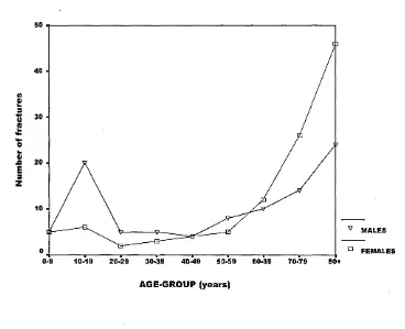
of the fracture incidence pattern (fig. 3) in the upper humerus as U-shaped’ (gradual
increase followed by a sudden rise in incidence) with its increasing gradient over 50
years of age. The male fracture pattern clearly shows a bimodal distribution (Melton
III 1988) with a higher male incidence in early years due to contact sport and road
traffic accidents (Donaldson et al. 1990) and then a rising in later years as a
consequence of falling. This apparent increase in the older population, Buhr and
Cook (Buhr and Cooke 1959) attributed to the ‘bones becoming thin and brittle’.
Many studies since have reported similar patterns, the only difference being the
Figure 3: Incidence of Proximal Humeral Fractures describing the T shaped
exponential growth in fracture incidence with increasing age (adapted from Burh &
Cook, 1959).
so
40
-10 0)
k3 *•0
1
•kO
l_
0)
41E 3
z
30 •
20 .
10 ■
MALES
FEMALES
0-9 10-19 20-29 30-39 40-49 50-59 60-69 70-79
AGE'GROUP (years)
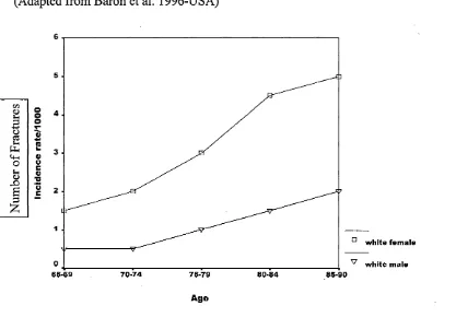
Absolute fracture rates vary as several factors (fig.4) are thought to influence fracture
incidence: gender (Baron et al. 1996b) geographic (Karagas et al. 1996; Ismail et al.
2002), seasonal (Lauritzen et al. 1993), demographic (Turner et al. 1998) and
ethnicity (Baron et al. 1994). Compounding these difficulties are variations in data
collection and small sample sizes, although in the only National survey (Kannus et al.
1996) conducted in Finland over a 23 year period, the results showed a 13% increase
in proximal humerus fractures each year between 1970 to 1993. Additionally, the
mean age of the fracture incidence also increased from 72.1 to 76.2 years over the 23
year period, thus reinforcing the idea that the consequences of PH fractures are mainly
Figure 4: Incidence of Proximal Humeral Fractures by Gender
(Adapted from Baron et al. 1996-USA)
6 5 4 3 2 1
white fem ale
0
65-69 70-74 75-79 80-84 85-90 white male
Age
VI
<DJ-H oo
a T”o o3Vi *1fl)n
Ph u
U-i 0)
O 0 S-H 0 *5 <U O c B 3 £
Bengner (Bengner et al. 1988) reported similar findings as Kannus and based their 30
year (1950-81) review of PH fractures around Malmo, Sweden. The same precautions
as those expressed in the Kannus study are applied to this study i.e. higher fracture
rates in Scandinavian countries, nevertheless their results clearly show an increase in
the PH fracture incidence from 1950 to 1980 (fig.5). In Tottori, Japan (Hagino et al.
1999), the fracture incidence rate increased over a period of nine years in a population
over the age of 35 years, however Asian populations have lower fracture rates (Ho et
al. 1989; Rowe et al. 1993).
The reason for the difference in fracture incidence between Asian and Caucasian
between genetic and environmental factors (Nguyen et al. 2004). No published work
exists that has specifically investigated PH fractures between different ethnic groups,
but hip fracture incidence is lower in African (Solomon 1979) and Asian (Xu et al.
1996; Yan et al. 1999) populations compared with Caucasians. The explanation for
the difference in fracture incidence is not simply related to BMD as studies have
demonstrated that Asian populations have lower or equal bone density compared with
Caucasians. Yan and colleagues (Yan et al. 2004) compared the BMD (upper femoral
shaft) of Chinese and Caucasian populations and the Chinese had lower bone density,
but still had lower incidence of hip fractures. The authors noted that the Chinese
population were significantly shorter and lighter than their Caucasian controls and
suggested that better neuromuscular function was in some way protective in the
Chinese subjects. To highlight the importance of neuromuscular status in fracture
prevention, work by Suriwongpaisal et al. (Suriwongpaisal et al. 2001) demonstrated
that higher levels of activity were associated with a reduced risk of hip fracture in a
Thai population. Interestingly, the incidence of hip fracture is not consistently lower
in all Asian countries when compared to Caucasian populations. Hong Kong and
Singapore have a similar incidence of hip fracture compared with Caucasians (Lau et
al. 2001), but Malaysia and Thailand are approximately 50% lower. This difference
between Asian countries is thought to reflect the increasing economic changes and
Figure 5: Age-specific annual incidence of Proximal Humerus fractures between
1950 and 1980 (From Bengner et al. 1988).
1950 1980
o
o V)
20
-1
80 20 60
age age
cn
d> O i Oo
ocd »h '35
P-l
O <D
Sh o <D au
6
3 ’oa1—1
Only one study has not shown an increase in PH fracture rates over time (Horak and
Nilson 1975). However, the results, as previously mentioned, are unreliable when
taken in isolation. The work of Kannus et al. (Kannus et al. 1996) and Hagino et al.
(Hagino et al. 1999) is probably a more accurate representation of the fracture rate as
they measured the same population over different time periods. If it is accepted and
the results are extrapolated to the UK, what are the implications? Proximal humerus
fractures constitute approximately 7.4% (Doherty et al. 2001) to 10% (Baron et al.
1996b) of all fractures. Population figures based on 1994 (Johansen et al. 1997)
estimated that 1.1 million fractures occurred in the UK. If 10% of these people
(Baron et al. 1996b) fractured their PH, this would represent an annual rate of 110 000
fractures. When considering risk of fracture, Barrett et al. (Barrett et al. 1999)
age of 90. Forty-two per cent (Doherty et al. 2001) of women aged 50 years or over
will have at least one fracture and of these, 50% will have multiple fractures.
As previously discussed, this has important implications for the author’s research as
PH fractures are increasingly seen as part of a continuum with people who fracture
their PH more likely to sustain a hip fracture in the future (Lauritzen et al. 1993).
Therefore, the proposed research, with its aim to determine the optimum rehabilitation
following a PH fracture, may prevent future fractures and, thus, directly influence
morbidity and long-term disability. With the recently published results of the 2001
population census (Statistics 2001) showing a five-fold increase in the population over
85 years since the 1950s, PH fracture rates (Baron et al. 1996) (Barrett et al. 1999) are
probably underestimating the impact of an ageing population.
2.2.5 Prevalence of Shoulder Disability in the Population
Several studies suggest that much of the population live with shoulder symptoms and
associated disability. The exact figures vary, but range from 21% (Chard et al. 1991)
to 34% (Chakravarty and Webley 1993). All studies sample people over the age of
50 and some evidence exists that the older population have higher levels of shoulder
dysfunction (Badley and Tennant 1992), but other studies refute this claim (Van der
Windt et al. 1995). However, Van der Windt et al. (1995) only included General
Practitioner (GP) patients and many elderly people who are symptomatic stop seeking
further medical help. Two recent studies by Badcock et al. (Badcock et al. 2002) and
Pope et al. (Pope et al. 1997) that surveyed a younger population (aged 18 to 75 years
et al. stresses the importance of defining case definition and its possible influence on
the final result.
What is not in doubt is that many people in the community have some level of
shoulder dysfunction, many of whom do not seek help. This has relevance to
planning research trials and long-term monitoring of shoulder problems. For example,
in North America a third of all acute injuries involve the upper limb (Kelsey et al.
1992) and many of these probably have pre-existing shoulder dysfunction. This pre
existing morbidity may probably influence their rate of recovery and possibly change
the disablement process.
This has important implications for PH fracture patients, especially if older adults
already have high levels of shoulder dysfunction before their fracture. Pre-existing
shoulder morbidity may influence recovery and eventual functional status. No
previous research has considered this fact as all assume, incorrectly, that the person
had good shoulder function pre-fracture and attribute any resulting dysfunction to the
injury. This complicates the interpretation of the results, but does give a realistic
representation of fracture recovery and is another reason why the proposed research
questions current thinking. Thus, pre-fracture shoulder status must be considered
when interpreting the outcome of interventions that aim to return patients to ‘normal’
function. Many people have reduced shoulder function before the fracture and this
may influence the recovery process. Thus, an important reason to record shoulder
2.2.6 Summary
The evidence suggests that the rate of osteoporotic fractures is increasing and the rate
of change appears to be accelerating. Humerus fractures, as a common osteoporotic
fracture, are also increasing, but the lack of large epidemiological studies makes
confirmation of these trends difficult. Osteoporotic fractures will continue to be a
major problem for a large section of the ageing population and limit their quality of
life (Wildner et al. 2002) whilst placing additional fiscal strain on the health and
social services (Torgerson and Dolan 2000). Thus, the proposed research is timely
and important in view of the problems facing society. The next part of the literature
review considers the risk factors, for example, gender, age, osteoporosis, neuro
muscular status, that predispose a certain person to having a PH fracture as this may
have significant implications to the findings of this study.
Following a PH fracture, it is often stated that full function is regained within one year
or less (Mills and Home 1985; Young and Wallace 1985) of the initial injury, but
serious methodological limitations and poor outcome measures of the research casts
doubt on this view. These studies reported ‘excellent’ or ‘very good’ results in terms
of shoulder function in over 90% of cases by 6 or 12 months, but reassessed the
patients with the Neer outcome measure (Neer 1970) which has not been validated
and no reliability data exists.
In fact, evidence suggests that patients have ongoing shoulder problems for many
years after an upper limb fracture (Wildner et al. 2002). Furthermore, an older
common shoulder problems many years after the initial presentation (Vecchio et al.
1995). Most orthopaedic research evaluates PH fractures after one year of the initial
injury and states that most make a full recovery (Clifford 1980; Mills and Home
1985; Kristiansen and Christensen 1987). Many might, but, especially in older adults,
patients never fully recover and they are forced to adapt to limited shoulder function.
Previous PH fracture research in this area has tended to use outcome measures that
have not undergone rigorous evaluation and this has possibly over estimated patient
recovery. This is the first study to evaluate patients using a battery of outcome
measures that will accurately assess recovery after a PH fracture.
2.
3 Epidemiology: Risk Factors for Proximal Humerus Fractures
2.3.1 Introduction
Risk factors are highly relevant to this research as they identify those patients who
will fracture their PH and this has important implications for rehabilitation an