Original Article
A simple method of isolation, identification, and
culturing retinal microglia
Wenmin Jiang1,2, Wenjie Li1,2, Xianghui Deng1,2, Luosheng Tang1,2, Tantai Zhao1,2
1Department of Ophthalmology, The Second Xiangya Hospital of Central South University, Changsha 410011,
Hunan, China; 2Hunan Clinical Research Center of Ophthalmic Disease, Changsha 410011, Hunan, China
Received January 19, 2017; Accepted October 12, 2018; Epub March 15, 2019; Published March 30, 2019
Abstract: The present study aimed to investigate a simple method for isolating, culturing, and identifying growth conditions and the culturing environment of retinal microglia. Eyeballs were obtained from newborn Sprague-Dawley rats (postnatal day 1). The retinas were then collected aseptically. Retinal vessels were carefully removed from the retinas. After digestion and centrifugation of the retinas, the obtained mixed cell suspensions were cultured for 12 days. The culturing flask was placed on a 37°C constant temperature oscillation shaker at 100 rpm for 1 hour. The supernatant was then collected, transferred to uncoated dishes, andincubated for 30 minutes. The dishes were gently shaken andthe supernatant was removed. After three times, oscillating separation was performed for purifi-cation. The cells were purified byoscillating separation and were identified by double immunofluorescence staining withmicroglia-specific marker CD11b and Isolectin-B4. This modified method was easily carried out.
Keywords: Retinal microglia, mixed culture, CD11b, IB4
Introduction
The neural retina mainly consists of ordered neurons and surrounding glial cells. Retinal glial cells are further divided into large glial cells (astrocytes or Müller cells) and microglia. In 1919, Del Rio-Hortega used a silver carbon-ate staining method to distinguish microglia from neurons and other glial cells. The microg-lia is the smallest group of gmicrog-lial cells, account-ing for about 10-20% of all glial cells. They are
specific in terms of structure, characteristics,
and cell lineage. They belong to a separate group of nervous system migratory macro-phages characterized by very active function [1]. Microglia are immune cells in the nervous retinal tissue which help to comprise the cen-tral nervous system (CNS) [2-4].
Because of the close relationship between the
retina and CNS, more and more attention has been paid to the potential roles of retinal microglia in the pathogenesis of multiple types of retinopathy [5]. However, separation and
purification of microglia has remained very dif
-ficult [2]. Therefore, establishment of a simple
in vitro purification and culturing system for pri
-mary retinal microglia is an important step in studying the function of these cells.
In 1932, Rio-Hortega first recognized that
microglia were a special group of cells in the CNS [6, 7]. It was found that when the brain was damaged, the microgliareactively migrated to the injured region, going through morphological changes and proliferation. Previously, it was considered that the retina is an immune-privi-leged organ. However, with more in-depth research, it has gradually become recognized that the microglia are, in fact, the only antigen-presenting cells in the retinaand are quite active. Therefore, application of cell culture technology to obtain high quality retinal microg-lia at a large quantity would be an important step for further research.
and microglia. Oscillating separation was per-formed after the formation of growing layers. This method resulted in obtaining microglia and oligodendrocytes with relatively high purity. Oligodendrocytes could then be removed according to differences in the ability and speed of oligodendrocytes and microglia to attach to the surfaces of culturing containers. The present study aimed to introduce a simple
method by refining the steps and improving specific steps.
Materials and methods
Materials
Premium fetal bovine serum and Dulbecco’s
modified Eagle medium/F12 were obtained
from Gibco. Trypsin, bovine insulin, glutamine,
and fluorescence secondary antibodies were
purchased from Sigma. Mouse anti-CD11b antibodies were obtained from Abcam. Mouse
IB4 antibodies were purchased from Sigma.
Penicillin and streptomycin were obtained from
North China Pharmaceutical Factory. Petri
dishes, centrifuge tubes, and 25-cm2 flasks were acquired from Corning-Costar. Newborn Sprague-Dawley (SD) rats were provided by the Animal Laboratory of the Second Xiangya Hospital of Central South University within 24 hours of birth.
Culturing methods
Mixed culture of glial cells: In a sterile environ-ment, eyeballs were obtained from newborn SD rats (within 24 hours of birth) and washed with cold D-Hanks solution (containing 1% tobramy -cin). The sclera of the eyeballs was cut open along the limbus and the retina was obtained aseptically, with the retinal vessels carefully stripped off. Retinal tissue was incubated and
digested in 0.125% trypsin at 37°C for 25 min -utes, then gently blown into a single cell sus-pension. The same amount of pre-cooled serum-contained medium was added into the reaction system to stop digestion. Undigested
tissue mass was then filtered out using a 200-mesh cell sieve. The filtrate was placed in a
sterile centrifuge tube and centrifuged at 1000 rpm for 5 minutes. The supernatant was dis-carded, then 10% fetal bovine serum was added, generating a cell suspension. The cell suspension was placed into a 25-cm2 culturing
flaskand incubated at 37°C in an incubator with
5% CO2. The next day, the medium was refreshed once with an equal volume to remove the dead cell debris. The medium was replaced every 4 days. The cells were observed for growth and survival over time under a microscope.
Isolation and purification of retinal microglia: After culturing for 12 days, the cells were observed to be growing in a complete layer. The
culturing flask was then placed on a 37°C con
-stant temperature oscillation shaker and shak -en at 100 rpm for 1 hour. The cells in the super-natant were collected, transferred to uncoated
dishes, and incubated in a 37°C incubator for
about 30 minutes. The dishes were gently
shakenand the supernatant was aspirated to
remove any non-attached cells and cells that were not tightly attached. The cells were then
seeded in a new flask. After the first oscillating
separation, fresh medium was added to the
flask with the original mixed cell culture to con -tinue cultivating the mixture for 6-8 days, when
the second oscillating separation would take
place. Cells were then cultured for another 5-6 days, after which the third oscillating
separa-tion was performed for purificasepara-tion (Schematic
1).
Immunocytochemical staining of retina microg-lia: Coverslips with the cell culture were washed
thoroughly with normal saline, fixed in 4% para
formaldehyde for 20 minutes, and washed with
0.01 M phosphate-buffered saline (PBS, pH
7.4) three times for 5 minutes each. Standard steps for immunocytochemical staining were
carried out as follows. First, 10% goat serum + 0.25% Triton X-100 (diluted with 0.01 M PBS)
was dropped onto the coverslips with the cell culture and the coverslips were incubated in a
wet box at 37°C for 30 minutes. Next, mouse
anti-CD11b monoclonal antibodies (1:100) were added onto the coverslips and the
cover-slips were kept at 4°C in a wet box overnight. PBS, instead of the primary antibodies, was
used for negative control. The coverslips were
washed with 0.01 M PBS (pH 7.4) three times
for 5 minutes each. Secondary antibodies (1:100; Sigma) were added and the coverslips
were incubated at 37°C for 1 hour. The cover
-slips were then washed again with 0.01 M PBS (pH 7.4) three times for 5 minutes each. Finally,
the coverslips were sealed, then observed and
photographed under a fluorescence
micro-scope (Figure 2).
Results
Morphology of the purified and cultured retina microglia
The mixed cultures grew in layers after being cultured for 7 days. The layers became clearer and more distinct at days 11-13. Cells in the upper layer were microglia while cells in the bottom layer were astrocytes.
The first and second generations of retinal
microglia after separation by oscillation were
small in volume, round, scattered, floating, and
[image:3.612.92.525.72.216.2]showed strong refraction (Figure 1A, 1B). After the third cell passage, the retinal microg-lia showed attachment and began to aggregate (Figure 1C).
Figure 1. A. The first generation of retinal microglia was small in volume, round in shape, and was growing in a scattered manner; B. The second passage of the retinal microglia formed a cluster; C. The third passage become.
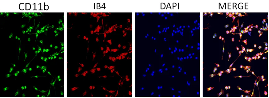
[image:3.612.92.524.263.420.2]Identification of retinal microglia
After successful cell passages, microglia,
marked with CD11b and IB4 monoclonal anti
-bodies, showed strong fluorescence staining.
Some of the cells were elongated. CD11b-positive cells and CD11b-negative cells were
counted in five randomly sampled fields of view
under a 20× microscope. The percentage of CD11b-positive cells was calculated for each
field of view (Figure 2).
Discussion
The present study observed that, during the
first 1-2 days of the mixed culturing stage, the
number of neurons decreased gradually. At days 3-4, the death rate of neurons accelerat-ed, the number of large glial cells increasaccelerat-ed, and layers gradually formed. At days 5-6, the astrocytes formed an apparent bottom-layer growing pattern, the number of microglia
increased significantly although scattered, and the morphology of retinal microglia kept chang
-ing. Present results were in accord with find -ings reported previously [9].
The method developed in this study was based on the method established by Giulian et al.,
with an improvement of some specific steps. First, the centrifugation of cells when process -ing the retinal tissue was minimized by reduc-ing centrifugation speed and time. This effec-tively reduced cell damage and improved the density of cells in initial seeding. Second, reti-nal blood vessels were removed as much as possible. All separation operations were car-ried out under a microscope since
immunohis-tochemical identification cannot distinguish
microglia from macrophages. Third, multiple steps of oscillating separation were performed to obtain retinal microglia with higher purity (as high as 95%).
At the same time, through experiments, it was discovered that trypsin digestion was not required for the separation of microglia, in con-trast to the requirement for general cell separa-tion. Trypsin digestion may easily cause dam-age to the microglia and lead to the
simultaneous shedding of large blocks of astro -cytes and microglia, resulting in low product purity.
Microglia belongs to monocytes. Therefore,
immunofluorescence was applied using CD11b (OX42) as a microglia-specific marker.
In conclusion, the present study improved the classic microglia culturing methods with an objective of obtaining retinal microglia of high purity and high quantity, aiming to provide a foundation for further investigation into their role and function.
Acknowledgements
This study was supported by the National
Natural Scienc Foundation of China (Grant NO:
81700838), and the Department of Science and Technology, Hunan, China (Grant NO: 2017JJ3452).
Disclosure of conflict of interest
None.
Address correspondence to: Dr. Tantai Zhao, De- partment of Ophthalmology, The Second Xiangya Hospital of Central South University, 139 Renmin Middle Road, Changsha 410011, Hunan, China. Tel: +86-13574897769; Fax: +86-21-57643271; E-mail:
zhaotantai@csu.edu.cn
References
[1] Wohl SG, Schmeer CW, Friese T, Witte OW and Isenmann S. In situ dividing and phagocy-tosing retinal microglia express nestin, vimen-tin, and NG2 in vivo. PLoS One 2011; 6: e22408.
[2] Devarajan G, Chen M, Muckersie E and Xu H. Culture and characterization of microglia from the adult murine retina. ScientificWorldJournal 2014; 2014: 894368.
[3] Chen L, Yang P and Kijlstra A. Distribution, markers, and functions of retinal microglia. Ocul Immunol Inflamm 2002; 10: 27-39. [4] Barron KD. Microglia: history, cytology, and
re-actions. J Neurol Sci 2003; 207: 98.
[5] Li L, Qu C and Wang F. A novel method for co-culture with muller cells and microglia in rat retina in vitro. Biomed Rep 2015; 3: 25-27. [6] Rock RB, Gekker G, Hu S, Sheng WS, Cheeran
M, Lokensgard JR and Peterson PK. Role of mi-croglia in central nervous system infections. Clin Microbiol Rev 2004; 17: 942-964; table of contents.
editors. Microglia. Hoeber, New York: 1932. p. 483-534.
[8] Giulian D and Baker TJ. Characterization of ameboid microglia isolated from developing mammalian brain. J Neurosci 1986; 6: 2163-2178.