Platelet-rich plasma, bone marrow and chitosan
in minimally invasive plate osteosynthesis of canine
tibia fractures – a randomized study
Fabricia Geovania Fernandes Filgueira
1*, Bruno Watanabe Minto
2,
Denise Granato Chung
2, Tiago Carmagni Prada
2, Natalie Massaro
Rosa-Ballaben
2, Maria Gabriela Nogueira Campos
31Veterinary Hospital Adílio Santos de Azevedo, Federal Institute of Education, Science
and Technology of Paraíba, Sousa, Paraiba, Brazil
2Department of Veterinary Clinic and Surgery, School of Agricultural and Veterinary
Sciences of the Universidade Estadual Paulista, Jaboticabal, São Paulo, Brazil
3Institute of Science and Technology, Federal University of Alfenas, Campus Poços
de Caldas, Alfenas, Minas Gerais, Brazil
*Corresponding author: fabriciavet@outlook.com
Citation: Filgueira FGF, Minto BW, Chung DG, Prada TC, Rosa-Ballaben NM, Campos MGN (2019): Platelet-rich plasma, bone marrow and chitosan in minimally invasive plate osteosynthesis of canine tibia fractures – a randomized study. Veterinarni Medicina 64, 309–316.
Abstract: The goal of this study was to analyse the effects of percutaneous application of platelet rich plasma (PRP), autologous bone marrow concentrate (BM) and chitosan gel (CHI) on bone consolidation following mini-mally invasive plate osteosynthesis (MIPO) of the fractures of the tibia in dogs. Client-owned dogs (n = 30) with tibial fracture were divided into four treatment groups – Group 1 (control), Group 2 (BM), Group 3 (PRP) and Group 4 (CHI). The biomaterial specific to each group was injected at the fracture site immediately after the MIPO procedure. Serial radiographs were used to determine the fracture line and the development of periosteal callus immediately after surgery and at 15, 30, 60, 90 and 120 days post-surgery. There was no significant difference (P > 0.05) in the degree of oedema or grade of lameness between the groups. Grade 4 (minimum) or 5 lameness (absent) was observed in 70% of animals from all groups at 15 days post-surgery. The biomaterials PRP, BM and CHI combined with MIPO contribute to bone consolidation of tibial fractures in dogs and do not cause adverse reactions or fracture complications. Bone marrow concentrate results in shorter bone consolidation time.
Keywords: bone consolidation; surgery;biomaterials; dogs; percutaneous application
Minimally invasive plate osteosynthesis (MIPO) enables greater preservation of the biological environ-ment and, thus, maximizes the healing potential of the bone and the damaged soft tissue (Boero Baroncelli et al. 2012). It results in fast recovery of limb
func-tion and reduces post-operative pain in long bone fractures in cats and dogs (Pozzi et al. 2013).
The biomaterials used in veterinary orthopaedics can help greatly in the consolidation as adjunct ap-plied in the fracture gap during the surgical
dure (Azevedo et al. 2014). In recent years, there has been an increase in research studies on bone marrow concentrate (Vaz et al. 2010), platelet-rich plasmaand chitosan (Guzman et al. 2014) as os-teostimulating biomaterials. These materials are easy to obtain, simple to use and do not require specialized equipment (Tan and Marra 2010). Furthermore, they can be applied percutaneously, as their consistency is compatible with the use of a syringe and needle. Their application can be minimally invasive and of low morbidity to the pa-tient (Connolly and Shindell 1986).
Complications of orthopaedic surgeries are often difficult to resolve and there has been a continuous search for ways to enable and promote the process of bone healing. The objective of this study was to analyse the percutaneous application of platelet enriched plasma, bone marrow concentrate and chitosan in minimally invasive plaque osteosynthe-sis in dogs. We hypothesized that the use of bio-materials would be to provide bone healing faster than osteosynthesis without the percutaneous ap-plication of the biomaterial.
MATERIAL AND METHODS
Specimens and groups. This study was approved by the Ethics Committee in the Use of Animals (CEUA) of the institution (Protocol No. 017930/12) and all owners signed a consent form to include the animal in the study.
The study was randomised in the period of four years (2012–2015) and used thirty dogs with tibi-al fractures that had been admitted to the Smtibi-all Animal Surgical Service of the School Hospital. The inclusion criteria were: young or adult dogs with fractures of the tibia, in conjunction or with-out fractures of the fibula, preferably comminuted or with more than one fractured segment. Animals that showed signs of systemic disorders that could compromise bone consolidation were excluded from the study.
All animals were subjected to minimally invasive plate osteosynthesis (MIPO) and divided into four experimental groups of eight animals (n = 8), with the exception of Group 4 (n = 6). The groups were classified according to the biomaterial to be injected: Group 1 – control (only MIPO), Group 2 – bone marrow concentrate, Group 3 – platelet-rich plas-ma, and Group 4 – chitosan gel. The group 4 had
eight animals, but two died for various reasons be-fore the end of the postoperative follow-up.
In this study we used straight stainless steel plates (Caomedica, Campinas, Sao Paulo, Brazil) and locking screws 2, 2.7, 3.5 and 4.5 mm long. The size was chosen based on the weight and bone structure of the patient.
Biomaterial preparation. Bone marrow con-centrate (BM):Bone marrow (10 ml) was extract-ed from the greater tubercle of the humerus using heparinized syringes and centrifuged (Centrifuge Baby – Model 206, Fanem, Sao Paulo, Brazil) at 1500 rpm for 10 min in order to concentrate the mononuclear cells(Del Carlo et al. 2004). The plas-ma supernatant was discarded and the bone The plas-marrow concentrate taken to the surgical theatre to be used in the osteosynthesis. Nucleated cells were counted before and after centrifugation, using a Neubauer chamber and bone marrow (40 µl) diluted in Turk’s solution (800 µl). This procedure was performed at the time of MIPO.
Platelet-rich plasma: Blood from each patient (4.5 ml) was collected into tubes containing sodium citrate (Vacuum blood collection tubes with 3.2% sodium citrate, Labor Import, Osasco, Sao Paulo, Brazil) and centrifuged twice. The samples were centrifuged at 1200 rpm for 10 min, the plasma and buffy coat layer aspirated and transferred to a ster-ile tube with no additive and further centrifuged at 1600 rpm for 10 minutes. Approximately 80% of the supernatant was discarded, leaving only the concentrated platelet portion and the platelet button at the bottom of the tube. Platelets were activated by gentle agitation and calcium chlo-ride (Calcium chlochlo-ride 10%, Injectcenter, Ribeirão Preto, São Paulo, Brazil).
Chitosan gel: The gel was obtained according to the protocol by Chenite et al. (2000), in which 200 mg chitosan (high molecular weight and degree of deacetylation higher than 75%; Sigma-Aldrich, CAS No. 9012-76-4) was dissolved in 9 ml of 0.1 M chlorhydric acid solution. Subsequently, 560 mg of disodium glycerol phosphate, dissolved in 1 ml of distilled water, was slowly added under gentle ag-itation (Glycerol phosphate disodium salt hydrate – Sigma-Aldrich, CAS No. 55073-41-1), resulting in a white gel. The gel was sterilised in an autoclave and kept at room temperature (25 ºC) until needed.
was injected. The control group did not receive any injection. The 2 ml volume was standardised as the amount that was obtained after preparation BM and PRP processes regardless of weight of the animal.
Post-operative care. The animals were prescribed Cephalexin (Cefalexina, Eurofarma Laboratorios Ltda, Sao Paulo, Brazil) (25 mg/kg every 12 h, for ten days), meloxicam (Maxicam injetavel 0.2%, Ouro Fino Saude Animal Ltda., Cravinhos, Sao Paulo, Brazil) (0.2 mg/kg on the first day and 0.1 mg/kg on subse-quent days, every 24 h, for three days) and dipyrone (Dipirona sodica, Laboratorio Geyer, Porto Alegre, Rio Grande do Sul, Brazil) (25 mg/kg every eight hours, for seven days) and tramadol chlorhydrate (Tramal 5%, Hipolabor Farmaceutica Ltda, Sabara, Minas Gerais, Brazil) (3 mg/kg every eight hours, for five days) for pain management. The wound was cleaned with saline solution and rifampicin (Rifocina spray, Sanofi Aventis Farmaceutica Ltda, Suzano, São Paulo, Brazil) was applied topically once a day, for 15 days. Skin sutures were removed on the 15th day post-surgery.
Post-operative evaluations. The surgeon and the specialist who evaluated the post-operative condition did not participate in the surgical pro-cedure and therefore did not know which treatment was received by which animal (a blinded study).
Patients were radiographically evaluated by the same veterinary specialist in imaging diagnosis before the surgery (Time 0 – T0) and at 15 (T15),
30 (T30), 60 (T60), 90 (T90) and 120 days
post-sur-gery (T120).
The surgeon evaluated the lameness at each time point (T0, T15, T30, T60, T90 and T120) graded
accord-ing to the adapted scores(Scott and Witte 2011) as follows: grade 1 represents severe lameness with no weight bearing on the affected limb at stance; grade 2, lameness present at walk and trot; grade 3, mild lameness at walk but worsens at trot; grade 4, lameness is present but only evident at trot and, grade 5, absence of lameness at walk or trot.
Serial craniocaudal and mediolateral radiogra-phies obtained during the immediate post-operative period and at 15, 30, 60, 90 and 120 days post-sur-gery. In order to analyse the process of bone con-solidation, the following variables were evaluated: radiopacity of the fracture line (RFL), localization of the periosteal callus (LC), presence of bone bridge (PBB), reestablishment of the cortices (RC), rees-tablishment of the medullary canal (RMC), callus remodelling (CR), callus volume (CV) and charac-Quimicos Farmaceutica Ltda, Itapira, Sao Paulo,
Brazil) (0.3 mg/kg) and morphine (Dimorf 10 mg/ml, Cristalia, Produtos Químicos Farmaceutica Ltda, Itapira, Sao Paulo, Brazil) (0.25 mg/kg). After 20 min, anaesthesia was induced with Propofol (Profol 1%, Cristalia, Produtos Quimicos Farmaceutica Ltda, Itapira, São Paulo, Brazil) (4 mg/kg, i.v.) and the ani-mals were intubated with an orotracheal tube. Anaesthesia was maintained by a mixture of iso-fluorane (Forane, Abbott, Sao Paulo, Brazil) and 100% oxygen. Subsequently, the animals were posi-tioned for local epidural anaesthesia with 2% lido-caine (Xylocaina 2%, Hipolabor Farmaceutica Ltda, Sabara, Minas Gerais, Brazil) without vasoconstric-tor (4 mg/kg) combined with 0.75% bupivacaine (Neocaina 0.75%, Cristalia, Produtos Quimicos Farmaceutica Ltda, Itapira, Sao Paulo, Brazil) with-out vasoconstrictor (2 mg/kg) and tramadol chlo-rhydrate (Tramal 5%, Hipolabor Farmaceutica Ltda, Sabara, Minas Gerais, Brazil) (0.5 mg/kg) in the space between the 7th lumbar and the 1st sacral
vertebrae (L7–S1).
Surgical procedure. Fracture reduction was per-formed using a closed and indirect method, with minimum manipulation. Bone alignment and length were achieved based on anatomical refer-ences of the limb and by comparison with the con-tralateral limb. No intra-operative image analysis was used. Surgical access was restricted to two small incisions on the medial face of the tibia, one proximal and the other distal to the fracture site. The plate was inserted at the proximal incision and slid through the tunnel of soft tissue adjacent to the bone surface, over the fracture site. Once the plate was adequately positioned on the bone surface, holes were drilled for the insertion of lock-ing screws with the aid of a perforation guide and a long screw bit. The number of screws used was determined based on each fracture. After inser-tion of the screws, the surgical wound was sutured in three layers. Poliglecaprone 2-0 suture was used for stitching the muscle layers and reduction of the dead space (subcutaneous) using simple continuous suture pattern. Nylon 3-0 was used for the simple interrupted suture of the skin.
teristics of the fracture line (Scale of Radiographic Evaluation – SRE – scores 1 to 6 (Souza et al. 2011).
Statistical analysis. Analysis was performed using the General Linear Model (GLM) of the Sta-tistical Analysis System software (SAS 9.1, SAS Institute, Cary NC, USA) and profile multivariate analysis cause it simultaneously analyses relationships be-tween the multiple temporal measures of the time variable for each animal with some other variable, in order to compare the treatments over time.
The data (groups, time, radiographic variables and degree of claudication) underwent the nor-mality test of Shapiro-Wilk. After, this study used with analysis of time-repeated measures with the split-plot design, in which the factor treatment (4 levels – groups) was tested on the plots (degree of lameness × radiographics variables) and the fac-tor time (6 levels – T0, T15, T30, T60, T90 and T120)
on the subplots, with 5 repetitions per treatment. If there were significant differences (P < 0.05) be-tween the means, they were compared by Tukey test at 5% significance.
RESULTS
Out of the 30 animals studied 40% were younger than 12 months, 57% were crossbreeds; 43% were of specific breeds (Blue Heeler, Border Collie, Dachshund, Fila Brasileiro, Labrador Retriever, Pinscher and Poodle); and 46% were males and 54% females. The diaphysis was the most affected section of the tibia, corresponding to 85% of the fractures. There were 63% transverse or oblique, 10 % spiral and 27% comminuted fractures. Approximately 47% of the animals were small (1 kg to 10 kg), 26.5% medi-um (11 kg to 20 kg) and 26.5% large (21 kg to 50 kg). Age was homogeneous in Group 1 (control) and Group 2 (BM), in which 50% of animals were younger than 12 months old. Group 3 (PRP) and Group 4 (CHI) had 75% and 67% of animals older than 12 months, respectively.
The types of fractures within each group were heterogeneous, with the exception of Group 4, and classified according to Piermattei et al. (2006). Group 1 had 37.5% of type A, 25% B and 37.5% C fractures; Group 2 had 75% A, 12.5% B and 12.5% type C fractures; Group 3 showed 50% A and 50% type C; and Group 4 had 100% type A fractures.
There was no significant difference (P > 0.05) in the degree of oedema or grade of lameness
be-tween the groups. In the present study, 70% of the animals showed lameness grade 4 or grade 5 at 15 days post-surgery. At T30, there were no signs
of lameness (grade 5).
No significant difference (P > 0.05) was observed in use of the limb between the treatment groups (Figure 1); however, Group 2 showed the highest percentage of weight bearing (60% of grade 5 – un-restricted use of the limb) at 15 days post-surgery. There was no significant difference (P > 0.05) in the radiopacity of the fracture line (RFL), lo-calization of the periosteal callus (LC), presence of bone bridge (PBB), reestablishment of cortices (RC), remodelling of the callus (RC) or volume of callus (VC) between the different treatment groups. However, the variables reestablishment of the medullary canal (RMC) and scale of radio-graphic evaluation (SRE) were significantly differ-ent (P < 0.05) in Group 2 and occurred earlier (T30) than in other groups.
The radiopacity of the fracture line was initially absent; in other words, the fracture line became apparent as time progressed (T90 and T120) and
the callus developed, increasing the radiodensity of the fracture line (Figure 1). Similarly, the re-establishment of the medullary canal was absent at first (T0 and T15) in all groups; at T30 it
was pre-sent in Group 2 (BM) and in half of the animals from Group 3 (PRP) but absent in Groups 1 and 4, with significant differences (P < 0.05) between them. At T60, T90 and T120 it was present in all
groups, with no significant difference (P > 0.05) being observed.
There was no significant difference (P > 0.05) in the scale of radiographic evaluation between the groups. However, Group 2 (BM) showed sig-nificantly shorter (P < 0.05) bone consolidation time (46.87 days) than Groups 1, 3 and 4 (69.37, 67.50 and 57.50 days, respectively).
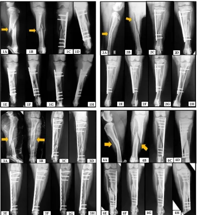
Figure 1. Photographic images of the radiographs of a patient group 1, 2, 3 and 4 in the experimental period
Patient group 1: (1A) – comminuted fracture in middle third of the tibia (arrow) in mediolateral projection; (1B) – cranio-caudal projection indicating a comminuted fracture (arrow); (1C) – immediate postoperative period; (1D) – 15 days post-operatively with early bone callus formation; (1E) – 30 days postoperatively with clinical fracture union; (1F) – 60 days after surgery; (1G) – 90 days after surgery; (1H) – 120 days postoperatively
Patient group 2: (2A) – long oblique fractures in the middle third of the tibia (arrow) in mediolateral projection; (2B) – cra-niocaudal projection indicating the long oblique fracture (arrow); (2C) – postoperative immediate with good alignment of the bone shaft; (2D) – 15 days postoperatively with early bone callus formation; (2E) – 30 days postoperatively with clini-cal fracture union; (2F) – 60 days after surgery; (2G) – 90 days after surgery; (2H) – 120 days postoperatively
Patient group 3: (3A) – comminuted fracture in middle third of the tibia (arrow) in mediolateral projection; (3B) – craniocaudal projection indicating the comminuted fracture (arrow); (3C) – Immediate postoperative period with good alignment of the bone shaft; (3D) – 15 days postoperatively with early bone callus formation; (3E) – 30 days postoperatively with clinical fracture union; (3F) – 60 days after surgery; (3G) – 90 days after surgery; (3H) – 120 days postoperatively with bone remodeling Patient group 4: (4A) – Long oblique fracture in middle third of the tibia (arrow) in mediolateral projection; (4B) – cranio-caudal projection indicating the long oblique fracture (arrow); (4C) – Immediate postoperative period; (4D) – 15 days after surgery; (4E) – 30 days after surgery; (4F) – 60 days after surgery; (4G) – 90 days postoperatively with clinical fracture union; (4H) – 120 days postoperativel
1A 1B 1C 1D
1E 1F 1G 1H
2A 2B 2C 2D
2E 2F 2G 2H
3A 3B 3C 3D
3E 3F 3G 3H
4A 4B 4C 4D
DISCUSSION
The principles of MIO (minimally invasive osteosyn-thesis) are better applied to non-reducible fractures, especially those that are complex and comminuted, many of which are accompanied by extensive soft tis-sue damage. However, its use is not contraindicated in other types of fractures as long as adequate spa-tial alignment is obtained(Beale and McCally 2012). In the present study, MIPO was used to successfully correct complex comminuted fractures as well as sim-pler oblique ones, corroborating with Schmokel et al. (2007), Beale and McCally(2012), Boero Baroncelli et al. (2012), Hudson et al. (2012) and Adelina et al. (2013) who reported the use of MIPO in simple line fractures and stressed that care must be taken when inserting screws close to the fracture line.
The present study did not use trans-operative ra-diography or fluoroscopy for the visualization of the fragments during the surgical procedure; neverthe-less, bone consolidation was obtained in all types of fractures, even in the simple ones. Many studies consider intra-operative imagine analysis indispen-sable(Hudson et al. 2012; Adelina et al. 2013) as it enables quick visualization of several exact projec-tions of the bone surface and is thus considered the most useful method in evaluating alignment (Peirone et al. 2012).
Young animals have shorter bone consolida-tion time(Piermattei et al. 2006), however, as the groups in the present study were composed of ani-mals of homogeneous age, age was not considered a relevant factor. Boero Baroncelli et al. (2012) com-pared MIPO to open reduction and, even though there were discrepancies in age within the groups (i.e. MIPO group had only 25% of animals younger than 12 months), there was no influence of age on bone consolidation time.
In the present study, MIPO was combined with the percutaneous application of bone marrow concentrate, platelet-rich plasma and chitosan gel at the reduced fracture site to minimize bone con-solidation time, and thus reduce patient morbidity. These biomaterials stimulate bone and cartilage regeneration through a minimally invasive proce-dure(Connolly and Shindell 1986) of low cost and that only requires a syringe and needle for appli-cation. Furthermore, studies on the combined use of MIPO and biomaterials are limited.
Hernigou et al. (2005) reported the percutane-ous use of biomaterials in cases of non-fusion
of the bone, which was considered effective and safe, especially when bone marrow concentrate was used. Differently from the study mentioned above, the biomaterials in this study were used to evaluate their influence on bone consolidation of recent fractures and on the clinical results, with the aim of reducing the post-operative complica-tions that may occur in tibial fractures. This study did not analyse signs of bone infection and only one animal developed complications in fracture reduction, in which delayed fusion was observed in a patient of the control group.
MIPO enables a quick recovery of the function of the limb, as it minimizes the damages to soft tissues and is thus more advantageous than open reduction internal fixation (ORIF; 1). There was no significant difference on the use of the limb between the groups; however, Group 2 showed greater per-centage (60%) of grade 5 lameness (no lameness) at 15 days post-surgery. This may be due to the osteogenic and osteoinduction properties of the bone marrow, as it is composed of mesenchymal stem cells that can differentiate into osteoblasts and act on bone formation and repair(Kraus and Kirker-Head 2006). However, no significant differ-ence was observed between the groups, attributing the lack of lameness to the stability obtained with the technique and implants used.
PRP did not contribute negatively to fracture consolidation as the consolidation time observed for that group was similar to the control (67.50 and 69.37 days, respectively). On the other hand, Batista et al. (2011) who used PRP and BM combined to β-tricalcium-phosphate in damaged rabbit tibia, concluded that PRP resulted in better consolida-tion by enabling greater formaconsolida-tion of compact bone than BM. In the current study, BM resulted in low-er consolidation time than the othin low-er treatments. In humans, percutaneous injection of concentrated bone marrow aspirate has been successfully used in promoting pseudoarthrosis in cases of exposed fractures of the tibia with non- or delayed fusion(Le Nail et al. 2014). Furthermore, these authors report that due to its efficacy, low rates of post-surgical complications, preservation of bone stock and low cost, the concentrated BM should be considered as an alternative therapy to the management of com-plicated fractures of long bones.
the ability to carry therapeutic agents and the ab-sence of residual substances (Naderi et al. 2011). The number of randomized studies with its use in fractures is scarce and only a few studies in ten-dons(Santana et al. 2014) and cartilage can be found (Martins et al. 2013).
Chitosan resulted in bone consolidation time similar to the control and PRP, indicating its poten-tial use in fractures. Chitosan activates macrophag-es, which in turn modulate bone consolidation and release cytokines and growth factors that aid in tis-sue healing(Gorzelanny et al. 2010). Furthermore, chitosan did not cause any inflammatory reaction nor did it negatively contribute to bone consolida-tion time.
There was no significant difference on the scale of radiographic evaluation between the groups; however, Group 2 (BM) showed significantly shorter bone consolidation time, at 46.87 days. This consoli-dation time is considered fast and is a result of the association of minimally invasive techniques (MIPO combined with percutaneous application of bone marrow concentrate), which preserved the biologi-cal environment of the fracture and provided undif-ferentiated mesenchymal stem cells that increased the chances of faster tissue healing with reduced complication risks. This bone consolidation time is similar to those reported by Guiot and Dajardin (2011), who observed clinical fusion within 45 days post-surgery when using MIPO without the use of adjuvants to stimulate healing.
The biomaterials platelet-enriched plasma, bone marrow concentrate and chitosan contributed to bone consolidation when combined with minimally invasive osteosynthesis of the tibia in dogs, with bone marrow concentrate resulting in lower con-solidation time. The radiography scores are essen-tial in determining the time of clinical fusion and consolidation of the fracture, especially the scale of radiographic evaluation. No significant difference was observed in clinical evaluation or bone callus between the groups. The MIPO technique can be adequately performed without trans-operative im-age analysis and chitosan does not cause inflamma-tory reaction or undesired complications.
Acknowledgement
The authors extend thanks to Dr. Michele Avante for helping with the radiography analysis.
REFERENCES
Adelina P, Dascalu R, Schuszler L, Sala A, Igna C (2013): Assessment of long bone fractures healing outgoing minimally invasive plate osteosynthesis in dogs. Veteri-nary Medicine 70, 293–300.
Azevedo AS, Sa MJC, Fook MVL, Nobrega Neto PI, Sousa OB, Azevedo SS, Teixeira MW, Costa FS, Araujo AL (2014): Use of chitosan and b-tricalcium phosphate, alone and in combination, for bone healing in rabbits. Journal Materials Science: Materials in Medicine 25, 481–486. Batista MA, Leivas TP, Rodrigues CJ, Arenas CG, Belitardo
DR, Guarniero R (2011): Comparison between the effects of platelet-rich plasma and bone marrow concentrate on defect consolidation in the rabbit tibia. Clinics 66, 1787–1792.
Beale BS, McCally R (2012): Minimally invasive plate os-teosynthesis: tibia and fibula. Veterinary Clinics of North America: Small Animal Practice 42, 1023–1044. Boero Baroncelli AB, Peirone B, Winter MD, Reese DJ, Pozzi
A (2012): Retrospective comparison between minimally invasive plate osteosynthesis and open plating for tibial fractures in dogs. Veterinary and Comparative Orthopae-dics and Traumatology 25, 410–417.
Chenite A, Chaput C, Wang D, Combes C, Buschmann MD, Hoemann CD, Leurox JC, Atkinson BL, Binette F, Selm-ani A (2000): Novel injectable neutral solutions of chi-tosan form biodegradable gels in situ. Biomaterials 21, 2155–2161.
Connolly J, Shindell R (1986): Percutaneous marrow injec-tion for no ununited tibia. The Nebraska Medical Journal 71, 105–107.
Del Carlo RJ, Monteiro BS, Daibert APS, Pinheiro LCP (2004): Bone marrow autografts a veterinary orthopaedics alternative. Revista Ceres 51, 411–418.
Gorzelanny C, Poppelmann B, Pappelbaum K, Moersch-bacher BM, Schneider SW (2010): Human macrophage activation triggered by chitotriosidase mediated chitin and chitosan degradation. Biomaterials 31, 8556–8563. Guiot LP, Dejardin LM (2011): Prospective evaluation
of minimally invasive plate osteosynthesis in 36 nonar-ticular tibial fractures in dogs and cats. Veterinary Sur-gery 40, 171–182.
Guzman R, Nardecchia S, Gutierrez MC, Ferrer ML, Ramos V, Del Manto F, Abarrategi A, Lopez-Lacomba JL (2014): Chitosan scaffolds containing calcium phosphate salts and rhbmp-2: in vitro and in vivo testing for bone tissue regeneration. PLoS ONE 9, doi: 10.1371/journal.pone. 0087149.
transplantation in nonunion and avascular necrosis of bone. The Journal of Bone and Joint Surgery 87, 896–902. Hudson CC, Lewis DD, Pozzi A (2012): Minimally invasive
plate osteosynthesis in small animals radius and ulna fractures. Veterinary Clinics of North America: Small Animal Practice 42, 983–996.
Kraus KH, Kirker-Head C (2006): Mesenchymal stem cells and bone regeneration. Veterinary Surgery 32, 232–242. Le Nail L, Stanovici J, Fournier J, Splingard M, Domenech
J, Rosset P (2014): Percutaneous grafting with bone mar-row autologous concentrate for open tibia fractures: analysis of forty-three cases and literature review. Inter-national Orthopaedics (SICOT) 38, 1845–1853.
Martins EAN, Invernizzi MS, Campos MGN, Teodoro PA, Contieri MB, da Silva LCLC (2013): Chitosan film in horses with experimentally induced skin wound. Ciencia Rural 43, 1824–1830.
Naderi H, Matin MM, Bahrami AR (2011): Review paper: critical issues in tissue engineering: biomaterials, cell sources, angiogenesis, and drug delivery systems. Journal of Biomaterials Applications 26, 383–419.
Peirone B, Rovesti GL, Boero Baroncelli A, Piras L (2012): Minimally invasive plate osteosynthesis fracture reduc-tion techniques in small animals. Veterinary Clinics of North America: Small Animal Practice 42, 873–895. Piermattei DL, Flo GL, Decamp CE (2006): Brinker,
Piermat-tei and Flo’s Handbook of Small Animal Orthopedics and Fracture Repair. St Louis, Elsevier Saunders. 718–749 p.
Pozzi A, Hudson CC, Gauthier CM, Lewis DD (2013): Retro-spective comparison of minimally invasive plate osteosyn-thesis and open reduction and internal fixation of radius-ulna fractures in dogs. Veterinary Surgery 42, 19–27.
Santana CC, Nobrega Neto PI, Sa MJC, Oliveira LM, Fook MVL, Azevedo AS, Souza OB (2014): Use of the film of chi-tosan in the repair of tendons in rabbits. Arquivo Brasileiro de Medicina Veterinaria e Zootecnia 66, 995–1002. Schmokel HG, Stein S, Radke H, Hurter K, Schawalder
P (2007): Treatment of tibial fractures with plates using minimally invasive percutaneous osteosynthesis in dogs and cats. Journal of Small Animal Practice 48, 157–160. Scott H, Witte P (2011): Investigation of lameness in dogs:
1. Forelimb. In Practice 33, 20–27.
Souza TFB, Ferreira GTNM, Sakamoto SS, Albuquerque VB, Bomfim SRM, Andrade AL (2011): Radiographic and den-sitometric aspects of experimental radial fractures of dogs treated with platelet-rich plasma. Ars Veterinária 27, 1–6. Tan H, Marra KG (2010): Injectable, biodegradable hydro-gels for tissue engineering applications. Materials 3, 1746–1767.
Vaz CES, Guarniero R, Santana PJ (2010): Effect of use of bone-marrow centrifugate on muscle injury treatment: experimental study on rabbits. Acta Ortopedica Brasileira 18, 321–326.