Original Article
Downregulation of TMEM176A suppresses cell growth
and proliferationin non-small cell lung cancer
Anquan Shang1,2*, Fujun Shen3*, Jun Wang1, Weiwei Wang4,5, Chunbin Wang3, Guoling Liao1
1Clinical Medicine School, Ningxia Medical University, Yinchuan, Ningxia, China; Departments of 2Laboratory
Medicine, 5Pathology, The Sixth People’s Hospital of Yancheng City, Yancheng, Jiangsu, China; 3Department of
Oncology, Yancheng Hospital Affiliated to Medical College of Southeast University & The Third People’s Hospital of Yancheng, Yancheng, Jiangsu, China; 4Department of Pathology, The First People’s Hospitalof Yancheng City,
Yancheng, Jiangsu, China. *Equal contributors.
Received December 12, 2016; Accepted December 27, 2016; Epub March 1, 2017; Published March 15, 2017
Abstract: As a member of membrane-spanning 4A family, transmembrane protein 176A (TMEM176A) has been recently reported to be overexpressed in various cancers. However, the biological functions of TMEM176A in non-small cell lung cancer (NSCLC) remain unclear. In this study, we investigated the expression of TMEM176A in pub-licly available Oncomine database and found it was upregulated in lung cancer tissues compared with normal lung tissues. Similar results were also found in fresh primary NSCLC tumor tissues and corresponding normal tissues
using Western blot analysis.Then the expression of TMEM176A was efficiently silenced in human NSCLC cell lines,
A549 and 95D by RNA interference. Knockdown of TMEM176A obviously inhibited cell growth and colony formation, induced cell arrest at G0/G1 phase and promoted cell apoptosis in NSCLC cells, as determined by MTT, clonogenic
assay and flow cytometry assays. Mechanistically, inhibition of TMEM176A significantly reduced the expression of
CDK4 and Cyclin D1, but increased the expression of caspase-3, PARP cleavage and bad. In conclusion, these
re-sults firstly demonstrated that TMEM176A might serve as a potential oncogene that involved in the cell proliferation
of NSCLC.
Keywords: Non-small cell lung cancer, TMEM176A, cell growth, apoptosis, caspase-3
Introduction
Non-small cell lung cancer (NSCLC), account for approximately 85% of all lung cancer, is the leading cause of cancer related death in the world [1]. Recently, despite lower toxicity and
higher efficiency in epidermal growth factor
receptor tyrosine kinase inhibitors compared with traditional postoperative chemotherapy [2], patients with NSCLC exhibited worse 5-year survival rate of only 15% [3]. The main factors associated with NSCLC are tumor uncontrolled
proliferation. Therefore, identification of new
molecular targets regarding the cellular and molecular mechanisms involved in the patho-genesis of NSCLC might provide a novel thera-peutic strategy for this malignancy.
Transmembrane proteins (TMEMs) familyis a group of ca. 310 different proteins and predict-ed as components of cellular membranes, including lysosomes and mitochondrial
mem-branes [4]. At present, members of TMEMs have been reported to be abnormally expressed in various tumors. TMEM7, acts as a tumor sup-pressor gene, was downregulated in renal cell carcinoma (RCC) tissues in comparison to nor-mal human kidney [5]. Additionally, TMEM22 is overexpressed and likely play a crucial role in growth of renal cell carcinoma (RCC) [6]. TMEM16A is highly expressed and contributes to tumor invasion in human gastric cancer [7]. TMEM45A and TMEM158 have been demon-strated to play a positive role in cell prolifera-tion and invasion in human ovarian cancer cells [8, 9]. Similarly, TMEM176A, belongs to mem-brane-spanning 4A family of proteins [10], was
first identified as tumor-associated protein in
hepatocellular carcinoma (HCC) [11]. Previously, the expression of TMEM176A was shown to be
significantly increased in lung cancer [12].
TMEM176A promotes non-small cell lung cancer in vitro
To investigate the role of TMEM176A in NSCLC,
we firstly compared its expression between
NSCLC tissues and normal tissues. The effects of TMEM176A silencing on cell growth of NSCLC cells, as well as the underlying mechanisms, were then determined. Our findings suggest
that TMEM176A might be a promising molecu-lar target for NSCLC therapy via inhibition of TMEM176A.
Materials and methods
Clinical samples and cell lines
Total 6pairs of fresh primary NSCLC tumor tis-sues and adjacent normal tistis-sues were collect-ed from patients undergoing surgery at Depart- ment of Thoracic Surgery, Union Hospital, Tongji Medical College between 2013 and 2015, whi- ch were subjected to compare the expression of TMEM176A between NSCLC tissues and paired normal lung tissues. All the participants were required to sign informed consent and approved by the ethics committee of Union Hospital, Tongji Medical College. All the patients
were pathological diagnosed and confirmed
received none of preoperative chemotherapy or radiotherapy.
Cell culture and transfection
NSCLC cell lines (A549 and 95D) were pur-chased from Cell Bank of the Chinese Academy of Sciences, Shanghai, China and maintained in RPMI-1640 (Hyclone, Biowest) supplement-ed with 10% fetal bovine serum (FBS). All cells
were incubated in a humidified atmosphere
containing 5% CO2 at 37°C. To further investi-gate the function of TMEM176A in NSCLC in cel-lular level, cells were cultured in six-well plates and transfected with 100 nmol/l of TMEM176A siRNA or control siRNA (Invitrogen, Carlsbad, CA, USA) using Lipofectamine 2000 transfec-tion reagent kit (Invitrogen, Carlsbad, California, USA). After 48 h transfection, cells were
under-went knockdown efficiency determination by
western blotting.
Western blot analysis
Total protein was obtained from tissue samples and cell lines using 2 mL ice-cold RIPA lysis buf-fer (Beyotime, Shanghai, China). Equal amount
of proteins (30 μg) were separated on 10%
sodium dodecyl sulfate-polyacrylamide gel
(SDS-PAGE) and transferred to a PVDF mem-brane (Millipore, Billerica, MA, USA) using Bio-Rad semidry transfer system. Then the mem-brane was blocked with TBST (Tris-buffered saline, 0.1% Tween-20) containing 5% non-fat dry milk for 1h at room temperature, and probed with the corresponding primary anti-bodies, including anti-TMEM176A, CDK4, Cyclin D1, Bad, caspase-3 and PARP overnight. After washed by TBST, the membrane was then incu-bated with appropriated horseradish peroxi-dase-conjugated secondary antibodies (Santa Cruz Biotechnology, Santa Cruz, CA) for 2 h at room temperature. The membranes were wash- ed and immunoreactive bands were visualized using super ECL detection reagent (Pierce Bio- technology, USA).
MTT assay
The effect of TMEM176A silencing on cell viabil-ity was determined in A549 and 95D cells using MTT assay. After 48 h of infection, cells (3,000 cells per well) were seeded into 96-well plates
in triplicates and then added into20 μL MTT
solution (5 mg/mL) at 1, 2, 3, 4, and 5 days after transfection. After another 4 h incubation,
200 μL dimethyl sulfoxide (DMSO) was added
into each well. The OD value was measured using anELISA reader (Bio-Rad, USA) at wave-length of 595 nm. Each experiment was per-formed in triplicate.
Clonogenic assay
For the clonogenic assays, A549 and 95D cells (500 cells/well) were reseeded in 6-well plates after infection for 48 h. Then cells were allowed to grow for an additional 7 days to form natural colonies. The adherent cells were washed twice with PBS and then stained with 0.5% crystal violet solution to visualize the colonies. Colonies
that contain > 50 cells were counted under flu -orescence microscope.
Flow cytometry analysis
Theeffects of TMEM176A silencing on cell cycle distribution and apoptosis were measured
using flow cytometry analysis. In brief, cells
apopto-sis assay according to the manufacturer’s ins- tructions. The stained cells were analyzed using
flow cytometer (FACS Calibur, BD Biosciences).
Statistical analysis
Statistical analyses on cellular level were per-formed by SPSS software version 10.0 (SPSS Inc, United States) and expressed as mean ± standard deviation (SD) of three independent experiments. Paired Student’s t test was used to compare differences between siCon and siT-MEM176A groups and P<0.05 was considered
as significant.
Results
TMEM176A was overexpressed in NSCLC tis-sues
To investigate the association of TMEM176A
with NSCLC progression, we firstly determined
the TMEM176A mRNA levelsusing the datasets from the publicly available Oncomine database
(www.oncomine.org) and found it was signifi -cantly upregulated in human lung cancer tis-sues compared with that in normal tistis-sues using Okayama Lung (Figure 1A, P = 3.14E-5) and Hou Lung (Figure 1B, P = 0.008) datasets.
To further confirm this result, Western blot
analysis were used to determine the expres-sion of TMEM176A in fresh primary NSCLC tumor tissues and corresponding normal
tis-sues. As shown in Figure 1C, the protein level of
TMEM176A was significantly elevated in 6 pairs
of NSCLC tissues compared with the adjacent normal tissues (P<0.001). These data suggest-ed that TMEM176A might play an important role in the development of NSCLC.
Downregulation of TMEM176A remarkably suppressed cell growth
Considering the expression TMEM176A was
significantly upregulated in NSCLC, we further
investigated the cellular functions of TMEM- 176A in NSCLC cell lines by loss-of-function assays. NSCLC cell lines, A549 and 95D were selected to knock down the expression of TMEM176A by siRNA transfection. As shown in
Figure 2A, the expression levels of endogenous TMEM176A proteins were obviously inhibited in
these two cell lines, as confirmed by Western
blot analysis. Subsequently, we investigated whether knockdown of TMEM176A inhibited uncontrolled cell proliferation in NSCLC cells. The results from the MTT assay demonstrated
that the cell growth rate was significantly
reduced in both A549 and 95Dcells transfect-ed with siTMEM176A compartransfect-ed with cells transfected with siCon (Figure 2B, P<0.001). In addition, clonogenic assay showed that the
numbers of colonies were significantly reduced
[image:3.612.93.525.73.280.2]TMEM176A promotes non-small cell lung cancer in vitro
speculated TMEM176A could promote cell growth in NSCLC.
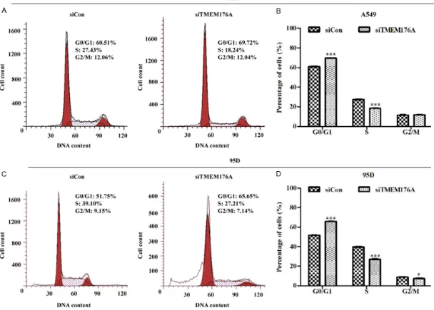
TMEM176A silencing induces cell cycle arrest and apoptosis
To further investigate the mechanism underly-ing the inhibition of cell proliferation, cell cycle
distribution and apoptosis were detected on A549 and 95D cells after lentivirus infection
using flow cytometric assay. As shown in Figure 3A and 3B, the percentage of cells in G0/G1
phase was significantly increased (P<0.001),
[image:4.612.93.520.72.535.2]whereas the number of cells in S phase was remarkably decreased in A549 cells following TMEM176A knockdown (P<0.001). Similarly, Figure 2. TMEM176A silencing induced by siRNA significantly inhibited NSCLC cell growth and proliferation. A. The protein level of TMEM176A was remarkably down regulated by specific siRNA targeting TEM176A (siTMEM176A)
compared with control siRNA (siCon) in A549 and 95 D cells. B. MTT assay indicated that the growth rate of A549
and 95 D cells was significantly decreased after TMEM176A silencing. C. Clonogenic assay showed knockdown of TMEM176A significantly impaired cell colony formation ability in A549 and 95 D cells. NSCLC, non-small cell lung
Figure 3. TMEM176A silencing induced cell cycle arrest at G0/G1 phase. The profile of cell percentages in G0/G1, S and G2/M phases in A549 (A) and 95D (C) cells
SiT-TMEM176A promotes non-small cell lung cancer in vitro
[image:6.792.96.709.82.485.2]3911 Int J Clin Exp Pathol 2017;10(3):3906-3914
knockdown of TMEM176A significantly elevated
the percentage of cells in G0/G1 phase (P<0.001), while reduced the cell counts in S (P<0.001) and G2/M (P<0.05) phases in 95D cells (Figure 3C and 3D).
What’s more, siTMEM176A did cause signifi
-cant change in the profile of Annexin
V/7-AAD-staining cell populations corresponding to via-ble and non-apoptotic (Annexin V-/7-AAD+), early (Annexin V+/7-AAD-) and late apoptotic (Annexin V+/7-AAD+) cells in A549 and 95D cells. Moreover, more cells presented Annexin V+/7-AAD- and Annexin V+/7-AAD+ signals in siTMEM176A than that in siCon groups in A549 and 95D cells (Figure 4A and 4C). Statistical analysis further demonstrated the overall apop-totic rate of siTMEM176A group cells was about 22.69% and 18.60% in A549 and 95D cells,
respectively, which was significantly higher
than that in corresponding siCon groups (Figure 4B and 4D). Collectively, TMEM176A might play an important role in regulating cell cycle pro-gression and apoptosis.
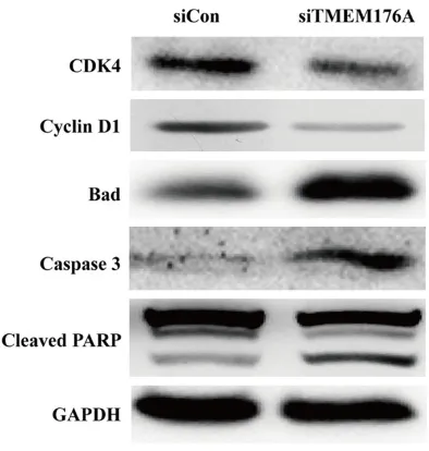
Downregulation of TMEM176A regulated cell cycle and apoptotic markers
Furthermore, we detected the expression alter-ations of some cell cycle regulators and
apop-totic markers. As shown in Figure 5, knockdown
of TMEM176A significantly downregulated the
expression levels of CDK4 and Cyclin D1, but upregulated the expression levels of bad, cas-pase-3 and cleaved PARP in xenografts. There-
fore, these findings suggest that TMEM176A
might be essential for cell proliferation of NSCLC.
Discussion
Recently, TMEMs have been identified to be
involved in tumor progression and develop-ment. TMEM176A, as a member of TMEM
fam-ily, has been reported to be significantly up
regulated in lung carcinoma [12], but limited functional data describing their detailed involvement in NSCLC is lack. In the present study, re-analysis of the data obtained from Oncomine database showed TMEM176A was commonly upregulated in lung cancer tissues. In line with this observation, we foundTME-M176A wasupregulated in NSCLC and adjacent normal tissues using Western blot analysis, which suggest TMEM176A might be a putative oncogene in NSCLC.
To further investigate the functions of TMEM- 176A in NSCLC, the expression of TMEM176A was suppressed in two NSCLC cell lines, A549 and 95D, by means of siRNA treatment. Loss-of-function assay indicated that knockdown of TMEM176A drastically suppressed cell growth and colony formation ability of NSCLC. It has been reported that cell cycle arrest and pro-moting cell apoptosis is an important mecha-nism in inhibiting tumor cell growth [13, 14]. Thus, we determined the effects of TMEM silencing on NSCLC cell cycle distribution and apoptosis. Based on our data, knockdown of TMEM176Acould arrest cell cycle at G0/G1 phase and promoted cell early and late apopto-sis, which suggested that TMEM176A plays an essential role in NSCLC cell growth. In line with
this finding, TMEM14A, also belongs to a mem -ber of TMEMs, has a pro-tumorigenic effect in ovarian cancer cells through increasing cell pro-liferation [15]. Overexpression of TMEM14A could inhibit N-(4-hydroxyphenyl) retinamide-induced cell apoptosis in glioma [16].
To further uncover the mechanisms underlying the inhibitory effects induced by TMEM176A knockdown, we detected the expression of found that TMEM176A knockdown downregu-Figure 5. Western blot analysis of proteins
[image:7.612.92.289.70.277.2]TMEM176A promotes non-small cell lung cancer in vitro
lated the expression of several cell cycle regula-tors and apoptotic markers. CDK4 and Cyclin D1 have been demonstrated to be responsible for initiation and completion of DNA replication [17, 18], which were both downregulated by knockdown of TMEM176A. Further data reve- aled that the TMEM176A silencing upregulated the expression of caspase-3, and cleaved PARP. Generally, cell apoptosis consists of two distinct signaling pathways, including the extrin-sic and intrinextrin-sic pathway, in which caspase family proteases can be activated [19, 20]. Caspase-3 is the key enzyme required in the caspase activation and execution [21]. PARP is reported to promote programmed cell death, in which it was cut by the caspase-3, leading to cell apoptosis [22, 23]. In addition, the activa-tion of caspase is also regulated by Bcl-2 family proteins [24]. Bad, as a member of Bcl-2 family, has been considered as a pro-apoptotic factor in promoting cell death [25]. Therefore, our results provide evidence implicate TMEM176A silencing as an essential inhibitor during NSCLC progression.
In summary, our study is the first to report that
TMEM176A is closely correlated with NSCLC tumorigenesis by regulating cell proliferation, as well as provide a mechanistic basis for the further exploration of TMEM176A as a diagnos-tic and therapeudiagnos-tic target for NSCLC. Further- more, it is still necessary to further investigate the potential application of TMEM176A-target- ed therapy using lentivirus-mediated shRNA approach in more preclinical and clinical stu- dies.
Acknowledgements
This work is support by 2015 Postgraduate Education Innovation Program in Ningxia (No. NXYC201511).
Disclosure of conflict of interest
None.
Address correspondence to: Weiwei Wang, Depart- ment of Pathology, the First People’s Hospital and the Sixth People’s Hospital of Yancheng City, Yan- cheng 224005, Jiangsu, China. Tel: +86-515-6882- 5356; E-mail: wangweiwei0601@yeah.net; Chunbin Wang, Department of Oncology, Yancheng Hospital
Affiliated to Medical College of Southeast University
&the Third People’s Hospital of Yancheng, Yancheng
224001, Jiangsu, China. Tel: +86-515-68825356; E-mail: yclvwenping@126.com
References
[1] Ma L, Huang Y, Zhu W, Zhou S, Zhou J, Zeng F, Liu X, Zhang Y and Yu J. An integrated analysis of miRNA and mRNA expressions in non-small cell lung cancers. PLoS One 2011; 6: e26502. [2] Ellis PM, Coakley N, Feld R, Kuruvilla S and Ung
YC. Use of the epidermal growth factor
recep-tor inhibirecep-tors gefitinib, erlotinib, afatinib,
dacomitinib, and icotinib in the treatment of non-small-cell lung cancer: a systematic re-view. Curr Oncol 2015; 22: e183-215.
[3] Spiro SG and Silvestri GA. One hundred years of lung cancer. Am J Respir Crit Care Med 2005; 172: 523-529.
[4] Wrzesiński T, Szelag M, Cieślikowski WA, Ida A, Giles R, Zodro E, Szumska J, Poźniak J, Kwias Z
and Bluyssen HA. Expression of pre-selected TMEMs with predicted ER localization as
po-tential classifiers of ccRCC tumors. BMC
Cancer 2015; 15: 1-18.
[5] Kholodnyuk ID, Kozireva S, Kost-Alimova M, Kashuba V, Klein G, Imreh S. Down regulation of 3p genes, LTF, SLC38A3 and DRR1, upon
growth of human chromosome 3-mouse fibro -sarcoma hybrids in severe combined
immuno-deficiency mice. Int J Cancer 2006; 119:
99-107.
[6] Dobashi S, Katagiri T, Hirota E, Ashida S, Daigo Y, Shuin T, Fujioka T, Miki T and Nakamura Y. Involvement of TMEM22 overexpression in the growth of renal cell carcinoma cells. Oncol Rep 2009; 21: 305-312.
[7] Liu F, Cao QH, Lu DJ, Luo B, Lu XF, Luo RC and Wang XG. TMEM16A overexpression contrib-utes to tumor invasion and poor prognosis of human gastric cancer through TGF-beta signal-ing. Oncotarget 2015; 6: 11585-11599. [8] Guo J, Chen L, Luo N, Yang W, Qu X and Cheng
Z. Inhibition of TMEM45A suppresses prolifera-tion, induces cell cycle arrest and reduces cell invasion in human ovarian cancer cells. Oncol Rep 2015; 33: 3124-3130.
[9] Cheng Z, Guo J, Chen L, Luo N, Yang W and Qu X. Overexpression of TMEM158 contributes to ovarian carcinogenesis. J Exp Clin Cancer Res 2015; 34: 75.
[10] Zuccolo J, Bau J, Childs SJ, Goss GG, Sensen CW and Deans JP. Phylogenetic analysis of the MS4A and TMEM176 gene families. PLoS One 2010; 5: e9369.
[11] Wang Y, Han KJ, Pang XW, Vaughan HA, Qu W, Dong XY, Peng JR, Zhao HT, Rui JA, Leng XS, Cebon J, Burgess AW and Chen WF. Large
scale identification of human hepatocellular
[12] Cuajungco MP, Podevin W, Valluri VK, Bui Q, Nguyen VH and Taylor K. Abnormal accumula-tion of human transmembrane (TMEM)-176A and 176B proteins is associated with cancer pathology. Acta Histochemica 2012; 114: 705-712.
[13] Abaza MS, Orabi KY, Quattan E and Al-Attiyah RJ. Growth inhibitory and
chemo-sensi-tization effects of naringenin, a natural flava
-none purified from Thymus vulgaris, on human
breast and colorectal cancer. Cancer Cell Int 2015; 15: 46.
[14] Gao Y, Teng J, Hong Y, Qu F, Ren J, Li L, Pan X, Chen L, Yin L and Xu D. The oncogenic role of EIF3D is associated with increased cell cycle progression and motility in prostate cancer. Med Oncol 2015; 32: 1-8.
[15] Zhang Q, Chen X, Zhang X, Zhan J and Chen J. Knockdown of TMEM14A expression by RNAi inhibits the proliferation and invasion of hu-man ovarian cancer cells. Biosci Rep 2016; 36: e00298.
[16] Woo IS, Jin H, Kang ES, Kim HJ, Lee JH, Chang KC, Park JY, Wan SC and Han GS. TMEM14A inhibits N-(4-hydroxyphenyl) retinamide-induc- ed apoptosis through the stabilization of mito-chondrial membrane potential. Cancer Lett 2011; 309: 190-198.
[17] Massague J. G1 cell-cycle control and cancer. Nature 2004; 432: 298-306.
[18] Cicenas J, Kalyan K, Sorokinas A, Jatulyte A, Valiunas D, Kaupinis A and Valius M. Highlights of the latest advances in research on CDK in-hibitors. Cancers (Basel) 2014; 6: 2224-2242.
[19] Degterev A and Yuan J. Expansion and evolu-tion of cell death programmes. Nat Rev Mol Cell Biol 2008; 9: 378-390.
[20] Denault JB and Boatright K. Apoptosis in bio-chemistry and structural biology. 3-8 February 2004, keystone, CO, USA. IDrugs 2004; 7: 315-317.
[21] Fan TJ, Han LH, Cong RS and Liang J. Caspase family proteases and apoptosis. Acta Biochim Biophys Sin (Shanghai) 2005; 37: 719-727. [22] Piskunova TS, Yurova MN, Ovsyannikov AI,
Se-menchenko AV, Zabezhinski MA, Popovich IG,
Wang ZQ and Anisimov VN. Deficiency in Poly
(ADP-ribose) Polymerase-1 (PARP-1) acceler-ates aging and spontaneous carcinogenesis in mice. Curr Gerontol Geriatr Res 2008; 754190. [23] Ghavami S, Hashemi M, Ande SR, Yeganeh B,
Xiao W, Eshraghi M, Bus CJ, Kadkhoda K, Wiechec E, Halayko AJ, Los M. Apoptosis and cancer: mutations within caspase genes. J Med Genet 2009; 46: 497-510.
[24] Brunelle JK and Letai A. Control of mitochon-drial apoptosis by the Bcl-2 family. J Cell Sci 2009; 122: 437-441.


![Crystal structure of di μ chlorido bis[dichlorido(L histidinium κO)cadmium(II)]](data:image/gif;base64,R0lGODlhAQABAIAAAP///wAAACH5BAEAAAAALAAAAAABAAEAAAICRAEAOw==)