Case Report
Combined squamous and small cell carcinoma of the
esophagus: a case report
Peng Zhou1, Dun Yang2, Yuyong Tan3, Shuyuan Xu1, Yinghuan Dai1
1Department of Pathology, Second Xiangya Hospital of Central South University, Changsha, Hunan Province,
China; 2Department of Pathology, Taoyuan County People’s Hospital, Changde, Hunan Province, China; 3
Depart-ment of Gastroenterology, Second Xiangya Hospital of Central South University, Changsha, Hunan Province, China Received March 4, 2016; Accepted May 24, 2016; Epub July 1, 2016; Published July 15, 2016
Abstract: The composite tumor of the esophagus is extremely rare. Here, we report an unusual case of combined squamous and small cell carcinoma of the esophagus. A 56-year-old woman was admitted to our hospital with dys-phagia and chest pain. Endoscopic examination showed a semi-annular mass in the middle esophagus. Because the lesion was limited, curative esophagectomy was then performed. In the resection specimen, the tumor revealed a mixture of squamous and small cell carcinoma. Pathologically, the squamous cell carcinoma component which showed focal keratinization and pearl formation was positive for CK5/6 and P40. While the small cell carcinoma component which was characterized by high nuclear-cytoplasmic ratio, hyperchromatic nuclei, scanty cytoplasm and absent nucleoli strongly expressed CD56, Syn, NSE, CgA. Both components were negative for TTF-1, Vimentin, LCA, CD99. A brief discussion of the clinical symptoms, etio-pathogenesis, histopathological features and treatment of this rare neoplasm is presented.
Keywords: Esophagus, combined carcinoma, small cell carcinoma, squamous cell carcinoma
Introduction
Esophageal carcinoma is one of the most com-mon malignancies worldwide. Of these esopha-geal carcinomas, approximately 90% or more are esophageal squamous cell carcinomas (SqCC) [1]. Primary small cell carcinoma of the esophagus (SCCE) is a relatively rare tumor, accounting for only 1% to 2.8% of all esopha-geal malignancies [2-4]. Combined squamous and small cell carcinoma of the esophagus, the so-called composite tumor of the esophagus, is even rarer. Here we report a case of squamous and small cell carcinoma of esophagus and a review of the literature.
Case report
A 56-year-old woman was admitted to our hos-pital with increasing dysphagia and chest pain for two months. Physical examination and rou-tine laboratory test were within normal limits. Endoscopic examination showed a semi-annu-lar mass in the middle esophagus. Histologic
examination of a biopsy specimen obtained from the lesion revealed a small cell carcinoma with hyperplasia of esophageal squamous epi-thelium, but no invasive squamous cell carci-noma was found. The computed tomography demonstrated abnormal thickening of the mid-dle esophageal wall, but no typical evidence of metastatic disease in the mediastinal lymph node or in distant organs. Because the lesion was limited, curative esophagectomy with lymph node dissection was then performed fol-lowed by adjuvant chemotherapy with etopo-side and cisplatin (EP) for 4 cycles. However the patient eventually died of systemic carcinoma-tosis 21 months after the surgery.
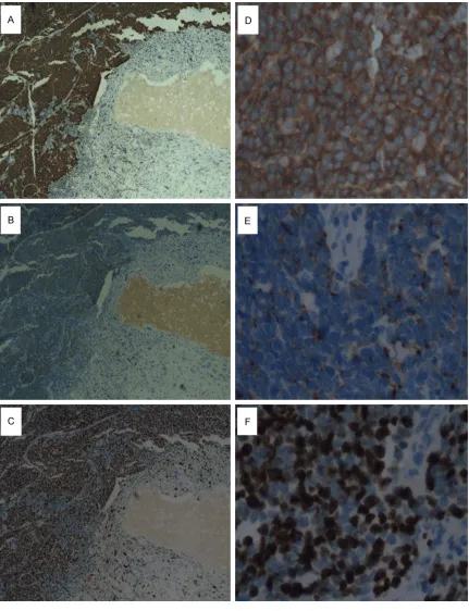
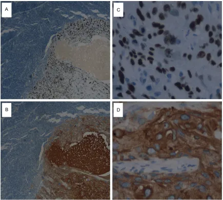
An immunohistochemical study was performed with the use of Dako Envision method as de- scribed previously [5]. The SCCE component was positive for synaptophysin (syn, Figure 2A, 2D), chromogranin A (CgA, Figure 2B, 2E), neuron-specific enolase (NSE), CD56, Ki67 (80%, Figure 2C, 2F), while being negative for P40 and cytokeratin 5/6 (CK5/6). Whereas the SqCC component strongly expressed P40 (Figure 3A, 3C) and CK5/6 (Figure 3B, 3D), but
it was negative for neuroendocrine markers. Both components were negative for LCA, thy-roid transcription factor (TTF-1), CD99, vimentin.
Discussion
The major risk factors and clinical symptoms of combined carcinoma of the esophagus are identical to other more common neoplasm of the esophagus. The patients had a significant history of poor nutritional status, low intake of fruits and vegetables, the use of tobacco or alcohol, and drinking beverages at high tem-peratures [1, 6, 7]. The presenting symptoms included rapidly progressive dysphagia, retro- sternal pain and weight loss.
At present, there are two viewpoints on the etio-pathogenesis of combined small carcino-ma of the esophagus. One is that the combined carcinomas are basically small cell carcinomas with squamous or adenocarcinomatous differ-entiation. And it is believed that the amine pre-cursor uptake and decarboxylase (APUD) cells of the submucosal gland or stratum basal is the histogenetic precursor of small cell carcinoma of the esophagus [8-10]. As APUD cells most commonly exist in the distal esophagus, the pri-mary lesions were often found in the middle and lower third of esophagus [3, 8-10]. The other is that biphasic neoplasm originates from pluripotential stem cells of the endoderm that can be partially differentiated into squamous cell, neuroendocrine cell or glandular cell because of the stimulation of different carcino-genic agent [11]. In the present case, the close merges between the squamous and small cell carcinoma elements histologically lend support to this hypothesis.
[image:2.612.89.289.66.520.2]In this case, the biopsy specimen showed a small cell carcinoma with hyperplasia of the surface epithelium. Otherwise, the combined carcinoma was demonstrated through multiple sampling of the tumor and the normal junction in the resection specimen. Therefore, more tis-sues and multipoint biopsies should be per-formed under an endoscope to establish a correct diagnosis. To make the diagnosis of combined esophageal carcinoma, it is crucial to ensure that the small cell carcinoma compo-nent is not a metastasis from either a broncho-genic or gastrointestinal origin through clinical workup. Sometimes it is difficult to differentiate
SCCE from poorly differentiated SqCC, malig-nant lymphoma, chronic inflammation and ma- lignant melanoma. In this setting, a panel of appropriate immunohistochemical stains will
[image:3.612.92.523.70.632.2]help for diagnosis. The expression of cytokera-tin demonstrates that the tumor is a carcino-ma. About 90% of SCCE has neuroendocrine features, so the most useful neuroendocrine
antigens such as CgA, syn, NSE and CD56 are best used as a pane. Other stains such as lym-phoid markers (LCA, CD3, CD20, CD45RO, PAX-5), squamous markers (CK5/6, P40, P63), mel-anoma markers (S100, HMB-45) and PENT marker (CD99) may be helpful in the differential diagnosis, respectively [12]. The present case showed that the small cell areas showed dif-fuse and intense immunoreactivity for neuroen-docrine markers, while being negative for CK5/6 and P40, whereas the squamous areas were positive for squamous markers and nega-tive for neuroendocrine markers.
The combined small cell carcinoma displays highly aggressive biologic behavior, early dis-tant metastasis and poor prognosis. The sur-vival rate is not clear owing to the paucity of
[image:4.612.89.524.70.459.2]cases. However, survival was thought to be similar to that of small cell lung carcinoma [8-11]. Nevertheless, no standard treatment of combined small cell carcinoma of the esopha-gus has been well defined because of only lim-ited reports. The significance of surgery is still controversial. Some reports have emphasized that surgery should be avoided for patients with advanced disease [13, 14]. On the other hand, some authors think surgery remains the prima-ry method in patients with locoregional disease [15, 16]. In recent reports, regimens including cisplatin and etoposide have achieved better response and radiotherapy is also effective [9]. Several cases suggested that the patients were treated with surgical resection, radiotherapy and chemotherapy in combination may result in
survival benefits [17, 18]. However, personal-ized treatment should be encouraged based on clinical features, pathologic diagnosis, the grading and staging classification.
Disclosure of conflict of interest
None.
Address correspondence to: Yinghuan Dai, Depart- ment of Pathology, Second Xiangya Hospital of Central South University, Changsha 410011, Hunan Province, China. Tel: 731-85292031; Fax: +86-731-85292031; E-mail: daiyhlucky@foxmail.com
References
[1] Torre LA, Bray F, Siegel RL, Ferlay J, Lortet-Tieu-lent J, Jemal A. Global cancer statistics, 2012. CA Cancer J Clin 2015; 65: 87-108.
[2] Zhu WU, Yang J, Shang Z. Primary small cell carcinoma of the esophagus. Journal of Tho-racic Oncology Official Publication of the Inter-national Association for the Study of Lung Can-cer 2008; 47: 477-80.
[3] Ku GY, Minsky BD, Rusch VW, Bains M, Kelsen DP, Ilson DH. Small-cell carcinoma of the esophagus and gastroesophageal junction: re-view of the Memorial Sloan-Kettering experi-ence. Ann Oncol 2008; 19: 533-7.
[4] Maru DM, Khurana H, Rashid A, Correa AM, Anandasabapathy S, Krishnan S, Komaki R, Ajani JA, Swisher SG, Hofstetter WL. Retrospec-tive study of clinicopathologic features and prognosis of high-grade neuroendocrine carci-noma of the esophagus. Am J Surg Pathol 2008; 32: 1404-11.
[5] Terada T. Ductal adenoma of the breast: Im-munohistochemistry of two cases. Pathol Int 2008; 58: 801-5.
[6] Hosokawa A, Shimada Y, Matsumura Y, Yama-da Y, Muro K, Hamaguchi T, Igaki H, Tachimori Y, Kato H, Shirao K. Small cell carcinoma of the esophagus: Analysis of 14 cases and literature review. Hepatogastroenterology 2005; 52: 1738-41.
[7] Bennouna J, Bardet E, Deguiral P, Douillard JY. Small cell carcinoma of the esophagus. Analy-sis of 10 cases and review of published data. Am J Clin Oncol 2000; 23: 455-9.
[8] Sun KL, He J, Cheng GY, Chai LX. Management of primary small cell carcinoma of the esopha-gus. Chin Med J 2007; 120: 355-8.
[9] Vos B, Rozema T, Miller RC, Hendlisz A, Van La-ethem JL, Khanfir K, Weber DC, El Nakadi I, Van Houtte P. Small cell carcinoma of the esophagus: a multicentre Rare Cancer Net-work study. Dis Esophagus 2011; 24: 258-64. [10] Chen SB, Yang JS, Yang WP, Weng HR, Li H, Liu
DT, Chen YP. Treatment and prognosis of limit-ed disease primary small cell carcinoma of esophagus. Dis Esophagus 2011; 24: 114-9. [11] Lu XJ, Luo JD, Ling Y, Kong YZ, Feng LL, Zhou J,
Wang F. Management of small cell carcinoma of esophagus in china. J Gastrointest Surg 2013; 17: 1181-7.
[12] Travis WD. Update on small cell carcinoma and its differentiation from squamous cell carcino-ma and other non-scarcino-mall cell carcinocarcino-mas. Mod Pathol 2012; 25 Suppl 1: S18-30.
[13] Casas F, Ferrer F, Farrus B, Casals J, Biete A. Primary small cell carcinoma of the esopha-gus: a review of the literature with emphasis on therapy and prognosis. Cancer 1997; 80: 1460-5.
[14] Isolauri J, Mattila J, Kallioniemi OP. Primary un-differentiated small cell carcinoma of the esophagus: clinicopathological and flow cyto-metric evaluation of eight cases. J Surg Oncol 1991; 46: 174-7.
[15] Yachida S, Matsushita K, Usuki H, Wanibuchi H, Maeba T, Maeta H. Long-term survival after resection for small cell carcinoma of the esophagus. Ann Thorac Surg 2001; 72: 596-7. [16] Dias AR, Sallum RA, Zalc N, Ctenas BB, Ribeiro
U, Cecconello I. Squamous cell carcinoma and neuroendocrine carcinoma colliding in the esophagus. Clinics 2010; 65: 114-7.
[17] Li J, Chen X, Shen Y, Hou Y, Zhang S, Wang H, Feng M, Tan L, Wang Q, Zeng Z. A rare collision tumor of squamous carcinoma and small cell carcinoma in esophagus involved with sepa-rate lymph nodes: a case report. J Thorac Dis 2013; 5: E203-6.