Original Article
MicroRNA-153 expression and prognosis in non-small
cell lung cancer
Wei-Jun Chen1,2*, En-Ning Zhang2*, Zhao-Kun Zhong2, Mao-Zhu Jiang3, Xi-Feng Yang2, Dong-Mei Zhou2, Xiu-Wen Wang4
1Shandong University, Jinan 250100, Shandong, China; 2Department of Medical Oncology, Yantaishan Hospital,
Yantai 264000, Shandong, China; 3Department of Radiotherapy, Yantaishan Hospital, Yantai 264000, Shandong,
China; 4Department of Chemotherapy, Qilu Hospital, Shandong University, Jinan 250117, Shandong, China. *Equal contributors.
Received February 26, 2015; Accepted April 14, 2015; Epub July 1, 2015; Published July 15, 2015
Abstract: Background: miR-153 has been found to be significantly decreased in non-small cell lung cancer (NSCLC) tissues; however, its clinical significance has not been investigated. Methods: The expression patterns of miR-153 in 137 pairs of human lung cancer tissues and adjacent normal lung tissues were analyzed using qRT-PCR. The relationships between miR-153 expression and clinicopathological parameters were examined by chi-square test. Kaplan-Meier method and the log-rank test were used to determine the difference in overall survival (OS) rates between two groups. Results: The expression of miR-153 was reduced significantly, compared with adjacent normal lung tissues (P<0.05). We observed that the expression level of miR-153 was positively correlated with the clinical stage (P=0.005), lymph node status (P=0.014), distant metastasis (P=0.004), and differentiated degree (P<0.001) in NSCLC patients. According to the Kaplan-Meier survival analysis, the patients with low miR-153 expression exhib -ited evidently poorer overall survival rates than those with high miR-153 expression (P=0.003). Multivariate analysis showed that the expression of miR-153 was an independent and significant factor associated with poor OS rates (P=0.002). Conclusion: Decreased expression of miR-153 might be a potential unfavorable prognostic factor for patients with NSCLC, and further studies would be needed to prove our findings.
Keywords: NSCLC, microRNA, miR-153, biomarker, prognosis
Introduction
Lung cancer is the most common cancer in the world. Approximately 1.6 million cases of lung cancer have occurred in 2008, of which 80% were non-small cell lung cancer (NSCLC) patients [1]. NSCLC is a slow-developing cancer with a complex pathogenesis; its progression involves several stages as well as activation of many oncogenes and inactivation of tumor sup-pressor genes.
MicroRNAs (miRNAs) are small non-coding RNAs of 20-22 nucleotides. It represses gene expression through interaction with 3’untrans-lated regions (3’-UTRs) of mRNAs [2]. miRNAs are predicted to target over 50% of all human protein-coding genes, enabling them to have numerous regulatory roles in many physiologi-cal and developmental processes, including
development, differentiation, apoptosis and proliferation, through imperfect pairing with tar-get mRNAs of protein-coding genes and the transcriptional or post-transcriptional regula-tion of their expression [3]. Many miRNAs are deregulated in cancer. They are involved in tumorigenesis and function as oncogenes or tumor suppressor genes [4, 5].
Materials and methods
Patients and samples
137 NSCLC samples resected between March 2007 and April 2013 were retrieved from the Yantaishan Hospital. Before the use of these clinical samples, prior consents from the patients and approval from the local Institutional Ethics Committee were obtained. The histo-pathological diagnosis of all samples was respectively diagnosed by two pathologists. TNM staging was based on the seventh edition of the AJCC TNM system. None of the patients had received chemotherapy, radiotherapy or immunotherapy prior to the surgery. The clinico-pathological data were retrospectively collect-ed by reviewing the patients’ mcollect-edical charts. Patients enrolled in the study were followed to obtain five-year survival data. Survival was defined as the time between the surgery of the
and clinicopathological parameters were exam-ined by chi-square test. Overall survival (OS) curves were calculated by the Kaplan-Meier method and the log-rank test was used to determine the difference in OS rates between two groups. P<0.05 was considered statistical-ly significant. All the statistical anastatistical-lyses were performed using SPSS18.0 for Windows (SPSS Inc., Chicago, IL, USA).
Results
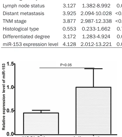
MiR-153 is down-regulated in NSCLC tissues
In order to explore the role of miR-153 in lung carcinogenesis, the expression patterns of miR-153 in 137 pairs of human lung cancer tis-sues and adjacent normal lung tistis-sues were analyzed using qRT-PCR. The result showed that the expression of miR-153 was reduced significantly, compared with adjacent normal
Table 1. Relationship between miR-153 expression and clinicopathologic features in NSCLC
miR-153 expression Characteristic numberCase (n=69)Low (n=68)High P value Age (years)
<60 53 28 25 0.726
≥60 84 41 43
Gender
Male 64 33 31 0.405
Female 83 36 47
Smoking history
Yes 107 56 51 0.415
No 30 13 17
TNM stage
I+II 104 45 59 0.005
III+IV 33 24 9
Lymph node status
Yes 31 22 9 0.014
No 106 47 59
Distant metastasis
Yes 12 11 1 0.004
No 125 58 67
Histological type
Adenocarcinoma 69 34 35 0.865
Squamous carcinoma 68 35 33 Differentiated degree
Low/middle 81 30 51 <0.001
High 56 39 17
primary tumor and mortality or final follow-up of the patient. The clinico-pathological data are summarized in
Table 1.
RNA isolation and quantitative real-time PCR
RNA was extracted from formalin- fixed tissues, using TRIzol reagent, PureLink™ FFPE RNA Isolation Kit and mirVana PARIS kit (Life Technology, California, USA). RNA was diluted in RNase-free water and stored at -80°C before use. For quantitative real-time PCR, the miRNA-specific TaqMan MicroRNA Assays (Applied Biosystems) for miR-153 was used as described by the manufacturer. U6 snRNA was used as an endogenous control for miRNA detection. The expression of miR-153 was quantified by measuring cycle threshold (Ct) values and normalized using the 2-ΔΔCt method relative to U6
snRNA.
Statistical analysis
lung tissues (shown in Figure 1, P<0.05). The median value of all 137 lung cancer samples was chosen as the cut-off point for separating tumors with low-level expression of miR-153 from high-level expression miR-153 tumors. Thus, 69 lung cancer patients had low-level expression of miR-153, while 68 lung cancer patients had high-level expression of miR-153.
Relationship between miR-153 expression and clinicopathological characteristics in NSCLC patients
[image:3.612.93.318.99.242.2]The relationships between miR-153 expression levels and clinicopathological characteristics in individuals with NSCLC are summarized in
Table 1. We did not find a significant associa-tion of miR-153 expression levels with patient’s gender (P=0.405), age (P=0.726), smoking sta-tus (P=0.415), and histological type (P=0.865) in 137 NSCLC cases. However, we observed that the expression level of miR-153 was positively correlated with the clinical stage
(P=0.003; shown in Figure 2). The five-year sur-vival rate for patients with low miR-153 expres-sion was 32.3%, compared with 71.6% for patients with high expression. Multivariate analysis was conducted using the Cox propor-tional hazards model to examine the impact of miR-153 expression and other clinicopathologi-cal parameters. The expression of miR-153 emerged as an independent and significant fac-tor associated with poor five-year survival rates (P=0.002, shown in Table 2).
Discussion
In humans, the miRNAs present in a genome harbor more than 500 experimentally cloned miRNAs, the total number of which could exceed 1000 [11]. Almost 30% of the human genome is estimated to be regulated by miR-NAs [12]. A number of reports have demon-strated that miRNAs control development, cell differentiation, apoptosis and proliferation. Moreover, the studies of miRNA expression pro-files of human tumors have reported phenotyp-ic signatures of partphenotyp-icular cancer types [13, 14]. The prognostic potential of miRNAs has been demonstrated for several types of cancer, including lung cancer. For example, Zhu et al found that miR-224 expression levels were sig-nificantly down-regulated in NSCLC compared to the corresponding noncancerous lung tis-sues. In addition, decreased miR-224 expres-sion was significantly associated with lymph node metastasis, advanced TNM stage, and shorter overall survival. Multivariate regression analysis corroborated that down-regulation of miR-224 was an independent unfavorable prog-nostic factor for patients with NSCLC [15]. Zhang et al found that miR-10b was
significant-Table 2. Multivariate Cox’s hazards model analysis for prognostic factors
Variable Hazard ratio 95% CI P value
Sex 1.056 0.378-2.677 0.461
Age 1.728 0.682-3.125 0.287
Smoking history 2.192 0.654-2.182 0.322 Lymph node status 3.127 1.382-8.992 0.009 Distant metastasis 3.925 2.094-10.028 <0.001 TNM stage 3.877 2.987-12.338 <0.001 Histological type 0.553 0.233-1.662 0.762 Differentiated degree 3.172 1.283-4.924 0.041 miR-153 expression level 4.128 2.012-13.221 0.002
(P=0.005), lymph node status (P=0.014), distant metastasis (P=0.004), and differen-tiated degree (P<0.001) in NSCLC patients (shown in Table 1).
Correlation between miR-153 expression and overall survival
[image:3.612.88.297.158.392.2]The prognostic value of miR-153 expression for overall survival in NSCLC patients was evaluated by comparing the patients with high and low miR-153 expression. According to the Kaplan-Meier survival analysis, the patients with low miR-153 expression exhib-ited evidently poorer overall survival rates than those with high miR-153 expression
[image:3.612.90.289.267.401.2]ly upregulated in NSCLC tissues as well as in A549 cell line. The relative miR-10b expression levels were significantly positively correlated with TNM stage and regional lymph node involvement. Kaplan-Meier analysis showed that patients with higher levels of miR-10b had significantly poorer survival than those with lower expression of this miRNA in patients, with a 5-year disease-specific survival (DSS) of 29.5 and 63.8%, respectively [16].
Several studies revealed that miR-153 played an important role in various types of cancer. Xu et al found that miR-153 was markedly down-regulated in the cells that underwent an epithe-lial-mesenchy maltransition. Ectopic expres-sion of miR-153 in mesenchymal-like cells resulted in an epithelial morphology change with decreased cellular invasive ability by sup-pressing SNAI1 and ZEB2 [7]. miR-153 induced apoptosis in a glioblastoma cell line DBTRG-05MG by downregulation of B-cell lymphoma 2 and myeloid cell leukemia sequence 1 [17]. However, there remain some studies showing that miR-153 promotes the development of cancer. Zhang et al. demonstrated that miR-153 upregulation increased colorectal cancer invasiveness and resistance to oxaliplatin and cisplatin both in vitro and in vivo by inducing MMP9 expression [18]. Wu et al found that in human prostate cancer upregulation of
miR-geting ADAM19 [19]. However, the clinical sig-nificance of miR-153 has not been inves- tigated.
In the present study, our results showed that the expression of miR-153 was reduced signifi-cantly, compared with adjacent normal lung tis-sues. We did not find a significant association of miR-153 expression levels with patient’s gender, age, smoking status, and histological type. However, we observed that the expres-sion level of miR-153 was positively correlated with the clinical stage, lymph node status, dis-tant metastasis, and differentiated degree in NSCLC patients. According to the Kaplan-Meier survival analysis, the patients with low miR-153 expression exhibited evidently poorer overall survival rates than those with high miR-153 expression. The five-year survival rate for patients with low miR-153 expression was 32.3%, compared with 71.6% for patients with high expression. Multivariate analysis was con-ducted using the Cox proportional hazards model to examine the impact of miR-153 expression and other clinicopathological pa- rameters. The expression of miR-153 emerged as an independent and significant factor asso-ciated with poor five-year survival rates.
In conclusion, decreased expression of miR-153 might be a potential unfavorable
prognos-Figure 2. MiR-153 as a prognostic factor in NSCLC patients. Patients with low miR-153 expression had poorer overall survival probability (P<0.05).
[image:4.612.90.377.69.305.2]tar-tic factor for patients with NSCLC. Further stud-ies would be needed to prove our findings and to find the role of miR-153 as a creditable clini-cal predictor for the outcome of NSCLC patients because of the limited sample size of patients in our investigation.
Disclosure of conflict of interest
None.
Address correspondence to: Dr. Xiu-Wen Wang, Department of Chemotherapy, Qilu Hospital, Shandong University, 107 West Wenhua Road, Qilu Hospital, Shandong University, Jinan 250012, Shandong, China. Tel: 086-531-82169114; Fax: 086-531-82169114; E-mail: dr_wangxiuwen@126. com
References
[1] Siegel R, Ma J, Zou Z and Jemal A. Cancer sta-tistics, 2014. CA Cancer J Clin 2014; 64: 9-29. [2] Bartel DP. MicroRNAs: genomics, biogenesis,
mechanism, and function. Cell 2004; 116: 281-297.
[3] Schratt GM, Tuebing F, Nigh EA, Kane CG, Sabatini ME, Kiebler M and Greenberg ME. A brain-specific microRNA regulates dendritic spine development. Nature 2006; 439: 283-289.
[4] van Kouwenhove M, Kedde M, Agami R. MicroRNA regulation by RNA-binding proteins and its implications for cancer. Nat Rev Cancer 2011; 11: 644-656.
[5] Calin GA and Croce CM. MicroRNA signatures in human cancers. Nat Rev Cancer 2006; 6: 857-866.
[6] Zhao S, Deng Y, Liu Y, Chen X, Yang G, Mu Y, Zhang D, Kang J and Wu Z. MicroRNA-153 is tumor suppressive in glioblastoma stem cells. Mol Biol Rep 2013; 40: 2789-2798.
[7] Xu Q, Sun Q, Zhang J, Yu J, Chen W and Zhang Z. Downregulation of miR-153 contributes to epithelial-mesenchymal transition and tumor metastasis in human epithelial cancer. Carcinogenesis 2013; 34: 539-549.
[8] Liu L, Chen R, Huang S, Wu Y, Li G, Zhang B, Liu Q, Yin D and Liang Y. miR-153 sensitized the K562 cells to As2O3-induced apoptosis. Med Oncol 2012; 29: 243-247.
[9] Wu Z, He B, He J and Mao X. Upregulation of miR-153 promotes cell proliferation via down-regulation of the PTEN tumor suppressor gene in human prostate cancer. Prostate 2013; 73: 596-604.
[10] Yuan Y, Du W, Wang Y, Xu C, Wang J, Zhang Y, Wang H, Ju J, Zhao L, Wang Z, Lu Y, Cai B and Pan Z. Suppression of AKT expression by miR-153 produced anti-tumor activity in lung can-cer. Int J Cancer 2015; 136: 1333-40. [11] Bentwich I, Avniel A, Karov Y, Aharonov R, Gilad
S, Barad O, Barzilai A, Einat P, Einav U, Meiri E, Sharon E, Spector Y and Bentwich Z. Identification of hundreds of conserved and nonconserved human microRNAs. Nat Genet 2005; 37: 766-770.
[12] Lewis BP, Burge CB and Bartel DP. Conserved seed pairing, often flanked by adenosines, in -dicates that thousands of human genes are microRNA targets. Cell 2005; 120: 15-20. [13] Iorio MV, Ferracin M, Liu CG, Veronese A,
Spizzo R, Sabbioni S, Magri E, Pedriali M, Fabbri M, Campiglio M, Ménard S, Palazzo JP, Rosenberg A, Musiani P,Volinia S, Nenci I, Calin GA, Querzoli P, Negrini M and Croce CM. MicroRNA gene expression deregulation in hu-man breast cancer. Cancer Res 2005; 65: 7065-7070.
[14] Lu J, Getz G, Miska EA, Alvarez-Saavedra E, Lamb J, Peck D, Sweet-Cordero A, Ebert BL, Mak RH, Ferrando AA, Downing JR, Jacks T, Horvitz HR and Golub TR. MicroRNA expres -sion profiles classify human cancers. Nature 2005; 435: 834-838.
[15] Zhu D, Chen H, Yang X, Chen W, Wang L, Xu J and Yu L. Decreased microRNA-224 and its clinical significance in non-small cell lung can -cer patients. Diagn Pathol 2014; 9: 198. [16] Zhang J, Xu L, Yang Z, Lu H, Hu D, Li W, Zhang
Z, Liu B and Ma S. MicroRNA-10b indicates a poor prognosis of non-small cell lung cancer and targets E-cadherin. Clin Transl Oncol 2015; 17: 209-14.
[17] Xu J, Liao X and Wong C. Downregulations of B-cell lymphoma 2 and myeloid cell leukemia sequence 1 by microRNA 153 induce apopto-sis in a glioblastoma cell line DBTRG-05MG. Int J Cancer 2010; 126: 1029-1035.
[18] Zhang L, Pickard K, Jenei V, Bullock MD, Bruce A, Mitter R, Kelly G, Paraskeva C, Strefford J, Primrose J, Thomas GJ, Packham G and Mirnezami AH. miR-153 supports colorectal cancer progression via pleiotropic effects that enhance invasion and chemotherapeutic re-sistance. Cancer Res 2013; 73: 6435-6447. [19] Shan N, Shen L, Wang J, He D and Duan C.