Original Article
Gli1, a potential cancer stem cell marker, is strongly
associated with prognosis in prostate cancer
Lili Lv1, Zhaoting Yang2, Tonghui Ma3, Yanhua Xuan2
1Department of Oncology and Hematology, The Second Hospital of Jilin University, Changchun 130041, P. R. Chi-na; 2Department of Pathology, Yanbian University College of Medicine, Yanji 133002, P. R. China; 3Jilin Provincial Key Laboratory on Molecular and Chemical Genetics, The Second Hospital of Jilin University, Changchun 130041, P. R. China
Received August 8, 2018; Accepted August 29, 2018; Epub October 1, 2018; Published October 15, 2018
Abstract: Background: Although glioma-associated oncogene homolog 1 (Gli1) is a key mediator of the Hedgehog pathway, Gli1 involvement in the maintenance of cancer stem-like cells (CSCs) in prostate cancer (PCa) is unclear. Methods: Herein, we assessed the expression of Gli1 and its relationship with cancer stemness genes, cell cycle
markers, epithelial-mesenchymal transition (EMT), and signaling pathway genes in 145 paraffin-embedded PCa tis
-sue samples using immunohistochemistry. In addition, we further confirmed the correlation between Gli1 and CSC marker in PC3 cells using immunofluorescence imaging. Results: High Gli1 expression was significantly associated with advanced primary tumor stage, positive lymph node metastasis, advanced clinical stage, and HIF-1α expres
-sion. The microvessel density was significantly higher in the Gli1 positive-cases than in the negative-cases. Further -more, Gli1 expression was positively correlated with stemness markers CD44. Survival analysis demonstrated that Gli1 and CD44 were strongly associated with the worse clinical outcome and an independent poor prognostic factor for overall survival. The enrichment analysis revealed that Gli1 was not correlated with E-cadherin, while positively correlated with Snail and vimentin. Notably, Gli1 expression was positively associated with the expression of cell cycle regulating genes such as cyclin D1, p21 and CDK4. Additionally, Gli1 expression was positively correlated
with pPI3K p85, pAkt-Ser473 and NF-κB p65 expression. Conclusions: Our results indicate that Gli1 is a potential
diagnostic marker of CSCs and that Gli1 expression is strongly associated with epithelial-mesenchymal transition in
PCa via PI3K/Akt/NF-κB signaling.
Keywords: Glioma-associated oncogene homolog 1, prostate cancer, prognosis, cancer stem-like cells, PI3K/Akt/
NF-κB signaling
Introduction
In patients with prostate cancer (PCa), metas-tasis is the most serious complication and the major cause of death [1]. Most patients with PCa ultimately develop invasive and drug-resis-tant metastatic cancer [2-4]. A study reported that PCa is the second leading cause of cancer-related death among males in the United States [5]. Therefore, studying the progression mecha-nisms of PCa is crucial to improve the survival rate of patients with PCa through effective treatment.
Cancer stem-like cells (CSCs) theory provides a new direction regarding the recurrence mecha-nism in tumors. Various human tumors contain CSCs which are positively correlated with the initiation, progression, and recurrence of
can-cer [6]. Therefore, the identification of novel tar-gets of CSCs or diagnostic markers of PCa to develop highly potent cancer molecular targets might be useful to improve the prognosis of PCa.
the association of Gli1 with CSCs markers in PCa. A positive correlation between Gli1 expres-sion and CSCs might provide a promising target to develop novel and efficient strategies to treat PCa. In addition, we explored whether Gli1 expression is associated with epithelial-mesen-chymal transition (EMT) in PCa via the phos-phatidylinositol-3-kinase (PI3K)/Akt/nuclear fa- ctor kappa B (NF-κB) signaling.
Materials and methods
Tissue specimens
A total of 145 cases of PCa tissue microarray paraffin-embedded samples were obtained from the Tissue Bank of the second hospital of Jilin University. The Institutional Review Board of Jilin University Medical College approved the study protocol and conducted in accordance with the 1996 Declaration of Helsinki. All patients provided written informed consent according to institutional guidelines. No patient received preoperative chemotherapy or radio-therapy. Pathologic parameters were carefully reviewed in all 145 PCa patients, including age, Gleason scores, tumor differentiation, primary tumor (pT), lymph node metastasis, and clinical stage. Follow-up survival data were collected retrospectively through medical-record analy-ses. The pTNM classification was applied
[image:2.612.89.375.71.285.2]Serial 4 μm tissue sections were deparaffinized with xylene, hydrated using a diluted alcohol series, and immersed in 3% H2O2 in methanol to quench endogenous peroxidase activity. Then sections were heated with TE buffer (1 mM EDTA and 10 mM Tris, pH 9.2) at 98°C for 30 minutes. To reduce non-specific staining, each section was blocked with 4% bovine serum albumin in PBS for 30 min. The sections were incubated with anti-Gli1 (1:100, Abcam, Cambridge, UK), anti-CD44 (1:100, Abcam, Cambridge, UK), anti-Sox2 (1:100, R&D, Min- neapolis, MN, USA), anti-LSD1 (1:250, Sigma, St. Louis, MO, USA), anti-Sox9 (1:100, Abnova, Walnut, CA, USA), anti-HIF-1α (1:80, Proteintech, Chicago, IL, USA), anti-CD68 (1:100, Abcam, Cambridge, UK), anti-cyclin D1 (1:100, Abcam, Cambridge, UK), anti-p21 (1:100, Abcam, Ca- mbridge, UK), anti-CDK4 (1:100, Abcam, Ca- mbridge, UK), anti-p27 (1:100, Abcam, Ca- mbridge, UK), anti-p16 (1:100, Abcam, Cam- bridge, UK), anti-E-cadherin (1:250, Abcam, Cambridge, UK), anti-Snail (1:100, Abcam, Cambridge, UK), anti-vimentin (1:80, Millipore, Bedford, MA, USA), anti-pPI3K p85 (1:30, Abcam, Cambridge, UK), anti-pAkt-Thr308 (1:80, Millipore, Bedford, MA, USA), anti-pAkt-Ser473 (1:80, Millipore, Bedford, MA, USA) and anti-NF-κB p65 (1:100, CST, Danvers, MA, USA), antibodies in PBST containing 3 mg/ml goat globulin (Sigma, St. Louis, MO, USA) for 2 hours
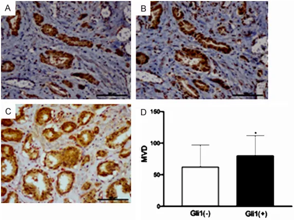
Figure 1. IHC staining of Gli1 (A) and HIF-1α (B) expression in the serial sec -tion of PCa tissues. (C) IHC double staining showed that Gli1 (brown reac-tion product) in the cancer cell and CD105 (red reaction product) indicated the
micro-vessel, and MVD was significantly higher in Gli1 positive cases than in
the negative cases (D). *P<0.05. (original magnification ×200).
according to guidelines from the 2010 American Joint Com- mittee on Cancer staging ma- nual (AJCC 7th edition) [15]. Cell lines
PC3 cells (PCa cell line), were maintained in RPMI-1640 with high glucose (Life Technolo- gies, Grand Island, NY) sup-plemented with 10% heat-inactivated fetal bovine serum (Life Technologies), 100 mg/ ml penicillin G, and 50 mg/ml streptomycin (Life Technolo- gies) at 37°C in a humidifi- ed atmosphere containing 5% CO2. All cell lines were purchased from the ATCC (Manassas, USA).
at room temperature, followed by three washes with PBST buffer. Then sections were incubat-ed with an anti-mouse/rabbit antibody (Envision plus, Dako, Denmark) for 30 minutes at room temperature. Next, the sections were immersed in chromogenic reagent 3, 3’-diaminobenzidine (Dako, Denmark) and then counterstaining with Meyer’s hematoxylin.
The double immunostaining procedure of Gli1/ CD105 in PCa tissues were the same as before. First, for the Gli1 protocols, except that the chromogen with the 3, 3’-diaminobenzidine
results were semi-quantitatively scored as neg-ative and positive [16].
Immunofluorescence (IF) analysis
PC3 cells were plated (50,000/well) on cover-slips in 6-well plates. We fixed the cells with 4% paraformaldehyde in PBS for 10 min at room temperature and permeabilized with 0.2% Triton X-100 in PBS for 5 min at room tempera-ture. To reduce non-specific staining, each sec-tion was blocked with 2% FBS in PBS for 30 min. The sections were then incubated with
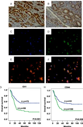
pri-Figure 2. IHC staining revealed Gli1 (A) and CD44 (B) in PCa tissues. IF stain-ing for Gli1 and CD44 in the PCa cells (C-F). Blue for DAP1; green for Gli1; red for CD44; double labeling for Merge. Kaplan-Meier analysis of OS that
the positive expression of Gli1 (G) and CD44 (H) in PCa was significantly
associated with a shortened OS compared to the negative groups. (original
magnification ×200).
(Dako) for 10 minutes, all steps were the same as before. Then, the subsequent staining of the same section was performed after incubat-ing the samples with an anti-body to CD105 by ImmPACT AEC Peroxidase Substrate for 20 minutes [14].
Evaluation of the IHC analysis
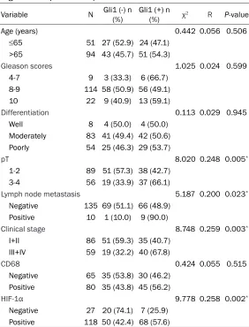
[image:3.612.93.368.69.489.2]Table 1. Comparison of clinicopathological characteristics accord-ing to Gli1 expression in prostate cancer
Variable N Gli1 (-) n (%) Gli1 (+) n (%) χ2 R P-value
Age (years) 0.442 0.056 0.506
≤65 51 27 (52.9) 24 (47.1)
>65 94 43 (45.7) 51 (54.3)
Gleason scores 1.025 0.024 0.599
4-7 9 3 (33.3) 6 (66.7)
8-9 114 58 (50.9) 56 (49.1)
10 22 9 (40.9) 13 (59.1)
Differentiation 0.113 0.029 0.945
Well 8 4 (50.0) 4 (50.0)
Moderately 83 41 (49.4) 42 (50.6)
Poorly 54 25 (46.3) 29 (53.7)
pT 8.020 0.248 0.005*
1-2 89 51 (57.3) 38 (42.7)
3-4 56 19 (33.9) 37 (66.1)
Lymph node metastasis 5.187 0.200 0.023*
Negative 135 69 (51.1) 66 (48.9)
Positive 10 1 (10.0) 9 (90.0)
Clinical stage 8.748 0.259 0.003*
I+II 86 51 (59.3) 35 (40.7)
III+IV 59 19 (32.2) 40 (67.8)
CD68 0.424 0.055 0.515
Negative 65 35 (53.8) 30 (46.2)
Positive 80 35 (43.8) 45 (56.2)
HIF-1α 9.778 0.258 0.002*
Negative 27 20 (74.1) 7 (25.9)
Positive 118 50 (42.4) 68 (57.6)
*Statistically significant.
mary antibodies against anti-Gli1 and anti-CD44 (1:50), overnight at 4°C. The next day, cells were incubated wi- th Alexa Fluor 568 goat an- ti-mouse IgG (Invitrogen, A12- 380) and Alexa Fluor 488 go- at anti-rabbit IgG (Invitrogen, A11008) secondary antibod-ies (1:150 dilution) for 1 hour. Nuclei were stained with DAPI and sections were mounted with vector shield mounting medium with DAPI for fluores-cence detection (vector lab, H-1200). Fluorescence detec-tion was performed with the Axiovert200II (Carl-Zeiss) and the intensity of IF cells was measured using Meta-Morph software.
Statistical analysis
Statistical analysis was per-formed by IBM SPSS17.0 sta-tistical software (SPSS Inc, Chicago, IL, USA). Correlations were assessed using Pear- son’s chi-square test as appro-priate. Overall survival (OS) rates were determined using the Kaplan-Meier method and compared using the log-rank test. Univariate and multivari-ate survival analyses were performed on all characteris-tics using the Cox proportional hazards model. Hazard ratios (HRs) and 95% confidence intervals (CI) were assessed for each factor. All tests were two sided, and P<0.05 was considered significant.
Results
Expression level of Gli1 and its association with clinico-pathologic characteristics in PCa
Gli1 expression in PCa was significantly associated with
Table 2. Correlation of Gli1 expression with cancer stem cell maker expression in prostate cancer
Variable N Gli1 (-) n (%) Gli1 (+) n (%) χ2 R P-value
Sox2 0.000 0.001 0.989
Negative 43 20 (46.5) 23 (53.5) Positive 102 50 (49.0) 52 (51.0)
Sox9 1.721 0.111 0.190
Negative 8 2 (25.0) 6 (75.0) Positive 137 68 (49.6) 69 (50.4)
LSD1 0.002 0.004 0.968
Negative 21 10 (47.6) 11 (52.4) Positive 124 60 (48.4) 64 (51.6)
CD44 17.363 0.356 <0.001*
[image:4.612.91.373.523.706.2]advanced pT (P = 0.005), positive lymph node metastasis (P = 0.023), and advanced clinical stage (P = 0.003), as these manifestations were highly common in patients with PCa. Additionally, we found that Gli1 expression (P = 0.002) was significantly higher in the 1α-positive group (57.6%, 68/118) than in the HIF-1α-negative group (25.9%, 7/27). Moreover, IHC staining revealed that Gli1 co-localized with HIF-1α in the serial sections of tissue samples from patients with PCa (Figure 1A, 1B). In the results of double staining using Gli1 and CD105 antibodies, MVD (P<0.05) was significantly higher in the Gli1-positive cases (79.82±32.119) than in the Gli1-negative cases (62.17±34.777) (Figure 2C, 2D). However, significant associa-tion was not observed between Gli1 expression and clinicopathologic features, such as age, Gleason score, tumor differentiation, and CD68 expression (Table 1).
Gli1 is a potential diagnostic marker of pros-tate CSCs
IHC staining revealed that Gli1 (Figure 2A) expression positively correlated with CD44 (P<0.001) (Figure 2B) expression in PCa tis-sues (Table 2). However, Gli1 expression did not correlate with the expression of other can-cer stemness-related factors such as Sox2,
Sox9, and LSD1. In addition, double IF staining also revealed that the Gli1 positive cell popula-tion mostly has a high overlap with CD44 posi-tive cell population in PC3 cells (Figure 2C-F). Kaplan-Meier survival analysis revealed that the positive expression of Gli1 (P<0.001) and CD44 (P = 0.009) in patients with PCa was associated with a shortened OS rate compared with that of the Gli1- and CD44-negative patients (Figure 2G, 2H). Furthermore, in the univariate as well as multivariate Cox regres-sion analyses, the Gli1 (Univariate: HR = 2.191, P<0.001; Multivariate: HR = 1.963, P = 0.002) and CD44 (Univariate: HR = 1.83, P = 0.01; Multivariate: HR = 1.654, P = 0.034) expres-sion levels in addition to the status of lymph node metastasis (Univariate: HR = 2.13, P = 0.002; Multivariate: HR = 1.687, P = 0.041) were an adverse independent prognostic factor of the OS rate in patients with PCa (Table 3). Gli1 regulates EMT and enhances the expres-sion of cell cycle regulating factors via PI3K/
Akt/NF-κB signaling in PCa
We estimated the variations in the levels of EMT markers in tissue samples by using IHC. Snail was localized in the cytoplasm and nuclei, and E-cadherin was present in the cytoplasm of cancer cells, whereas vimentin was detected in
Table 3. Univariate and multivariate analyses for prognostic variables of overall survival in prostate cancer patients using Cox proportional-hazards regression
Characteristic Univariate analyses Multivariate analyses
HR 95% CI p-value HR 95% CI p-value
Age (years) 0.097 0.205
≤65 1.00 1.00
>65 1.493 0.931-2.397 1.382 0.838-2.278
pT 0.002* 0.054
1-2 1.00 1.00
3-4 5.933 1.879-18.733 3.177 0.979-10.307
Lymph node metastasis 0.002* 0.041*
Negative 1.00 1.00
Positive 2.13 1.326-3.422 1.687 1.021-2.788
CD44 0.01* 0.034*
Negative 1.00 1.00
Positive 1.83 1.153-2.904 1.654 1.038-2.635
Gli1 <0.001* 0.002*
Negative 1.00 1.00
Positive 2.191 1.442-3.329 1.963 1.289-2.991
Figure 3. (A) IHC double staining showed that Gli1 (brown reaction product) in cancer cells and vimentin (red
reac-tion product) in the stromal fibroblasts; IHC staining of E-cadherin (B), Snail (C), cyclinD1 (D), CDK4 (E), p21 (F), pPI3K p85 (G), pAkt-Ser473 (H) and NF-κB p65 (I) in prostate cancer tissues. (original magnification ×200).
Table 4. Correlation of Gli1 expression with epithelial-mesenchy-mal transition maker expression in prostate cancer
Variable N Gli1 (-) n (%) Gli1 (+) n (%) χ2 R P-value
E-cadherin 5.842 0.201 0.016*
Negative 36 11 (30.6) 25 (69.4) Positive 109 59 (54.1) 50 (45.9)
vimentin 4.544 0.176 0.033*
Negative 44 27 (61.4) 17 (38.6) Positive 101 43 (42.6) 58 (57.4)
Snail 11.261 0.283 0.001*
Negative 117 65 (55.6) 52 (44.4)
Positive 28 5 (17.9) 23 (82.1) *Statistically significant.
the cytoplasm of stromal fibroblasts (Figure 3A-C). Furthermore, IHC double staining for Gli1 and vimentin were performed (Figure 3A). The results demonstrated that Gli1 was nega-tively correlated with E-cadherin (P = 0.016), while positively correlated with Snail (P = 0.001) and vimentin (P = 0.033) (Table 4).
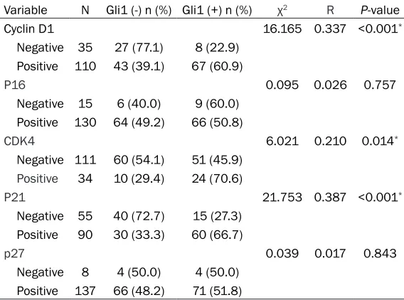
IHC staining of PCa patient tis-sues revealed that Gli1 expres-sion was positively associated with the cell cycle regulating factors such as cyclin D1 (P<0.001), p21 (P<0.001), and CDK4 (P = 0.014) expression (Table 5). Positive expression signals were localized mainly in the nuclei of cancer cells (Figure 3D-F). However, Gli1 expression did not correlate with p16 and p27 expression (Table 5). In addition, Gli1 was positively correlated with the pPI3K (P<0.001) and pAkt-Ser473 (P = 0.001) as well as NF-κB p65 (P<0.001) expression (Table 6; Figure 3G-I).
Discussion
corre-Table 5. Correlation of Gli1 expression with cell-cycle related gene expression in prostate cancer
Variable N Gli1 (-) n (%) Gli1 (+) n (%) χ2 R P-value
Cyclin D1 16.165 0.337 <0.001*
Negative 35 27 (77.1) 8 (22.9) Positive 110 43 (39.1) 67 (60.9)
P16 0.095 0.026 0.757
Negative 15 6 (40.0) 9 (60.0) Positive 130 64 (49.2) 66 (50.8)
CDK4 6.021 0.210 0.014*
Negative 111 60 (54.1) 51 (45.9)
Positive 34 10 (29.4) 24 (70.6)
P21 21.753 0.387 <0.001*
Negative 55 40 (72.7) 15 (27.3) Positive 90 30 (33.3) 60 (66.7)
p27 0.039 0.017 0.843
[image:7.612.90.374.97.308.2]Negative 8 4 (50.0) 4 (50.0) Positive 137 66 (48.2) 71 (51.8) *Statistically significant.
Table 6. Correlation of Gli1 expression with PI3K/Akt/NF-ΚB sig-naling in prostate cancer
Variable N Gli1 (-) n (%) Gli1 (+) n (%) χ2 R P-value
pPI3K p85 13.720 0.306 <0.001*
Negative 71 46 (64.8) 25 (35.2)
Positive 74 24 (32.4) 50 (67.6)
pAkt-Ser473 11.319 0.288 0.001*
Negative 31 23 (74.2) 8 (25.8) Positive 114 47 (41.2) 67 (58.8)
pAkt-Thr308 0.693 0.071 0.405
Negative 10 3 (30.0) 7 (70.0) Positive 135 67 (49.6) 68 (50.4)
NF-ΚB p65 24.204 0.409 <0.001*
Negative 54 40 (74.1) 14 (25.9) Positive 91 30 (33.0) 61 (67.0) *Statistically significant.
lated with the initiation, progression, and recur-rence of cancer [6]. The novel finding in our study was that Gli1 might be a potential diag-nostic marker of prostate CSCs and strongly associated with EMT through PI3K/Akt/NF-κB signaling in PCa.
Increasing evidence proves that Gli1 expres-sion is positively correlated with the degree of malignancy and poor survival rate in several cancers including ESCC, LSCC, and DBC [12-14]. Consistent with our previous studies, Gli1
expression was very high in aggressive cases of PCa with advanced pT, positive lymph node metastasis and ad- vanced clinical stage, as Gli1 promotes tumor progression in PCa. In this study, survival analysis demonstrated that Gli1 was strongly associated with worse clinical outcome and independent poor prog-nostic factors of OS rate in PCa. As the formation of new blood vessels is necessary for the proliferation and metasta-sis of PCa cells, we assessed MVD and found that it was sig-nificantly higher in the Gli1-positive cases than that in the Gli1-negative cases. This re- sult suggested that the role of Gli1 as a prognostic factor is associated with the induction of angiogenesis. The statisti-cal significance indicated that Gli1 is an important predictive factor to detect tumor aggres-siveness and prognosis in patients with PCa.
[image:7.612.91.373.366.550.2]EMT is a process that stimulates cancer cell invasion and metastasis [20]. It is essential to study cancer metastasis to establish the molecular mechanisms of EMT, for the develop-ment of novel therapeutic strategies [21]. Gli1 enhances invasion and EMT by promoting CSCs-like properties in the ER-positive breast cancers [22]. The high Gli1 expression pro-motes EMT by reducing E-cadherin expression and enhancing the Snail and vimentin expres-sion in ESCC [23]. To identify Gli1 as a regulator of EMT process in PCa, we examined the cor-relation between Gli1 and EMT-related markers by IHC. Consistent with the aforementioned studies, our study demonstrated that Gli1 expression was strongly associated with EMT as it was negatively correlated with E-cadherin expression, while positively correlated with Snail and vimentin expression in PCa. These results suggested that Gli1 might be a master regulator of EMT and in turn regulates cancer cell invasion and metastasis in PCa.
Cell proliferation and cell cycle progression lead to the clonal expansion of tumor cells dur-ing tumor development [24]. Sun et al. indicat-ed that Gli1 regulates the cyclin D1 expression and in turn modulates the cell cycle or prolifera-tion via the Hh signal pathway [25]. Further, Cui et al. revealed that Gli1 co-localizes with cyclinD1, CDK4, and p21 in LSCC tissue sam-ples by using IHC multi-staining [13]. Sun et al. reported that the inhibition of Gli1 suppresses cell growth and cell cycle progression [26]. Consistent with these results, we found that Gli1 expression was significantly correlated with cyclinD1, p21, and CDK4 expression in PCa. Our results indicated that the up-regula-tion of Gli1 was associated with the regulaup-regula-tion of tumor cell proliferation which induces cell cycle progression in PCa.
PI3K/Akt/mTOR/NF-κB signaling is involved in cell survival, growth, migration, differentiation, and maintenance of self-renewal in the tumor cells [27, 28]. Sonic Hh-Gli1 signals promote the EMT process by modulating PI3K/Akt path-way in ovarian cancer [29]. Colavito et al. reported evidence of crosstalk between Gli1 signaling and NF-κB pathway in EMT [30]. In ESCC, Gli1 interacts with NF-κB signaling and regulates the cell cycle [12]. Various studies have reported that activation of the PI3K/Akt
pathway is involved in NF-κB activation [31]. Our results show that Gli1 goes along with pPI3K p85, pAkt-Ser473 and NF-κB p65 expres-sion. Moreover, Gli1 plays a critical role in regu-lating the EMT and cell cycle process in PCa. This suggested that Gli1 regulates EMT process to promote tumor proliferation and metastasis probably via the activation of PI3K/Akt/NF-κB signaling and exhibits the potential as a promis-ing target for PCa CSCs therapy. However, fur-ther studies are necessary to determine the specific regulatory mechanism through Gli1 in PCa.
Conclusions
In conclusion, the over-expression of Gli1 might act as a potential prostate CSCs marker and effectively predict poor prognosis in PCa. Moreover, Gli1 might be used as a promising target for therapeutic strategies to prevent the progression of EMT via PI3K/Akt/NF-κB sig- naling.
Acknowledgements
This study was supported by grants from the National Natural Science Foundation of China (No. 81460390, 81760531, 81560400).
Disclosure of conflict of interest
None.
Address correspondence to: Dr. Tonghui Ma, Jilin Pro- vincial Key Laboratory on Molecular and Chemical Genetics, The Second Hospital of Jilin University, Changchun 130041, P. R. China. Tel: +86-411-86110278; Fax: +86-411-86110378; E-mail: tong-huima@dlmedu.edu.cn; Dr. Yanhua Xuan, Depart- ment of Pathology, Yanbian University College of Medicine, No. 977, Gongyuan Road, Yanji 133002, P. R. China. Tel: +86-433-2435107; Fax: +86-433-2732456; E-mail: xuanyh1@ybu.edu.cn
References
[1] Liu Q, Jernigan D, Zhang Y, Fatatis A. Implica-tion of platelet-derived growth factor receptor alpha in prostate cancer skeletal metastasis. Chin J Cancer 2011; 30: 612-619.
[3] van den Hoogen C, van der Horst G, Cheung H,
Buijs JT, Lippitt JM, Guzmán-Ramírez N, Hamdy
FC, Eaton CL, Thalmann GN, Cecchini MG, Pel-ger RC, van der Pluijm G. High aldehyde
dehy-drogenase activity identifies tumor-initiating
and metastasis-initiating cells in human pros-tate cancer. Cancer Res 2010; 70: 5163-5173. [4] Dubrovska A, Elliott J, Salamone RJ, Kim S,
Aimone LJ, Walker JR, Watson J, Sauveur-Mi-chel M, Garcia-Echeverria C, Cho CY, Reddy VA,
Schultz PG. Combination therapy targeting
both tumor-initiating and differentiated cell populations in prostate carcinoma. Clin Cancer Res 2010; 16: 5692-5702.
[5] Siegel RL, Miller KD, Jemal A. Cancer statistics, 2016. CA Cancer J Clin 2016; 60: 7-30. [6] Visvader JE, Lindeman GJ. Cancer stem cells in
solid tumours: accumulating evidence and un-resolved questions. Nat Rev Cancer 2008; 8: 755-768.
[7] Clement V, Sanchez P, de Tribolet N, Radova
-novic I, Ruiz i Altaba A. HEDGEHOG-GLI1 sig -naling regulates human glioma growth, cancer stem cell self-renewal and tumorigenicity. Curr Biol 2007; 17: 165-172.
[8] Palma V, Ruiz i Altaba A. Hedgehog-GLI signal -ing regulates the behavior of cells with stem cell properties in the developing neocortex. De-velopment 2004; 131: 337-345.
[9] Palma V, Lim DA, Dahmane N, Sánchez P, Bri
-onne TC, Herzberg CD, Gitton Y, Carleton A, Al
-varez-Buylla A, Ruiz i Altaba A. Sonic hedgehog
controls stem cell behavior in the postnatal and adult brain. Development 2005; 132: 335-344.
[10] Lai K, Kaspar BK, Gage FH, Schaffer DV. Sonic hedgehog regulates adult neural progenitor proliferation in vitro and in vivo. Nat Neurosci 2003; 6: 21-27.
[11] Machold R, Hayashi S, Rutlin M, Muzumdar
MD, Nery S, Corbin JG, Gritli-Linde A, Dellovade T, Porter JA, Rubin LL, Dudek H, McMahon AP, Fishell G. Sonic hedgehog is required for pro-genitor cell maintenance in telencephalic stem cell niches. Neuron 2003; 39: 937-950. [12] Yang Z, Cui Y, Ni W, Kim S, Xuan Y. Gli1, a
po-tential regulator of esophageal cancer stem
cell, is identified as an independent adverse
prognostic factor in esophageal squamous cell carcinoma. J Cancer Res Clin Oncol 2017; 143: 243-254.
[13] Cui Y, Cui CA, Yang ZT, Ni WD, Jin Y, Xuan YH. Gli1 expression in cancer stem-like cells pre-dicts poor prognosis in patients with lung squa-mous cell carcinoma. Exp Mol Pathol 2017; 102: 347-353.
[14] Ni WD, Yang ZT, Qi WB. Gli1 is a potential can-cer stem cell marker and predicts poor
progno-sis in ductal breast carcinoma. Hum Pathol 2017; 69: 38-45.
[15] Edge SB, Byrd DR, Compton CC, et al. AJCC cancer staging manual. 7th edition. New York, NY: Springer; 2010. pp. 103-111.
[16] Yang ZT, Yeo SY, Yin YX, Lin ZH, Lee HM, Xuan YH, Cui Y, Kim SH. Tenascin-C, a prognostic de-terminant of esophageal squamous cell carci-noma. PLoS One 2016; 11: e0145807. [17] Jaworska D, Szliszka E. Targeting apoptotic ac
-tivity against prostate cancer stem cells. Int J Mol Sci 2017; 18: 1648-1669.
[18] Korski K, Malicka-Durczak A, Bręborowicz J. Ex -pression of stem cell marker CD44 in prostate cancer biopsies predicts cancer grade in radi-cal prostatectomy specimens. Pol J Pathol 2015; 65: 291-295.
[19] Carnero1 A, Lleonart M. The hypoxic microenvi-ronment: a determinant of cancer stem cell evolution. Bioessays 2016; 38 Suppl 1: S65-74.
[20] Nieto MA, Huang RY, Jackson RA, Thiery JP. EMT: 2016. Cell 2016; 166: 21-45.
[21] Wang Y, Lin Z, Sun L, Fan S, Huang Z, Zhang D,
Yang Z, Li J, Chen W. Akt/Ezrin Tyr353/NF-κB
pathway regulates EGF-induced EMT and me-tastasis in tongue squamous cell carcinoma. Br J Cancer 2014; 110: 695-705.
[22] Sun Y, Wang Y, Fan C, Gao P, Wang X, Wei G, Wei J. Estrogen promotes stemness and inva-siveness of ER-positive breast cancer cells through Gli1 activation. Mol Cancer 2014; 13: 137-153.
[23] Min S, Xiaoyan X, Fanghui P, Yamei W, Xiaoli Y, Feng W. The glioma-associated oncogene ho-molog 1 promotes epithelial--mesenchymal transition in human esophageal squamous cell cancer by inhibiting E-cadherin via snail. Cancer Gene Ther 2013; 20: 379-85.
[24] Zhong ZQ, Song MM, He Y, Cheng S, Yuan HS.
Knockdown of ezrin by RNA interference re -verses malignant behavior of human pancre-atic cancer cells in vitro. Asian Pac J Cancer Prev 2012; 13: 3781-3789.
[25] Sun J, Wang D, Li X, Yan J, Yuan X, Wang W. Targeting of miR-150 on Gli1 gene to inhibit proliferation and cell cycle of esophageal carci-noma EC9706. Cancer Biomark 2017; 21: 203-210.
[26] Sun Y, Guo W, Ren T, Liang W, Zhou W, Lu Q, Jiao G, Yan T. Gli1 inhibition suppressed cell growth and cell cycle progression and induced apoptosis as well as autophagy depending on ERK1/2 activity in human chondrosarcoma cells. Cell Death Dis 2014; 5: e979-990. [27] McCubrey JA, Abrams SL, Stadelman K,
and cancer stem cells. Adv Enzyme Regul
2010; 50: 285-307.
[28] Bitting RL, Armstrong AJ. Targeting the PI3K/ Akt/mTOR pathway in castration-resistant prostate cancer. Endocr Relat Cancer 2013; 20: R83-99.
[29] Ke Z, Caiping S, Qing Z, Xiaojing W. Sonic hedgehog-Gli1 signals promote epithelial-mes-enchymal transition in ovarian cancer by medi-ating PI3K/AKT pathway. Med Oncol 2015; 32: 368-376.
[30] Colavito SA, Zou MR, Yan Q, Nguyen DX, Stern
DF. Significance of glioma-associated onco -gene homolog 1 (GLI1) expression in claudin-low breast cancer and crosstalk with the nucle-ar factor kappa-light-chain-enhancer of ac-
tivated B cells (NFκB) pathway. Breast Cancer
Res 2014; 16: 444-462.
[31] Dolcet X, Llobet D, Pallares J, Matias-Guiu X.