Original Article
Detection and genotyping of human papillomavirus
virus (HPV): a comparative analysis of clinical
performance in cervical and urine
samples in Chilean women
Kurt Buchegger1,2,3*, Tamara Viscarra1,2,3*, Alejandra Andana1,2,3, Carmen Ili1,2,3, Jaime López1,2,3, Louise
Zanella1,2,3, María Inés Carmona-López4, Juan José Fernández4, Irene Cartas Espinel1,2,3, Raúl Sánchez2,5,
Juan Carlos Roa6, Priscilla Brebi1,2,3
1Laboratory of Molecular Pathology, Department of Pathology, School of Medicine, Universidad de La Frontera, Temuco, Chile; 2Centro de Excelencia en Medicina Traslacional (CEMT), Universidad de La Frontera, Casilla 54-D, Temuco, Chile; 3Scientific and Technological Bioresource Nucleus (BIOREN), Universidad de La Frontera, Casilla 54-D, Temuco, Chile; 4Instituto de Ciencias Biomédicas (ICBM) y Centro de Investigaciones Multidisciplinares de
La Araucanía (CIMA), Universidad Autónoma de Chile; 5Department of Preclinical Science, School of Medicine, Universidad de La Frontera, Temuco, Chile; 6Department of Pathology, UC Centre for Investigational Oncology
(CITO), Advanced Centre for Chronic Diseases (ACCDiS), Pontificia Universidad Católica de Chile, Santiago de Chile, Chile. *Equal contributors.
Received May 15, 2018; Accepted June 22, 2018; Epub November 1, 2018; Published November 15, 2018
Abstract: Human papillomavirus (HPV) is the most common sexually transmitted infectious agent and is the main cause of cervical cancer (CC). In Chile, CC is the second leading cause of death by cancer in women aged 20-44 years, four times higher than in developed countries. Currently, the detection of HPV infection using a cervical brush is recommended; however, this is an invasive procedure that many women try to avoid. The aim of this study was to evaluate the clinical performance of a self-collected, urine-based HPV detection method using conventional PCR fol-lowed by a reverse line blot. A PCR-based HPV genotyping was performed on 190 paired cervical and urine samples from gynecological exams at public health centers in the Araucania Region, Chile. HPV DNA detection and genotyp-ing were performed by PCR and reverse line blot assay. Carcinogenic HPV types were present in 64.7% and 65.8% of the cervical and urine samples; the infection rates of HPV16 were 34.7% and 33.2%, respectively. The overall percent agreement between carcinogenic HPV detection in cervical and urine samples was 73.7%, with a moderate concordance rate of carcinogenic HPV detection (kappa = 0.42). Clinical sensitivities for cervical and urine-based sampling methods to diagnose cervical intraepithelial neoplasia 2/3 (CIN2/3) by histology were 93.4% and 90.2%, respectively. These results suggest that both cervical brush and urine-based sampling show a good clinical perfor-mance in the detection of HPV infection. The urine-based sampling method represents a valuable alternative with a great impact on public health, allowing increased cervical cancer screening coverage among women who do not undergo pelvic examinations.
Keywords: Human papillomavirus, cervical intraepithelial neoplasia, cervical cancer, urine-based sampling method
Introduction
Human papillomavirus (HPV) represents the most common sexually transmitted infectious agent throughout the world; the major risk fac-tors are behaviors associated with sexual activ-ity [1]. The etiological cause of nearly 100% of cervical cancers (CC) is being associated
sig-nificantly with vulvar and vaginal cancers in
shows similar sensitivity and specificity to cervi -cal-based sampling [15, 24-26]. Our study was performed in the Araucania Region, a high-risk population due to its sociodemographic fea-tures; one of the highest native populations in the country, 30% of women living in non-urban-ized areas, and high rates of incidence and mortality by CC [7, 27].
The aim of this study was to evaluate the clini-cal performance of a self-collected, urine-ba- sed HPV detection method using conventional PCR followed by reverse line blot that could be used as a non-invasive method for the early detection of CC, thereby allowing an increase in screening among populations of women at a high risk of the disease.
Material and methods
Study design
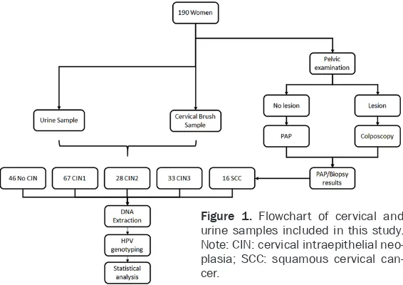
We analyzed a cohort of 190 women with cy- tology and histological diagnosis of cervical lesions conducted by specialized clinicians per-formed at the Unit of Pathological Anatomy and Cytology of the Hernán Henríquez Aravena Hospital (Temuco, Chile). To perform this study, three paired samples were collected from women referred for a gynecological exam at public health centers in the Araucania Region; one for diagnostic purposes and the other two for research purposes (cervical and urine
sam-ples). Subsequently, these were classified as
no cervical Intraepithelial neoplasia (No CIN), cervical intraepithelial neoplasia grade 1 (CIN1), grade 2 (CIN2), grade 3 (CIN3) and squa-mous cervical cancer (SCC), according to the results of the histological diagnosis. At the sa- me time - without knowing the diagnosis -DNA was extracted from each sample for HPV geno-typing (Figure 1).
Clinical sample collections
Cervical samples were collected by a physician using a cervical brush and placed in a Tris-EDTA buffer solution and transported immediately to the lab for further processing. On the same day, prior to undergoing a pelvic examination, 60 mL
of first-stream urine samples were collected at
home in a sterile container with crystal violet (10%) and then transported immediately to the lab.
Screening efforts are focused on preventing CC, primarily by Pap test, which can detect abnormal cellular changes or pre-malignant lesions in cervical tissues [9]. Since the Pap test came into use, mortality rates from CC have decreased dramatically [10]. Neverthele- ss, the PAP test has several disadvantages; it requires a pelvic examination, a procedure that is invasive and uncomfortable for the patient, time consuming for healthcare providers, and cannot be carried out easily [11]. In addition, cervical cytology is susceptible to technical limitations such as the inadequate transfer of cells to the slide with homogenous distribution of abnormal cells, the presence of obscuring
blood, and inflammation or thick areas of over
-lapping epithelial cells. Also, it is subject to interpretation errors, resulting in false negative results associated with relatively low sensitivity (less than 50%) [12, 13]. In the last two decades, interest in molecular techniques for identifying HPV DNA present in infected tissues
has increased, because it enables the identifi
-cation of women at risk of developing CC due to its higher sensitivity of detection with only a small amount of viral DNA [14-16].
DNA extraction from cervical and urine
samples
DNA extraction from the cervical and urine samples was performed using the Tissue DNA Kit E.Z.N.A (Zymo Research), according to the manufacturer’s instructions. DNA was
evaluat-ed by amplification of a 268 bp fragment of
the ß-globin gene using the GH20 and PCO4 primers and PCR conditions previously des- cribed [28, 29].
HPV genotyping
HPV genotyping was performed by
convention-al PCR amplification followed by non-radioac
-tive hybridization, which enables the detection of HPV carcinogenic genotypes (16, 18, 31, 33, 35, 39, 45, 51, 52, 56, 58, 59, 66) and HPV non-carcinogenic genotypes (6, 11, 42, 53, 70). The PCR reaction for the L1 gene was
per-formed using the specific primers GP5+ and biotinylated GP6+ as previously described [30].
Subsequently, a reverse line blot was per-formed to identify the HPV genotype, as
previ-ously reported [29]. The PCR product of GP5+/ biotinylated GP6+ was denaturalized at 96ºC
and cooled in ice before starting the hybridiza- tion step. Then, the PCR products were placed in a pre-treated Biodyne C membrane (Pall Bio-Support West Chester, USA) containing
non-radioactive labeled oligoprobes specific to each
viral genotype. A colorimetric reaction using anti-digoxigenin conjugate with alkaline phos-phatase was used to detect the presence/ absence of each HPV genotype. Finally, the membranes were developed using a NBT/BCIP
The overall percent agreement (OPA), positive and negative predictive agreement (PPA and NPA), and agreement beyond chance (Cohen’s kappa) were calculated as percentages with
95% confidence intervals (95% CI) for carcino
-genic HPV detection in the cervical and urine samples. The kappa concordance test was interpreted according to a previous study [25]. McNemar’s test was used to calculate the dif-ferences between paired proportions, with a p
value less than 0.05 being considered
statisti-cally significant.
In addition, we calculated the sensitivity,
speci-ficity, positive and negative predictive values,
and Youden’s index with a 95% CI in the cervi-cal and urine samples for the detection of car-cinogenic HPV DNA against two histologically
confirmed cervical disease endpoints: CIN2/3
and CIN3. Results
Comparison of carcinogenic HPV detection in cervical and urine samples
All 190 paired cervical and urine samples col-lected were suitable for analysis in this study,
and the β-globin gene was amplified in all of
[image:3.612.92.375.74.274.2]those. The median age of the study participants was 34 years (interquartile range: 16 years; range: 15-75 years). At least one of the detect-able HPV genotypes was present in 70.5% (134/190) of the cervical samples and 71.1% (135/190) of the urine samples. At least one of the detectable HR-HPV types was present in 65.8% (125/190) of the cervical samples and Figure 1. Flowchart of cervical and
urine samples included in this study. Note: CIN: cervical intraepithelial neo-plasia; SCC: squamous cervical can-cer.
substrate solution (Thermo Fisher), according to the man-ufacturer’s protocol.
Ethics statement
This study was approved by Ethics Committee of the Fa- culty of Medicine of the Uni- versity of La Frontera, Temuco, Chile (Resolution Number 246/006, august 25th 2010).
All the women were informed of the purposes of this study and written informed consent was obtained.
64.7% (123/190) of the urine samples (P-value = 0.89). The detection of LR-HPV type was practically equal in the cervical and urine sam-ples (4.7% and 6.3%; P-value = 0.63, respec-tively). The HPV16 frequency was 34.7% and 33.2% (P-value = 0.79) in cervical and urine samples, respectively (Figure 2). In summary,
there were no significant differences in HPV
detection and genotyping between the cervical and urine samples.
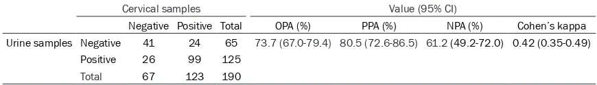
The OPA between HR-HPV detection in the cer-vical and urine samples was 73.7% (95% CI: 67.0%-79.4%). The PPA between the cervical and urine samples was 80.5% (95% CI: 72.6%-86.5%). A moderate concordance rate of HR- HPV detection in the two samples was observ-ed (kappa = 0.42; 95% CI: 0.35-0.49) (Table 1). There were 50/190 (26.3%) discrepancies in the results obtained for the two types of sam-ples. In 24 of them, the detection of carcino-genic HPV was positive in the cervical but nega-tive in the urine samples. Conversely, 26 samples were positive for HR-HPV detection in urine but negative in cervical samples (see Supplementary Table 1).
HR-HPV detection in cervical and urine sam -ples according to grade lesion endpoint
Histology results were available for all 190 women, which 46/190 (24.2%) were No CIN, 67/190 (35.3%) CIN1, 28/190 (14.7%) CIN2, 33/190 (17.4%) CIN3, and 16/190 (8.4%) SCC.
tive for CIN2 in urine samples was positive for HPV16 in the cervical samples. None of the negative cases, either cervical or urine sam-ples, were the same (Supplementary Table 1). No CIN was compared against histologically
confirmed CIN2/3 or CIN3 lesions. CIN1 was
not considered in this analysis because group-ing it with samples from normal epithelia is an error, due to it presenting an early cervical lesion; therefore, many of those cases were positive for HR-HPV infections (74.6% of the cervical and 70.1% of the urine samples), whi-
ch affects the specificity of methodology with
possible “false positives”. Compared to his-
tologically confirmed CIN2/3, cervical-based
detection of HR-HPV had a clinical sensitivity of 93.4% (95% CI: 84.1%-98.2%) and a clinical
specificity of 80.4% (95% CI: 66.1%-90.6%). The corresponding sensitivity and specificity
for urine specimens were 90.2% (95% CI: 79.8%-96.3%) and 71.7% (95% CI: 56.6%-84.0%), respectively. Moreover, the sensitivity estimated for CIN3 lesions compared to res- pective estimates for CIN2/3 was lower (cer- vical: 90.9%; 95% CI: 75.7%-98.1%; urine: 72.7%; 95% CI: 54.5%-86.7%); however, the
specificity estimates were higher (cervical:
82.6%; 95% CI: 68.6%-92.2%; urine: 78.3%; 95% CI: 63.6%-89.1%) (Table 3).
Discussion
[image:4.612.91.376.71.256.2]Cervical cancer (CC) has among the highest prevalence and incidence of neoplasia in Figure 2. Prevalence of HPV genotypes detected in cervical and urine
sam-ples. Note: HR-HPV: high risk HPV; LR-HPV: low risk HPV; HPV 16: HPV geno-type 16.
nega-women worldwide, mostly in developing coun-tries [31]. The main screening technique for cervical lesions is the Pap stain, which has a sensitivity that does not reach 60%. In recent years new HPV-based tests have emerged showing better sensitivity than the Pap test in CC diagnoses. It is known that almost all CCs are caused by HPV infections [32]. Conse- quently, in 2005, the IARC made recomm- endations that supported primary screening based on cytology and/or HPV-DNA testing [22]. The test for HR-HPV genotypes has recent-ly been shown to be a better indicator of cervi-cal cancer risk than cytology [33]. Co-testing of cytology and HPV detection at 5-year intervals is the preferred strategy for cervical cancer screening for women aged 30 to 64 years in the United States [34]. Nevertheless, the methods clinically validated for HPV detection, including the Pap test, usually require cervical brushing, a sampling method that is usually avoided in high-risk populations because of shame, reli-gious or sociocultural background, or lack of access to health care centers. On the other hand, the urine-sampling method has some advantages over cervical scraping such as: non-invasive collection method, self-sampling method, and highly accepted among women [35], and a low amount of DNA can be detected [36]. Many reports indicates that HPV infec-tions are very common and most women will clear HPV infections within 6-12 months, but in this sense, the presence of HR-HPV DNA does not mean that CC is present or will persist and progress to cancer [19, 37, 38]. Therefore, it is necessary for a HR-HPV test to be non-invasive, easy to use, and inexpensive, in order to increase the coverage of CC screening in high-risk populations, usually those in low-income regions.
In this framework, we evaluated the clinical per-formance of urine-based HPV DNA testing and its different genotypes detected in 190 paired cervical and urine samples of women refer-
red from a public health care center (Te- muco, Chile). The prevalence of HR-HPV infec-tion was increased with the grade of CIN dis-ease. This prevalence was higher in urine than cervical samples for CIN1, CIN2, and SCC. Nevertheless, the prevalence in CIN3 was high-er in the chigh-ervical than in the urine samples. Several reports indicate that HR-HPV detection in urine samples could be increased as the cer-vical lesion progresses due to the exfoliation of cervical cells infected with HPV DNA [24, 39-41].
[image:5.612.88.523.97.159.2]Also, we found an OPA of 73.7% with a moder-ate concordance rmoder-ate between HR-HPV infec-tions detected in tissue samples from cervical lesions and urine. These results are similar to those reported by Sahasrabuddhe et al., (OPA = 79.2%; k = 0.55) [24], Mendez et al., (OPA = 76.0%) [39], and Bisset et al., (k = 0.58). These three studies used the Linear Array HPV Genotyping Test. On the other hand, the results obtained in this study were slightly lower than those documented by Tanzi et al., with a Cohen’s kappa of 0.80 obtained in a population of women infected with HIV using an in-house PCR [15], and Bernal et al. (OPA = 88.0%; k = 0.76) using a Cobas 4800 HPV test in a population of women with an abnormal PAP test [25]. One possible explanation of this vari-ation could be the presence of DNA HPV from urethral and vulvar epithelial tissues, because these tissues are susceptible to different HPV genotype infections than cervical tissue, usu-ally non-carcinogenic genotypes [15, 35]. The method we used detects both oncogenic and non-oncogenic HPV types, unlike the COBAS 4800 methods detect only carcinogenic HPV types; this could likely affect the OPA and Cohen’s kappa value. Nevertheless, we de- monstrated genotype-level similarities betwe- en sampling approaches due to infection with HR-HPV, LR-HPV and/or HPV16 being uniform across either the cervical or the urine sampling methods. This is quite interesting because it Table 1. Comparative table of agreement between carcinogenic HPV detection in cervical and urine samples
Cervical samples Value (95% CI)
Negative Positive Total OPA (%) PPA (%) NPA (%) Cohen’s kappa Urine samples Negative 41 24 65 73.7 (67.0-79.4) 80.5 (72.6-86.5) 61.2 (49.2-72.0) 0.42 (0.35-0.49)
Positive 26 99 125
Total 67 123 190
shows that urine samples as well as cervical samples can be used with this method.
The clinical sensitivity and specificity of urinary
detection shows that 90.2% and 71.7% (cervi-cal: 93.4% and 80.4%, respectively) of women with CIN2/3 could be accurately detected with this type of sampling. In women with CIN3, the sensibility decreased slightly to 72.7% with
a specificity of 78.3% (cervical: 90.9% and
82.6%, respectively) for the urine samples. Sahasrabuddhe et al., [24] reported a
sensitiv-ity and specificsensitiv-ity of 80.8% and 53.3% for
women with CIN2/3 and 90.0% and 45.9% for women with CIN3 in urine samples. Another study conducted by Bernal et al. [25] showed a
sensitivity and specificity in women with CIN2/3
of 95.0% and 51.7% and for CIN3 100% and 54.1%, respectively. Sellors et al. [35] obtain-
ed a sensitivity and specificity of 44.8% and
69.7% in women with HSIL (CIN2/3).
A recent meta-analysis reported that urine-based testing is an alternative to cervical sam-pling [42]. In this regard, our study supports the use of urine-based sampling as a viable meth-od for screening and genotyping HPV infection in women who lack access to CC screening or that avoid pelvic examinations. Moreover, in Chile, especially in non-urbanized areas, a large number of women do not undergo CC screening due to the invasiveness of the sam-ple collection. Therefore, this method
repre-sents an important alternative to increase
cov-erage for those populations that are difficult to
include in CC screening programs. Acknowledgements
We would like to thank everyone from the par-ticipating healthcare institutions who collabo-rated in this study. This work was supported
by the National Fund for Scientific and
Te-chnological Development of Chile (FONDECYT) [11150802 to P.B., 11150622 to C.G.I., 318- 0550 to K.B.]; the Corporation for Production Development (CORFO) [12IDL2-18157, 09CN- 14-5960 to CEGIN]; Dirección de Investigación, Universidad de La Frontera (DIUFRO) [DI14-0072 to A.A and J.L., DI17-0079 to K.B.]; the
National Commission for Scientific and
Tech-nological Research - the Fund for Research Center in Priority Areas (CONICYT-FONDAP) [15130011 to JCR] and The Millennium In-
stitute on Immunology and Immunotherapy [Nº
P09-016-F to JCR]. The authors alone are responsible for the content and writing of this article.
Disclosure of conflict of interest
None.
[image:6.612.88.524.95.138.2]Address correspondence to: Priscilla Brebi, La- boratory of Molecular Pathology, Department of Pathology, School of Medicine, Universidad de La Table 2. HR-HPV prevalence in cervical and urine samples according different stages of the disease by histology
No CIN (%) CIN 1 (%) CIN 2 (%) CIN 3 (%) SCC (%) Overall (%)
Cervical 8/46 (17.4) 50/67 (74.6) 25/28 (89.3) 30/33 (90.9) 12/16 (75.0) 125/190 (65.8)
Urine 10/46 (21.7) 47/67 (70.1) 27/28 (96.4) 24/33 (72.7) 15/16 (93.8) 123/190 (64.7)
Note: CIN: Cervical intraepithelial neoplasia; SCC: Squamous cervical cancer.
Table 3. Comparison of test agreement between carcinogenic HPV detection in cervical and urine
samples for detection of histologically confirmed CIN2/3 and CIN3 lesions
Cervical disease endpoint
No. of samples % (95% CI)
Youden’s index (95% CI) Lesion
present Lesion absent Sensitivity Specificity PPV NPV CIN 2/3 Total TP FN FP TN
Cervical 107/190 57 4 9 37 93.4 (84.1-98.2) 80.4 (66.1-90.6) 86.4 (75.7-93.6) 90.2 (76.9-97.3) 0.739 (0.625-0.853) Urine 107/190 55 6 13 33 90.2 (79.8-96.3) 71.7 (56.6-84.0) 80.9 (69.5-89.4) 84.6 (69.5-94.1) 0.619 (0.491-0.747) CIN 3
Cervical 79/190 30 3 8 38 90.9 (75.7-98.1) 82.6 (68.6-92.2) 78.9 (62.7-90.5) 92.7 (80.1-98.5) 0.735 (0.577-0.893) Urine 79/190 24 9 10 36 72.7 (54.5-86.7) 78.3 (63.6-89.1) 70.6 (52.5-84.9) 80.0 (65.4-90.4) 0.51 (0.310-0.710)
[image:6.612.92.522.195.305.2]Frontera, Avenida Alemania #0478, 3th Floor, Temuco, Chile. E-mail: brebimieville@gmail.com
References
[1] Wheeler CM. The natural history of cervical hu-man papillomavirus infections and cervical cancer. Obstet Gynecol Clin North Am 2013; 40: 165-76.
[2] Alemany L, Saunier M, Alvarado-Cabrero I, Quirõs B, Salmeron J, Shin HR, Pirog EC, Gui-merà N, Hernandez-Suarez G, Felix A, Clavero O, Lloveras B, Kasamatsu E, Goodman MT, Hernandez BY, Laco J, Tinoco L, Geraets DT, Lynch CF, Mandys V, Poljak M, Jach R, Verge J, Clavel C, Ndiaye C, Klaustermeier J, Cubilla A, Castellsagué X, Bravo IG, Pawlita M, Quint WG, Muñoz N, Bosch FX, De Sanjosé S. Human papillomavirus DNA prevalence and type distri-bution in anal carcinomas worldwide. Int J Cancer 2015; 136: 98-107.
[3] Alemany L, Cubilla A, Halec G, Kasamatsu E, Quirós B, Masferrer E, Tous S, Lloveras B, Hernández-Suarez G, Lonsdale R, Tinoco L, Alejo M, Alvarado-Cabrero I, Laco J, Guimerà N, Poblet E, Lombardi LE, Bergeron C, Clavero O, Shin HR, Ferrera A, Felix A, Germar J, Mandys V, Clavel C, Tzardi M, Pons LE, Wain V, Cruz E, Molina C, Mota JD, Jach R, Velasco J, Carrilho C, López-Revilla R, Goodman MT, Quint WG, Castellsagué X, Bravo I, Pawlita M, Muñoz N, Bosch FX, De Sanjosé S. Role of human papil-lomavirus in penile carcinomas worldwide. Eur Urol 2016; 69: 953-61.
[4] De Sanjosé S, Alemany L, Ordi J, Tous S, Alejo M, Bigby SM, Joura EA, Maldonado P, Laco J, Bravo IG, Vidal A, Guimerà N, Cross P, Wain GV, Petry KU, Mariani L, Bergeron C, Mandys V, Sica AR, Félix A, Usubutun A, Seoud M, Hernán-dez-Suárez G, Nowakowski AM, Wilson G, Dal-stein V, Hampl M, Kasamatsu ES, Lombardi LE, Tinoco L, Alvarado-Cabrero I, Perrotta M, Bhatla N, Agorastos T, Lynch CF, Goodman MT, Shin HR, Viarheichyk H, Jach R, Cruz MO, Velasco J, Molina C, Bornstein J, Ferrera A, Do-mingo EJ, Chou CY, Banjo AF, Castellsagué X, Pawlita M, Lloveras B, Quint WG, Muñoz N, Bosch FX; HPV VVAP study group. Worldwide human papillomavirus genotype attribution in over 2000 cases of intraepithelial and invasive lesions of the vulva. Eur J Cancer 2013; 49: 3450-61.
[5] Brunner AH, Grimm C, Polterauer S, Hefler L,
Stani J, Heinze G, Horvat R. The prognostic role of human papillomavirus in patients with vagi-nal cancer. Int J Gynecol Cancer 2011; 21: 923-9.
[6] Ferlay J, Soerjomataram I, Ervik M, Dikshit R, Eser S, Mathers C, Rebelo M, Parkin DM,
For-man D, Bray F. Globocan 2012 v1.0, cancer incidence and mortality worldwide: IARC can-cerbase. No 11 [Internet]. Vol. 11, Lyon, France: International Agency for Research on Cancer 2013. p. http://globocan.iarc.f. [7] MINSAL. Series y Gráficos de Mortalidad [Inter
-net]. 2017. Available from: http://www.deis.cl/
series-y-graficos-de-mortalidad/.
[8] Forouzanfar MH, Foreman KJ, Delossantos AM, Lozano R, Lopez AD, Murray CJ, Naghavi M. Breast and cervical cancer in 187 countries between 1980 and 2010: a systematic analy-sis. Lancet 2011; 378: 1461-84.
[9] Peto PJ, Gilham PC, Fletcher O, Matthews FE. The cervical cancer epidemic that screening has prevented in the UK. Lancet 2004; 364: 249-56.
[10] Safaeian M, Solomon D, Castle PE. Cervical cancer prevention-cervical screening: science in evolution. Obstet Gynecol Clin North Am 2007; 34: 739-60.
[11] Cronjé HS. Cervical screening strategies in re-sourced and resource-constrained countries. Best Pract Res Clin Obstet Gynaecol 2011; 25: 575-84.
[12] Sherwani R, Khan T, Akhtar K, Zeba A. Conven-tional pap smear and liquid based cytology for cervical cancer screening-a comparative study. Journal of Cytology 2007; 24: 167-172. [13] Park IA, Lee SN, Chae SW, Park KH, Kim JW,
Lee HP. Comparing the accuracy of thinprep pap tests and conventional papanicolaou smears on the basis of the histologic diagno-sis: a clinical study of women with cervical ab-normalities. Acta Cytol 2000; 45: 525-31. [14] Ronco G, Giorgi-Rossi P, Carozzi F, Confortini
M, Palma PD, Del Mistro A, Gillio-Tos A, Minucci D, Naldoni C, Rizzolo R, Schincaglia P, Volante R, Zappa M, Zorzi M, Cuzick J, Segnan N. Re-sults at recruitment from a randomized con-trolled trial comparing human papillomavirus testing alone with conventional cytology as the primary cervical cancer screening test. J Natl Cancer Inst 2008; 100: 492-501.
[15] Tanzi E, Bianchi S, Fasolo MM, Frati ER, Mazza F, Martinelli M, Colzani D, Beretta R, Zappa A, Orlando G. High performance of a new PCR-based urine assay for HPV-DNA detection and genotyping. J Med Virol 2013; 85: 91-8. [16] Ronco G, Dillner J, Elfström KM, Tunesi S,
Sni-jders PJF, Arbyn M, Kitchener H, Segnan N, Gil-ham C, Giorgi-Rossi P, Berkhof J, Peto J, Meijer CJLM, Cuzick J, Zappa M, Carozzi F, Confortini M, Dalla Palma P, Zorzi M, Del Mistro A, Gillio-Tos A, Naldoni C, Rijkaart D, Van Kemenade F, Bulkmans N, Heideman D, Rozendaal R, Kenter G, Almonte M, Roberts C, Desai M,
screening for prevention of invasive cervical cancer: follow-up of four European randomised controlled trials. Lancet 2014; 383: 524-32. [17] Paavonen J. Human papillomavirus infection
and the development of cervical cancer and related genital neoplasias. Int J Infect Dis 2007; 11 Suppl 2: S3-9.
[18] Bouvard V, Baan R, Straif K, Grosse Y, Secre-tan B, El Ghissassi F, Benbrahim-Tallaa L, Guha N, Freeman C, Galichet L, Cogliano V. A review of human carcinogens--Part B: biological agents. Lancet Oncol 2009; 10: 321-2. [19] Schiffman M, Castle PE, Jeronimo J, Rodriguez
AC, Wacholder S. Human papillomavirus and cervical cancer. Lancet 2007; 370: 890-907. [20] Castle PE, Stoler MH, Wright TC, Sharma A,
Wright TL, Behrens CM. Performance of carci-nogenic human papillomavirus (HPV) testing and HPV16 or HPV18 genotyping for cervical cancer screening of women aged 25 years and older: a subanalysis of the ATHENA study. Lan-cet Oncol 2011; 12: 880-90.
[21] Agorastos T, Chatzistamatiou K, Katsamagkas T, Koliopoulos G, Daponte A, Constantinidis T, Constantinidis TC; HERMES study group. Pri-mary screening for cervical cancer based on high-risk human papillomavirus (HPV) detec-tion and HPV 16 and HPV 18 genotyping, in comparison to cytology. PLoS One 2015; 10: e0119755.
[22] IARC Working Group on the Evaluation of Can-cer. Cervix Cancer Screening (IARC Handbooks of Cancer Prevention, 10). In: Cervix Cancer Screening, Lyon: IARC; 2005. pp. 201-12. [23] MINSAL. Guías Clínicas AUGE Cáncer Cérvico
Uterino [Internet]. Santiago; 2015 [cited 2017 Mar 20]. p. 102. Available from: http://www. bibliotecaminsal.cl/wp/wp-content/uploads/ 2016/04/GPC-CaCU-Final.PLdocx.pdf
[24] Sahasrabuddhe VV, Gravitt PE, Dunn ST, Brown D, Allen RA, Eby YJ, Smith K, Zuna RE, Zhang RR, Gold MA, Schiffman M, Walker JL, Castle PE, Wentzensen N. Comparison of human pap-illomavirus detections in urine, vulvar, and cer-vical samples from women attending a colpos-copy clinic. J Clin Microbiol 2014; 52: 187-92. [25] Bernal S, Palomares JC, Artura A, Parra M, Ca-bezas JL, Robles A, Martín Mazuelos E. Com-parison of urine and cervical samples for de-tecting human papillomavirus (HPV) with the cobas 4800 HPV test. J Clin Virol 2014; 61: 548-52.
[26] Sahasrabuddhe VV, Gravitt PE, Dunn ST, Rob-bins D, Brown D, Allen RA, Eby YJ, Smith KM, Zuna RE, Zhang RR, Gold MA, Schiffman M, Walker JL, Castle PE, Wentzensen N. Evalua-tion of clinical performance of a novel urine-based HPV detection assay among women at-tending a colposcopy clinic. J Clin Virol 2014; 60: 414-7.
[27] INE. Compendio estadístico regional La Arau-canía, informe anual 2015 [Internet]. Compen-dio Estadístico Regional 2015- Región de La Araucanía. 2015 [cited 2017 Sep 15]. 1-92. Available from: http://www.inearaucania.cl/
archivos/files/pdf/SistemaEstadisticoRegion -al/Compendio Estadístico Regional 2015 - La Araucanía.pdf.
[28] Aedo AS, Melo AA, García P, Guzmán GP,
Ca-purro VI, Roa S JC. Detection and typification of
human papilloma virus in pre cancerous cervi-cal lesions. Rev Med Chil 2007; 135: 167-73. [29] Brebi P, Ili CG, Andana A, Menzel D, Lopez J,
Guzman P, Melo A, Buchegger K, Roa JC. Fre-quency of human papillomavirus in women at-tending cervical cancer screening program in Chile. BMC Cancer 2017; 17: 518.
[30] van den Brule AJ, Pol R, Fransen-Daalmeijer N,
Schouls LM, Meijer CJ, Snijders PJ. GP5+/6+
PCR followed by reverse line blot analysis
en-ables rapid and high-throughput identification
of human papillomavirus genotypes. J Clin Mi-crobiol 2002; 40: 779-87.
[31] Ferlay J, Shin HR, Bray F, Forman D, Mathers C, Parkin DM. Estimates of worldwide burden of cancer in 2008: GLOBOCAN 2008. Int J Cancer 2010; 127: 2893-917.
[32] Burd EM. Human papillomavirus and cervical cancer. Clin Microbiol Rev 2003; 16: 1-17. [33] Gage JC, Schiffman M, Katki HA, Castle PE,
Fetterman B, Wentzensen N, Poitras NE, Lorey T, Cheung LC, Kinney WK. Reassurance against future risk of precancer and cancer conferred by a negative human papillomavirus test. J Natl Cancer Inst 2014; 106.
[34] Guerrero-Preston R, Valle BL, Jedlicka A, Tura-ga N, Folawiyo O, Pirini F, Lawson F, Vergura A, Noordhuis M, Dziedzic A, Perez G, Renehan M, Guerrero-Diaz C, De Jesus Rodriguez E, Diaz-Montes T, Rodriguez Orengo J, Mendez K, Ro-maguera J, Trock BJ, Florea L, Sidransky D. Molecular triage of premalignant lesions in liquid-based cervical cytology and circulating cell-free dna from urine, using a panel of meth-ylated human papilloma virus and host genes. Cancer Prev Res (Phila) 2016; 9: 915-24. [35] Sellors JW, Lorincz AT, Mahony JB, Mielzynska
I, Lytwyn A, Roth P, Howard M, Chong S, Daya D, Chapman W, Chernesky M. Comparison of self-collected vaginal, vulvar and urine sam-ples with physician-collected cervical samsam-ples for human papillomavirus testing to detect high-grade squamous intraepithelial lesions. CMAJ 2000; 163: 513-8.
[36] Enerly E, Olofsson C, Nygård M. Monitoring hu-man papillomavirus prevalence in urine sam-ples: a review. Clin Epidemiol 2013; 67-79. [37] Bulkmans NW, Berkhof J, Bulk S, Bleeker MC,
HPV type-specific clearance rates in cervical
screening. Br J Cancer 2007; 96: 1419-24. [38] Schiffman M, Wentzensen N. Human
papillo-mavirus (HPV) infection and the multi-stage carcinogenesis of cervical cancer. Cancer Epi-demiol Biomarkers Prev 2013; 22: 553-60. [39] Mendez K, Romaguera J, Ortiz AP, Lopez M,
Steinau M, Unger ER. Urine-based human pap-illomavirus DNA testing as a screening tool for cervical cancer in high-risk women. Int J Gyne-col Obstet 2014; 124: 151-5.
[40] Gupta A, Arora R, Gupta S, Prusty BK, Kailash U, Batra S, Das BC. Human papillomavirus DNA in urine samples of women with or with-out cervical cancer and their male partners compared with simultaneously collected cervi-cal/penile smear or biopsy specimens. J Clin Virol 2006; 37: 190-4.
[41] Brinkman JA, Jones WE, Gaffga AM, Sanders JA, Chaturvedi AK, Slavinsky IJ, Clayton JL, Du-mestre J, Hagensee ME. Detection of human papillomavirus DNA in urine specimens from
human immunodeficiency virus-positive wom -en. J Clin Microbiol 2002; 40: 3155-61. [42] Pathak N, Dodds J, Zamora J, Khan K.
Supplementary Table 1. Characteristics of all patients include in this study
Histological-ly confirmed Lesion Age
Cervical-based sampling Urine-based sampling
HPV Genotyping
ANY HPV type (0: negative;
1: positive)
ANY HR-HPV type (0: negative;
1: positive)
ANY LR-HPV type (0: nega-tive; 1: positive)
HPV16 (0: negative; 1: positive)
HPV Genotyping
ANY HPV type (0: negative;
1: positive)
ANY HR-HPV type (0: negative;
1: positive)
ANY LR-HPV type (0: negative;
1: positive)
HPV16 (0: negative;
1: positive)
CIN 1 24 16 1 1 0 1 18 1 1 0 0
CIN 1 29 18 1 1 0 0 33, 18, 39,
53, 70
1 1 0 0
CIN 2 40 16 0 0 0 1 18, 45 0 0 0 0
CIN 1 24 58 0 0 0 0 58 0 0 0 0
CIN 2 41 56 0 0 0 0 56 0 0 0 0
CIN 1 47 18 0 0 0 0 33, 18, 45 0 0 0 0
CIN 2 29 45 1 1 0 0 18, 51, 45, 6,
42, 11, 70 1 1 0 0
CIN 2 22 58 0 0 0 0 59 0 0 0 0
CIN 2 27 16 0 0 0 1 16, 18 0 0 0 1
SCC 56 16 0 0 0 1 16, 18 0 0 0 1
CIN 1 61 16, 52 0 0 0 1 0 1 0 1 0
CIN 1 27 18 0 0 0 0 18, 45, 6, 11 0 0 0 0
CIN 1 50 16 0 0 0 1 0 0 0 0 0
CIN 1 37 83 0 0 0 0 16, 56, 59, 45 0 0 0 1
CIN 1 47 45 0 0 0 0 16, 18, 52,
35, 45, 11
0 0 0 1
CIN 3 32 18 0 0 0 0 16, 18, 35, 11 0 0 0 1
CIN 3 28 16, 18, 56 0 0 0 1 16, 56, 18 0 0 0 1
CIN 3 23 16 0 0 0 1 0 0 0 0 0
CIN 3 35 68 0 0 0 0 42 0 0 0 0
CIN 3 34 45 1 1 0 0 0 0 0 0 0
CIN 1 44 45 0 0 0 0 16, 18 0 0 0 1
CIN 2 31 16 1 1 0 1 31 0 0 0 0
CIN 3 43 45 0 0 0 0 31 0 0 0 0
CIN 3 34 16, 18, 52 0 0 0 1 16, 18, 35 0 0 0 1
CIN 1 32 66 0 0 0 0 52, 66 1 1 0 0
CIN 2 31 16 0 0 0 1 0 1 0 1 0
CIN 2 26 16 0 0 0 1 16 0 0 0 1
CIN 3 37 16 0 0 0 1 18 0 0 0 0
SCC 38 16 0 0 0 1 16 0 0 0 1
CIN 1 40 66 1 1 0 0 0 0 0 0 0
CIN 3 31 16 0 0 0 1 16, 18 1 1 0 1
CIN 2 38 66 0 0 0 0 18 1 1 0 0
CIN 1 61 16, 45 1 1 0 1 0 1 1 0 0
SCC 35 16 0 0 0 1 16, 42 0 0 0 1
CIN 1 52 16 0 0 0 1 0 0 0 0 0
CIN 3 40 16 0 0 0 1 16, 39, 53, 11 1 1 0 1
SCC 31 16 0 0 0 1 16, 58 1 0 1 1
CIN 3 40 52, 58 0 0 0 0 18, 53, 52,
45, 16, 35, 66, 11, 58, 56
0 0 0 1
CIN 1 42 58 0 0 0 0 18, 45, 16,
52, 11, 53 0 0 0 1
CIN 1 21 16 0 0 0 1 16, 18 0 0 0 1
CIN 2 29 16 1 1 0 1 16, 18 0 0 0 1
CIN 2 26 16 0 0 0 1 16, 52, 18,
35, 53, 11 1 1 0 1
CIN 3 35 16 0 0 0 1 59, 42 0 0 0 0
CIN 1 45 45 1 0 1 0 0 0 0 0 0
CIN 3 31 16 0 0 0 1 16 1 1 0 1
CIN 3 64 16 1 1 0 1 0 1 1 0 0
CIN 3 43 45 1 1 0 0 42 0 0 0 0
CIN 1 25 45 1 1 0 0 45 1 1 0 0
CIN 1 43 45 1 1 0 0 35 1 1 0 0
CIN 1 23 16 1 0 1 1 0 0 0 0 0
CIN 1 24 16 1 1 0 1 16 0 0 0 1
CIN 3 26 16 1 1 0 1 16, 42 0 0 0 1
CIN 3 41 46 1 1 0 0 56, 18, 31,
53, 11
1 1 0 0
CIN 2 23 58 1 1 0 0 58 0 0 0 0
CIN 1 27 16, 66 1 1 0 1 16, 52, 66,
53, 18, 42
1 1 0 1
CIN 2 57 16 0 0 0 1 58 0 0 0 0
CIN 1 34 16 1 1 0 1 16, 58 1 1 0 1
CIN 1 20 16 0 0 0 1 16, 33 0 0 0 1
CIN 2 32 45 1 1 0 0 16 0 0 0 1
CIN 2 33 45 0 0 0 0 31 1 1 0 0
CIN 1 25 52 1 1 0 0 16, 33, 18, 59 1 1 0 1
CIN 1 48 16 1 1 0 1 16, 18 1 1 0 1
CIN 1 38 0 1 1 0 0 0 0 0 0 0
CIN 3 49 16 1 0 1 1 16 1 0 1 1
CIN 1 23 0 1 1 0 0 59, 42 1 1 0 0
CIN 2 25 52 0 0 0 0 31 1 1 0 0
CIN 1 24 45, 56 1 1 0 0 56, 45, 6, 53 1 1 0 0
CIN 3 60 16 1 1 0 1 16 1 1 0 1
CIN 3 40 16 1 1 0 1 0 0 0 0 0
CIN 1 32 0 1 1 0 0 0 1 1 0 0
CIN 2 35 18 1 0 1 0 16, 58, 18, 31,
45, 11
1 0 1 1
CIN 2 19 31 1 1 0 0 18, 31, 66, 53 0 0 0 0
CIN 1 46 35 0 0 0 0 0 0 0 0 0
CIN 1 24 16 0 0 0 1 16, 59, 35,
45, 42, 53 1 1 0 1
CIN 1 49 0 1 1 0 0 16, 18, 52,
66, 45, 6, 53 1 0 1 1
CIN 1 20 18, 45 0 0 0 0 18, 45 1 1 0 0
CIN 2 30 17 1 1 0 1 16 1 1 0 1
CIN 1 22 16 1 1 0 1 18, 45 1 1 0 0
CIN 1 35 45.16 1 1 0 1 35 1 1 0 0
CIN 1 38 16 1 1 0 1 0 1 1 0 0
CIN 2 35 0 1 1 0 0 16, 56, 33,
58, 18, 35, 45, 6, 42
0 0 0 1
CIN 1 21 16 1 1 0 1 16 1 1 0 1
CIN 1 35 0 1 0 1 0 11 1 1 0 0
CIN 1 33 0 1 1 0 0 56, 51 1 1 0 0
CIN 2 30 16, 18, 37 1 1 0 1 16, 33, 35, 42 1 1 0 1
CIN 1 19 70 1 1 0 0 70 1 1 0 0
CIN 1 26 16, 52 1 1 0 1 16 1 1 0 1
CIN 1 25 0 1 1 0 0 16, 18, 31, 35,
45, 6, 70
0 0 0 1
CIN 1 42 52 1 1 0 0 31, 6, 70 1 1 0 0
CIN 1 19 16 1 1 0 1 56, 6 1 1 0 0
SCC 52 16 1 1 0 1 16, 11 1 1 0 1
CIN 1 61 45 1 1 0 0 0 1 1 0 0
CIN 1 18 51 1 1 0 0 0 1 1 0 0
CIN 1 35 18 0 0 0 0 18, 45, 35 1 1 0 0
CIN 1 27 33, 59 1 1 0 0 59, 42, 35 1 0 1 0
SCC 46 16 1 1 0 1 0 1 1 0 0
CIN 1 17 51 1 1 0 0 51 1 1 0 0
CIN 2 52 16-18 1 1 0 1 56, 18, 31, 53 1 1 0 0
CIN 3 35 16 0 0 0 1 16, 18 1 0 1 1
CIN 1 19. 70 0 0 0 0 70 1 1 0 0
CIN 3 33 16 1 1 0 1 16, 11 1 1 0 1
SCC 16 1 1 0 1 16, 18 1 1 0 1
SCC 56 16 1 1 0 1 18 1 1 0 0
CIN 1 40 16 1 0 1 1 59, 42, 53 1 1 0 0
CIN 1 23 0 1 1 0 0 0 1 1 0 0
CIN 3 55 31 1 1 0 0 59 1 1 0 0
CIN 2 50 52 1 1 0 0 52, 42, 53,
18, 16, 59, 45, 11
1 1 0 1
CIN 2 26 33 1 1 0 0 33, 42 1 1 0 0
CIN 3 33 52 1 1 0 0 42 1 1 0 0
CIN 3 45 31 0 0 0 0 16, 18, 52 1 1 0 1
SCC 35 0 1 1 0 0 58 1 1 0 0
CIN 3 58 0 1 1 0 0 42 1 1 0 0
CIN 3 36 0 1 1 0 0 0 1 1 0 0
CIN 1 30 0 1 1 0 0 52, 66 1 1 0 0
CIN 1 50 18 1 1 0 0 11 1 1 0 0
CIN 1 23 66 1 1 0 0 52, 66, 53 1 1 0 0
CIN 3 37 16 1 1 0 1 33, 18 0 0 0 0
CIN 3 21 16 1 1 0 1 16, 18, 11 1 1 0 1
CIN 1 23 0 1 0 1 0 58, 11 1 1 0 0
CIN 2 26 16 1 1 0 1 18 1 1 0 0
CIN 3 37 16 1 1 0 1 16, 11 1 1 0 1
CIN 1 20 0 1 1 0 0 56, 11 1 1 0 0
CIN 2 24 66 1 1 0 0 16, 18, 59,
52, 66, 42, 53, 11
1 1 0 1
CIN 3 60 33 1 1 0 0 11, 16, 18, 59 1 1 0 1
CIN 1 25 16 1 1 0 1 16 1 1 0 1
CIN 3 40 31,56,58 1 1 0 0 16, 18 1 1 0 1
CIN 3 34 0 1 1 0 0 16, 18, 59, 52,
35, 66, 45, 6, 42, 11
1 1 0 1
CIN 2 31 31 1 1 0 0 31, 35, 53,
18, 16
1 1 0 1
CIN 1 51 51 1 1 0 0 18, 53, 11, 16,
45, 35
1 1 0 1
CIN 1 40 16, 52 0 0 0 1 16, 52, 42 1 1 0 1
CIN 1 24 56 1 1 0 0 56, 35 1 1 0 0
CIN 1 55 58 1 1 0 0 16, 58, 18, 52,
31, 45, 11
1 1 0 1
CIN 2 33 16 1 1 0 1 16 1 1 0 1
CIN 1 25 0 1 1 0 0 58, 18, 51, 53 1 1 0 0
SCC 38 18 1 1 0 0 18 1 1 0 0
SCC 27 53 1 0 1 0 53 1 1 0 0
SCC 66 31 1 1 0 0 33, 58, 18, 31, 53
1 1 0 0
SCC 37 0 1 1 0 0 16, 18 1 1 0 1
SCC 75 0 1 1 0 0 16, 53 1 1 0 1
SCC 29 18 1 1 0 0 18, 42 0 0 0 0
No CIN 26 16 1 1 0 1 18 1 0 1 0
No CIN 47 16,45,58 1 1 0 1 58 0 0 0 0
No CIN 38 0 1 1 0 0 0 1 1 0 0
No CIN 39 0 1 1 0 0 0 1 1 0 0
No CIN 42 0 1 1 0 0 0 1 1 0 0
No CIN 32 0 1 1 0 0 0 1 1 0 0
No CIN 22 53 1 1 0 1 16 1 1 0 1
No CIN 27 0 1 1 0 0 0 1 1 0 0
No CIN 36 0 1 1 0 0 0 1 1 0 0
No CIN 38 0 1 1 0 0 0 1 1 0 0
No CIN 28 0 1 1 0 0 53 0 0 0 0
No CIN 49 0 1 1 0 0 0 1 0 1 0
No CIN 36 0 1 1 0 0 0 1 1 0 0
No CIN 22 0 1 1 0 0 0 1 1 0 0
No CIN 28 0 1 1 0 0 0 1 1 0 0
No CIN 43 0 1 1 0 0 0 1 1 0 0
No CIN 15 0 1 1 0 0 0 0 0 0 0
No CIN 41 0 1 1 0 0 0 1 1 0 0
No CIN 47 0 1 1 0 0 0 1 1 0 0
No CIN 32 11 1 1 0 0 0 1 1 0 0
No CIN 19 0 1 1 0 0 0 1 0 1 0
No CIN 28 56 1 1 0 0 0 1 1 0 0
No CIN 26 0 0 0 0 0 0 1 0 1 0
No CIN 20 0 0 0 0 0 0 0 0 0 0
No CIN 53 0 1 1 0 0 52 1 1 0 0
No CIN 40 0 1 1 0 0 42 1 1 0 0
No CIN 37 0 1 1 0 0 0 1 1 0 0
No CIN 34 0 1 1 0 0 0 1 1 0 0
No CIN 47 0 1 1 0 0 0 1 1 0 0
No CIN 27 58 0 0 0 0 0 1 1 0 0
No CIN 50 0 1 1 0 0 56 1 1 0 0
No CIN 21 0 1 1 0 0 16, 18, 51 1 1 0 1
No CIN 20 0 1 1 0 0 0 1 1 0 0
No CIN 21 35 1 1 0 0 16, 35 1 1 0 1
No CIN 24 0 1 1 0 0 0 1 1 0 0
No CIN 31 0 1 1 0 0 51 1 1 0 0
No CIN 53 0 1 1 0 0 53 1 1 0 0
No CIN 41 0 0 0 0 0 0 1 1 0 0
No CIN 42 0 1 1 0 0 0 1 1 0 0
No CIN 40 0 1 1 0 0 0 1 1 0 0
No CIN 56 16 1 0 1 1 0 1 1 0 0
No CIN 39 0 1 1 0 0 18 1 1 0 0
No CIN 24 0 0 0 0 0 0 1 1 0 0
No CIN 28 53 0 0 0 0 0 1 1 0 0
No CIN 28 0 1 1 0 0 18 1 1 0 0