Original Article
Bone marrow-derived mesenchymal stem cells
(BM-MSCs) inhibit apoptosis of spinal cord cells
in a kaolin-induced syringomyelia-associated
scoliosis rabbit model
Zhi Zhao1, Wei Xu2, Jingming Xie1, Yingsong Wang1, Tao Li1, Ying Zhang1, Daohong Zhao1, Ni Bi1, Zhiyue Shi1
Departments of 1Orthopaedics, 2Neurosurgery, The 2nd Affiliated Hospital of Kunming Medical University, Kun
-ming, Yunnan Province, P. R. China
Received January 28, 2018; Accepted March 12, 2018; Epub April 1, 2018; Published April 15, 2018
Abstract: The mechanisms and causes of scoliosis are believed to be multifactorial. Syringomyelia can often be found in scoliosis patients but the relationship between the two remains obscure. In this study, based on a rabbit model of syringomyelia-associated scoliosis, the involved pathological mechanism was explored in an attempt to further understand the relationship. This will also be helpful in determining how scoliosis occurred. In this study, a syringomyelia-associated scoliosis rabbit model was established by kaolin-injection technique. Spinal cell apoptosis following scoliosis and syringomyelia induction were analyzed. Furthermore, the effect of bone marrow-mesenchy-mal stem cell (BM-MSCs) transplantation on spinal cell apoptosis and on incidence of scoliosis and syringomyelia were assessed. Most of the experimental animals injected with kaolin developed progressive scoliotic curves and syringomyelia. Syrinx and scoliosis were found in 64.7% and 58.8% of the experimental animals. Syringomyelia-as-sociated scoliosis appeared in 41.2% of the animals. Syrinx size and scoliotic curves increased with time. Apoptosis was found on postoperative day 3 both in surgical segments and adjacent segments in the spinal cord, peaking at week 6. The number of apoptotic cells was significantly lower in BM-MSCs transplantation group compared with the saline-injection group. Fewer rabbits in the BM-MSCs injection group developed scoliosis or syringomyelia by the end of the experiment. Our findings indicate the potential value of kaolin-induced scoliotic animal models. For the first time, we studied features of apoptosis of spinal cells in a syringomyelia-associated scoliosis rabbit model. Our results demonstrate that BM-MSCs transplanted into the spinal cord decrease both apoptosis of spinal cells and incidence of scoliosis and syringomyelia.
Keywords: Animal model, scoliosis, syringomyelia, apoptosis, mesenchymal stem cell
Introduction
Scoliosis is a common spine disease. The prev-alence of scoliosis is approximately 1.02% among primary and middle school students [1]. The mechanisms and causes of scoliosis have not been identified, however, especially for idio-pathic patients. The cause of scoliosis is believed to be multifactorial because of known associations between development of scoliosis and growth, hormonal secretion, gravity, and other factors. The association between apopto-sis and scolioapopto-sis has been reported recently [2]. The mechanism of apoptosis in the patho-genesis of scoliosis and syringomyelia remains unclear, however, despite the number of stud-ies performed.
human disease has been widely investigated as a therapeutic strategy. Neural stem cells have been used for treatment of neurological diseases such as spinal cord injury, stroke, etc. In this study, we first developed and validated a rabbit model of syringomyelia-associated scoliosis. We then examined time-dependent changes of spinal cord cell apoptosis to prelimi-narily explore the relationship between apopto-sis and scolioapopto-sis occurrence. We further inves-tigated the effect of bone marrow-mesenchymal stem cells (BM-MSCs) treatment on apoptosis and on incidence of scoliosis and syringomy-elia. Our study sheds more light into the patho-genic mechanism of syringomyelia-associated scoliosis and may provide a new possible meth-od for treating the disease.
Materials and methods
Experimental animals
All animal experiments were performed acco- rding to guidelines approved by the Ethics Committee of Kunming Medical University (Approval No. SYDW20080125001). A total of 78 Japanese white rabbits (purchased from Hunan SJA Laboratory Animal Co., Ltd, Hunan, China) weighing 1.8-2.5 kg were used for this investigation. Each animal was kept in an indi-vidual cage and had free access to food and water. The room temperature range was 20-28°C, with relative humidity of 35-60% and a 12 hour light-dark cycle.
BM-MSCs isolation and culture
Primary BM-MSCs were isolated from the fetus of Japanese white rabbits following published
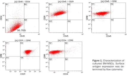
medium. Isolated BM-MSCs were defined as P0 and confluent cells were split 1:3 and passaged two times. Passage 2 BM-MSCs were used for transplantation after 7 days of cultivation. Before transplantation, cell surface markers CD34, CD45, CD29 and CD9 were checked by flow cytometry using specific antibodies (SC-7324PE-rcp, Santa Cruz; 561867, 562154, 561409, BD Biosciences) to be sure of their stem cell status [8]. Mesenchymal cells should be negative for CD34 and CD45 but should express CD29 and CD90. CD90 expression was used to estimate the purity of BM-MSCs. Purity of the BM-MSC preparations was > 90%.
Scoliosis/syringomyelia induction
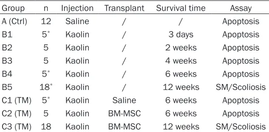
[image:2.612.91.359.84.216.2]Ten minutes before surgery, general anesthe-sia was induced by intravenous administration of 3% pentobarbital sodium (Sigma, America) through ear marginal vein at a dose of 30 mg/ kg. The skin was shaved and prepared with povidone iodine. Animals were then placed prone on a self-made frame to raise the cervi-cothoracic junction and avoid interruption from the scapula to facilitate next steps. A midline incision was made over the cervicothoracic junction from C7 to T1. At the base of C7 spi-nous process, a hole was drilled through the bone using a high-speed drill (diameter of 2.0 mm) but never injuring the dura. The flaval ligament was thin and easily cut off for the exposure of dura. A 100 microliter glass micro-injector (Qing Niu medical apparatus and instru-ments factory, Chengdu, China) was held in a stereotaxic apparatus (SR-6N, Narishige Gr- oup, Japan) and a 28-gauge needle was con-nected to puncture the dura for spinal cord injection. Site of puncture was along the drilled Table 1. Summary of experimental groups
Group n Injection Transplant Survival time Assay
A (Ctrl) 12 Saline / / Apoptosis
B1 5* Kaolin / 3 days Apoptosis
B2 5 Kaolin / 2 weeks Apoptosis
B3 5 Kaolin / 4 weeks Apoptosis
B4 5* Kaolin / 6 weeks Apoptosis
B5 18* Kaolin / 12 weeks SM/Scoliosis
C1 (TM) 5* Kaolin Saline 6 weeks Apoptosis
C2 (TM) 5 Kaolin BM-MSC 6 weeks Apoptosis C3 (TM) 18 Kaolin BM-MSC 12 weeks SM/Scoliosis n, number of animals per group; SM, syringomyelia; *one animal died and
failed to complete the assessment.
bony hole as well as to avoid dorsal vessels and the depth was 2.0 mm under the dura. Then, sixty microliters of 25% kaolin (Sigma-Aldrich) were injected into the center of the spi-nal cord. Wounds were closed with a single layer silk suture. After surgery, all rabbits were under close observation.
Animal grouping
The 78 experimental rabbits were randomly divided into group A (n = 12), group B (n = 38), and group C (n = 28) (Table 1). In group A, ster-ile saline was injected as these animals were used as control. Group B was further randomly subdivided into 5 subgroups (n = 5 for B1, B2, B3, and B4 subgroup, n = 18 for B5 subgroup). Rabbits were sacrificed and tissues were taken at postoperative day 3 (group B1), week 2 (group B2), week 4 (group B3), and week 6 (group B4) to investigate progression of apop-tosis of spinal cord cells. Group C was further
subdivided into 3 subgroups (n = 5 for C1 and C2 subgroup, n = 18 for C3 subgroup). Rabbits received injections of 10 ul of BM-MSCs cell (C1) or sterile saline (C2) into the epicenter of lesion site at postoperative week 2 and week 4. Radiographs and MRIs were taken at different time points after surgical induction.
Radiological observation
In a prone position, coronal full-length posteri-or-anterior radiographs of the spine were taken with heads and bodies straightened at postop-erative 4, 6, 8, 12 weeks under general anes-thesia. The Cobb method was used to measure and observe curve on the coronal plane. Progression was then recorded.
MRI observation
[image:3.612.92.520.73.331.2]MRI examinations were performed with a 1.5-T MR scanner (Sonata, Siemens) using cervical
Figure 1. Characterization of cultured BM-MSCs. Surface antigen expression was de-termined by flow cytometry.
Table 2. Summary of incidence of syringomyelia and scoliosis in rabbit models after kaolin injection Postop time
Week 4 Week 6 Week 8 Week 10 Week 12
SM 0 23.5% (4/17*) 47.1% (8/17) 64.7% (11/17) 64.7% (11/17)
Scoliosis 0 29.4% (5/17) 58.8% (10/17) 58.8% (10/17) 58.8% (10/17)
SM & scoliosis 0 11.8% (2/17) 29.4% (5/17) 41.2% (8/17) 41.2% (8/17)
[image:3.612.89.523.368.435.2]and spinal coils. MRI scans were performed preoperatively and at 4, 6, 8, 10 and 12 weeks postoperatively. Ten minutes before MRI scan-ning, general anesthesia was induced by intra-muscular injection of 3% pentobarbital sodium (Siagma, America) through the gluteus maxi-mus at a dose of 30 mg/kg.
investigated for each animal. TUNEL assay is described as follows.
TUNEL staining
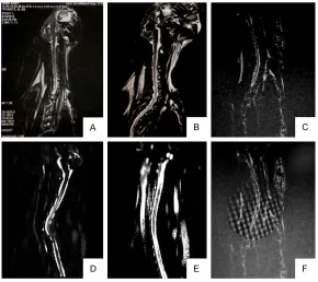
[image:4.612.89.379.71.265.2]TUNEL staining was performed using a com-mercial kit (Roche, Mannheim, Germany), acc- Figure 2. MRI (transverse) scan shows syrinx occurrence and progression.
A. Preoperative MRI Scan; B. 4 weeks Postoperative MRI scan; C. 6 weeks Postoperative MRI scan; D. 8 weeks Postoperative MRI scan; E. 10 weeks Postoperative MRI scan; F. 12 weeks Postoperative MRI scan.
BM-MSC transplantation
Two weeks after surgery, the rabbits were prepared for transplantation. Animals were assigned randomly into three major groups: one group of rabbits was injected with 5 µL saline as control group (n = 5). The second group of rabbits were transplanted with BM- MSCs (n = 23). All animals were anaesthetized with pen-tobarbitone sodium (40 mg/ kg body weight) and their spi-nal cords were exposed at the C7-T1 area, as described ab- ove in the primary operational section. About 1 × 10⁵ cells/ 5 µL BM-MSCs were trans-planted into the paracentral area of the spinal cord at a depth of 1 mm below the dor-sal surface, at a rate of 1 μl/ min. Subsequently, the mus-cles and skin were sutured. In experiment 1, treated rabbits were designed to test apopto-sis of spinal cells after trans-plantation (n = 5). In experi-ment 2, the treated animals were used to evaluate clinical therapeutic effects (n = 18).
Tissue preparation
At day 3, week 2, 4, and 6 after surgery, animals were sacrificed under anesthesia and spines cord were harvest-ed and post-fixharvest-ed in 4% para-formaldehyde in 0.1 M phos-phate buffer overnight. Tiss- ues surrounding spines were removed. Two spinal cord seg-ments (C6-C7, C7-T1) and two adjacent segments (C4-C5, T1-T2) were isolated and Figure 3. MRI (sagittal) scan shows syrinx occurrence and progression. A.
[image:4.612.89.379.334.592.2]ording to supplier’s instructions. Briefly, 5 um spinal tissue sections were de-waxed in xylene, rehydrated, and pretreated with proteinase K for 15 minutes at 37°C. After rinsing in 2% hydrogen peroxide for 5 minutes, they were washed with PBS. Sections were incubated with TUNEL reaction mixture for 60 minutes at 37°C in a humidified chamber. After washing with PBS, sections were incubated in a convert-er-POD solution at 37°C for an additional 30 minutes. Slides were washed again three times in PBS and then reacted with DAB substrate at room temperature for 10 minutes. Eventually, slides were counterstained with hematoxylin and dehydrated in a series of alcohols, mount-ed under coverslips, and analyzmount-ed under a light microscope. Five random visual fields (× 200) were examined on each side in one sample at a magnification of 200. Results are expressed as an apoptotic index (the average number of pos-itive cells per hundred spinal cord cells) to quantify apoptosis.
Statistical analysis
All data were analyzed by GraphPad prism (Graphpad 5.0). Two-tailed unpaired t-test was used to compare data within and between groups. For all tests, data are indicated by mean ± standard deviation and two-sided P < 0.05 was considered to be significant.
Results
Phenotype of rabbit BM-MSCs
After isolation, BM-MSCs adhered to the bot-tom of the flask and formed colonies. In culture, they displayed a fibroblast-like spindle-shaped morphology. No obvious morphological chang-es were observed during culture period. Cells from the second passage were analyzed and used for transplantation. Expression of cell sur-face antigens was examined using flow cytom-etry analyses (FACSCanto II, BD Biosciences, USA). The pattern of staining of the BM-MSC surface markers is shown in Figure 1. All cul-tured cells were CD45 and CD34 negative but positive for CD29 and CD90. The purity of BM-MSCs in culture was examined by CD90 expression. Purity of BM-MSCs was > 90%.
Characteristics of syringomyelia-associated scoliosis rabbit model
Postoperatively, most rabbits undergoing kaolin injection exhibited signs of lethargy and
anorex-ia. Four animals died after the operation and failed to complete the assessment (Table 2). Two of them died of intraoperative anesthesia. The other two were excluded due to postopera-tive infections resulting in death. We assessed incidence of syringomyelia and scoliosis in ani-mals at postoperative week 12.
Syrinx began to appear at postoperative week 6. Transverse and sagittal MRI scans showed syrinx in cervical-thoracic segments. Syrinx was found in 64.7% (11/17) of the experimental ani-mals by the end of the experiment (week 12). Cavity size and amount increased with time during the course of the experiment (Table 2 and Figures 2, 3).
Scoliosis also began to appear at postopera- tive week 6 and the apexes of curves were at cervical-thoracic or upper thoracic segments. Scoliosis was found in 58.8% (10/17) of the experimental animals by the end of the ex- periment (week 12). Follow up spinal radio-graphs showed a gradual increase of coronal curve during the continuous phase after sur- gical induction (Table 2 and Figure 4). More- over, syringomelia-associated scoliosis appea- red in 41.2% (8/17) of the experimental an- imals.
These findings indicate that our constructed model has typical syrinx and scoliosis represen-tation. Therefore, this constructed animal model was close to clinical cervical syringomy-elia accompanied with scoliosis.
Spinal cord cells apoptosis
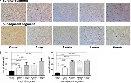
4.7) compared to controls (P = 0.001) but adja-cent segments remained with a similar apop-totic rate as our 4-week time point (P = 0.0004). Compared to adjacent sections, surgical sec-tions had fewer TUNEL-positive spinal cells. The mean number of TUNEL-positive spinal cells increased over this 6 weeks period.
BM-MSCs reduce apoptosis in treated rabbits
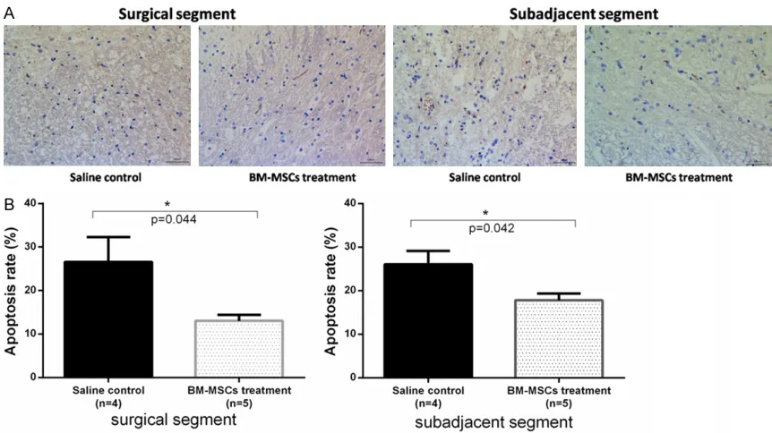
[image:6.612.92.524.72.213.2]At week 6, we evaluated the effect of BM-MSC transplantation on apoptosis of spinal cord cells. Apoptosis was rarely observed at the defective spinal cord region around transplant-ed BM-MSCs (Figure 6). Figure 6A shows the Figure 4.Posterior-anterior radiographs of the spine and scoliosis development (rabbit #21). A. Postoperative 4-week; B. Postoperative 6-week, scoliosis appeared and Cobb angle was 14°; C. Postoperative 8-week, scoliosis developed to 46°; D. Postoperative 10-week, scoliosis developed to 57°; E. Postoperative 12-week, scoliosis de-veloped to 59°.
Figure 5. TUNEL staining of (x200) of apoptotic cells in spinal cord surrounding the surgical site at postoperative day 3, week 2, 4, 6. A. TUNEL-positive cells in the spinal cord of the rabbit model. B. The statistical data are shown as
apoptotic index (the average number of TUNEL-positive cells per hundred spinal cord cells ± SD). Surgical (C6-C7, C7-C8) versus sub-adjacent (C4-C5, C9-C10) levels. *Significant difference was found between experimental group
[image:6.612.94.524.289.563.2]representative images of TUNEL assay in sp- inal tissues. Rate of apoptosis of spinal cells in sub-adjacent segments in BM-MSCs group and saline control group was 26.6 ± 5.5% and 13.0 ± 3.4%, respectively. Apoptosis rate of spinal cells in surgical segments in BM-MSC group and saline control group was 26.1 ± 6.1% and 17.8 ± 3.5%, respectively. Number of TU- NEL-positive cells was significantly lower in BM-MSCs transplantation group compared with saline-injection group (P = 0.04). These results suggest that MSCs have an impact on apoptosis of spinal cells in a rabbit model.
BM-MSCs administration reduces syringomy -elia and scoliosis occurrence
After the significant effects of BM-MSCs trans-plantation in spinal cell apoptosis, syringomy-elia and scoliosis occurrence was evaluated at postoperative weeks 4, 6, 8, and 12 (Table 3). Consistent with apoptosis results at week 6, occurrence of syringomyelia and scoliosis decreased (16.7% and 16.7%, respectively) in rabbits treated with BM-MSCs compared with those animals in the control group (23.5% and 29.4%, respectively). We also observed a
decrease at postoperative week 8. No evident differences of incidence rate of syringomyelia and scoliosis were observed between BM-MSC-treated or saline-rabbits by the end of our experiment (postoperative 12-week). These results suggest that BM-MSCs administration reduced incidence of syringomyelia as well as scoliosis in kaolin induced rabbit models. Discussion
[image:7.612.91.522.71.313.2]omy, unilateral tethering, or injecting botulinum toxin [13]. Methods that establish an experi-mental porcine model of early-onset scoliosis have also been developed by use of a radi-opaque ultra-high molecular weight polyethyl-ene posterior spinal tether [14]. Barrios et al. reported that temporary interpedicular tether-ing at the thoracic spine induces severe scoli-otic curves in pigs [15]. However, commonly used methods still have some disadvantages including complicated preparation and high mortality rates, etc. In 2005, Lee et al. attempt-ed to establish a rat model of syringomyelia by intraparenchymal injection of kaolin into the rat cervical spinal cord. They further noted that large numbers of macrophages were recruited from bone marrow in kaolin-induced rat syrin-gomyelia [16]. Wong et al. reported that they created a posttraumatic syringomyelia model using excitotoxic amino acid and kaolin-injec-tion [17]. More recently, in a study by Mohrman et al., rats injected with quisqualic acid and kaolin were also observed to develop syringo-myelia [18]. In our study, we developed an ani-mal model for scoliosis and syringomyelia in rabbits using kaolin administration. Twelve weeks after syrinx induction, a relatively higher incidence of scoliosis (58.8%) and syringomy-elia (64.7%) was observed. Syrinx and scoliosis were found in 64.7% and 58.8% of our experi-mental animals. Syringomyelia-associated sco-liosis appeared in 41.2% of the animals, sug-gesting that kaolin-administration could be an optimal choice for induction of scoliosis and syringomyelia.
A better understanding of underlying molecul- ar mechanisms associated with syringomyelia or scoliosis formation will unveil new targets for treatment and possibly the future preven-tion of morbidity [19]. The cause of scoliosis and syringomyelia is believed to be multifacto-rial [20]. Increased apoptosis has always been thought to be a driving factor in development
and progression of many bone diseases and has been reported in intervertebral discs of patients with adolescent idiopathic scolio- sis [21]. The role of apoptosis in scoliosis and syringomyelia remains unclear due to the relatively few number of studies performed. Bao et al. observed cell apoptosis and necrosis in herniation of the cerebellar tonsil in Chiari I malformation complicated with syringomyelia patients and suggested that apoptosis might play a role in development of syringomyelia [22]. Karner et al. reported that increased apoptosis suggests a common pathophysiolo-gy for adolescent idiopathic scoliosis [23]. These related studies have provided evidence indirectly supporting the idea that apoptosis might be involved in the pathogenesis of syrin-gomyelia and scoliosis. In our present study, apoptosis of spinal cord tissues from surgical segments and adjacent segments of the rabbit model were evaluated by TUNEL method. Apoptosis indexes of spinal cells in both the surgical and adjacent segments of rabbit mod-els were significantly higher than that of the control. Levels of apoptosis of spinal cells increased along the observation period (post-operative 6-week). Apoptosis of spinal cord tis-sues prior to manifestation of syringomyelia and scoliosis suggests a cellular etiology for both diseases.
[image:8.612.90.524.97.164.2]In past years, several potential treatments for syringomyelia or scoliosis have been evaluated by many research groups [24, 25]. MSCs can differentiate into different cell types such as osteoblasts, chondrocytes and myoblasts, fibroblasts, adipocytes, and oligodendrocytes [26]. Based on the results of Hanetal [27], about 115 differently expressed proteins were found in MSCs of patients with degenerative scoliosis and the abnormality of MSCs in DS may be associated with the pathophysiology of scoliosis. Application of MSC-based therapy to supplement traditional surgical therapy shows
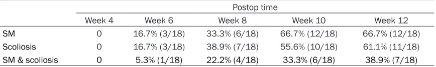
Table 3. Summary of incidence of syringomyelia and scoliosis in rabbit models after BM-MSCs
admin-istration
Postop time
Week 4 Week 6 Week 8 Week 10 Week 12
SM 0 16.7% (3/18) 33.3% (6/18) 66.7% (12/18) 66.7% (12/18)
Scoliosis 0 16.7% (3/18) 38.9% (7/18) 55.6% (10/18) 61.1% (11/18)
SM & scoliosis 0 5.3% (1/18) 22.2% (4/18) 33.3% (6/18) 38.9% (7/18)
great promise for treatment of spinal cord inju-ries and degenerative disc disease [28-30]. Vaquero et al. reported that injection of MSCs in the syrinx of posttraumatic syringomyelia is safe and is associated with clinical and neuro-imaging improvement. They concluded that cell therapy is a new approach to posttraumatic syringomyelia and even for idiopathic syringo-myelia [31]. In this study, we evaluated the ther-apeutic potential of BM-MSCs transplantation in treatment of scoliosis/syringomyelia in a rab-bit model. Our results demonstrate that, in addition to decreased levels of apoptosis of spi-nal cells, BM-MSCs administration could decline morbidity of syringomyelia/scoliosis when injected before occurrence of clinical symptoms (postoperative week 2), compared with control rabbits. This indicates that BM-MSCs may be attributable to a partial blocking of occurrence of syringomyelia/scolio-sis and that apoptosyringomyelia/scolio-sis might be a factor con-tributing to development of syringomyelia/ scoliosis.
One limitation to our study was that we failed to investigate effects of spinal cord cell apoptosis on syringomyelia/scoliosis occurrence in a rab-bit model. Association between apoptosis and incidence of syringomyelia/scoliosis remains unclear. This should be investigated in the future.
In summary, a kaolin-induced syringomyelia-associated scoliosis rabbit model was estab-lished. In postoperative periods, we noticed an increase of apoptosis in surgical segments and adjacent segments of spinal cord. We found that prenatal BM-MSCs transplantation could decrease spinal tissue apoptosis and reduce incidence of syringomyelia and scoliosis in models. This study supports the concept of using BM-MSCs transplantation to treat scolio-sis and syringomyelia before occurrence of clin-ical symptoms.
Acknowledgements
This work was supported by grants from the National Natural Science Foundation of China (No. 81360281, 81460347) and grants from the “Special and Joint Program” of Yunnan Provincial Science and Technology Department & Kunming Medical University (No. 2017FE467 (-064)).
Disclosure of conflict of interest
None.
Address correspondence to: Wei Xu, Department of Neurosurgery, The 2nd Affiliated Hospital of Kunming
Medical University, Kunming, Yunnan Province, P. R. China. E-mail: 1017469868@qq.com; Jingming Xie, Department of Orthopaedics, The 2nd Affiliated
Hospital of Kunming Medical University, Kunming, Yunnan Province, P. R. China. E-mail: xiejingming@ vip.163.com
References
[1] Zhang H, Guo C, Tang M, Liu S, Li J, Guo Q, Chen L, Zhu Y, Zhao S. Prevalence of scoliosis among primary and middle school students in mainland China: a systematic review and me-ta-analysis. Spine 2015; 40: 41-49.
[2] Sha S, Li Y, Qiu Y, Liu Z, Sun X, Zhu W, Feng Z, Wu T, Jiang J, Zhu Z. Posterior fossa decom-pression in Chiari I improves denervation of the paraspinal muscles. J Neurol Neurosurg Psychiatry 2017; 88: 438-444.
[3] Roth AK, Bogie R, Jacobs E, Arts JJ, van Rhijn LW. Large animal models in fusionless scolio-sis correction research: a literature review. Spine J 2013; 13: 675-688.
[4] de Seze M, Cugy E. Pathogenesis of idiopathic scoliosis: a review. Ann Phys Rehabil Med 2012; 55: 128-138.
[5] Kim HJ, Ahn HS, Nam Y, Chang BS, Lee CK, Yeom JS. Comparative study of the efficacy of transdermal buprenorphine patches and pro-longed-release tramadol tablets for postopera-tive pain control after spinal fusion surgery: a prospective, randomized controlled non-inferi-ority trial. Eur Spine J 2017; 26: 2961-2968. [6] Park WW, Suh KT, Kim JI, Kim SJ, Lee JS.
De-creased osteogenic differentiation of mesen-chymal stem cells and reduced bone mineral density in patients with adolescent idiopathic scoliosis. Eur Spine J 2009; 18: 1920-1926. [7] Ding Y, Yan Q, Ruan JW, Zhang YQ, Li WJ, Zeng
X, Huang SF, Zhang YJ, Wu JL, Fisher D, Dong H, Zeng YS. Electroacupuncture promotes the differentiation of transplanted bone marrow mesenchymal stem cells overexpressing TrkC into neuron-like cells in transected spinal cord of rats. Cell Transplant 2013; 22: 65-86. [8] Undale AH, Westendorf JJ, Yaszemski MJ,
Kho-sla S. Mesenchymal stem cells for bone repair and metabolic bone diseases. Mayo Clin Proc 2009; 84: 893-902.
[10] Zadeh HG, Sakka SA, Powell MP, Mehta MH. Absent superficial abdominal reflexes in chil-dren with scoliosis. An early indicator of syrin-gomyelia. J Bone Joint Surg Br 1995; 77: 762-767.
[11] Tanaka M, Sugimoto Y, Arataki S, Takigawa T, Ozaki T. A rare course of scoliosis associated with Chiari malformation and syringomyelia. Acta Med Okayama 2014; 68: 303-306. [12] Charry O, Koop S, Winter R, Lonstein J, Denis F,
Bailey W. Syringomyelia and scoliosis: a review of twenty-five pediatric patients. J Pediatr Or-thop 1994; 14: 309-317.
[13] Bobyn JD, Little DG, Gray R, Schindeler A. Ani-mal models of scoliosis. J Orthop Res 2015; 33: 458-467.
[14] Bogie R, Roth AK, Willems PC, Weegen VW, Arts JJ, van Rhijn LW. The development of a representative porcine early-onset scoliosis model with a standalone posterior spinal teth-er. Spine Deform 2017; 5: 2-10.
[15] Barrios C, Lloris JM, Alonso J, Maruenda B, Burgos J, Llombart-Blanco R, Gil L, Bisbal V. Novel porcine experimental model of severe progressive thoracic scoliosis with compensa-tory curves induced by interpedicular bent rigid temporary tethering. J Orthop Res 2018; 36: 174-182.
[16] Lee GY, Jones NR, Mayrhofer G, Brown C, Cle-land L. Origin of macrophages in a kaolin-in-duced model of rat syringomyelia: a study us-ing radiation bone marrow chimeras. Spine 2005; 30: 194-200.
[17] Wong J, Hemley S, Jones N, Cheng S, Bilston L, Stoodley M. Fluid outflow in a large-animal model of posttraumatic syringomyelia. Neuro-surgery 2012; 71: 474-480.
[18] Mohrman AE, Farrag M, Huang H, Ossowski S, Haft S, Shriver LP, Leipzig ND. Spinal cord tran-scriptomic and metabolomic analysis after ex-citotoxic injection injury model of syringomy-elia. J Neurotrauma 2017; 34: 720-733. [19] Grimes DT, Boswell CW, Morante NF,
Henkel-man RM, Burdine RD, Ciruna B. Zebrafish mod-els of idiopathic scoliosis link cerebrospinal fluid flow defects to spine curvature. Science 2016; 352: 1341-1344.
[20] Burwell RG, Clark EM, Dangerfield PH, Moulton A. Adolescent idiopathic scoliosis (AIS): a mul-tifactorial cascade concept for pathogenesis and embryonic origin. Scoliosis Spinal Disord 2016; 11: 8.
[21] Sitte I, Kathrein A, Pfaller K, Pedross F, Klosterhuber M, Lindtner RA, Zenner J, Ferraris L, Meier O, Koller H. Morphological differences in adolescent idiopathic scoliosis: a histologi-cal and ultrastructural investigation. Spine 2013; 38: 1672-1680.
[22] Bao CS, Liu L, Wang B, Xia XG, Gu YJ, Li DJ, Zhan SL, Chen GL, Yang FB. Craniocervical de-compression with duraplasty and cerebellar tonsillectomy as treatment for Chiari malfor-mation-I complicated with syringomyelia. Gen-et Mol Res 2015; 14: 952-960.
[23] Karner CM, Long F, Solnica-Krezel L, Monk KR, Gray RS. Gpr126/Adgrg6 deletion in cartilage models idiopathic scoliosis and pectus excava-tum in mice. Hum Mol Genet 2015; 24: 4365-4373.
[24] Theroux J, Stomski N, Losco CD, Khadra C, La-belle H, Le May S. Spinal manipulative therapy for adolescent idiopathic scoliosis: a system-atic review. J Manipulative Physiol Ther 2017; 40: 452-458.
[25] Czaprowski D. Manual therapy in the treat-ment of idiopathic scoliosis. Analysis of current knowledge. Ortop Traumatol Rehabil 2016; 18: 409-424.
[26] Aizman I, McGrogan M, Case CC. Quantitative microplate assay for studying mesenchymal stromal cell-induced neuropoiesis. Stem Cells Transl Med 2013; 2: 223-232.
[27] Han S, Zhu Y, Wu Z, Zhang J, Qiu G. The differ-ently expressed proteins in MSCs of degenera-tive scoliosis. J Orthop Sci 2013; 18: 885-892. [28] Steffen F, Smolders L, Roentgen A, Bertolo A,
Stoyanov J. Bone marrow-derived mesenchy-mal stem cells as autologous therapy in dogs with naturally occurring intervertebral disc dis-ease: feasibility, safety and preliminary results. Tissue Eng Part C Methods 2017; 23: 643-651.
[29] Yousefifard M, Nasirinezhad F, Shardi Mana-heji H, Janzadeh A, Hosseini M, Keshavarz M. Human bone marrow-derived and umbilical cord-derived mesenchymal stem cells for alle-viating neuropathic pain in a spinal cord injury model. Stem Cell Res Ther 2016; 7: 36. [30] Freeman BJ, Kuliwaba JS, Jones CF, Shu CC,
Colloca CJ, Zarrinkalam MR, Mulaibrahimovic A, Gronthos S, Zannettino AC, Howell S. Alloge-neic mesenchymal precursor cells promote healing in postero-lateral annular lesions and improve indices of lumbar intervertebral disc degeneration in an ovine model. Spine 2016; 41: 1331-1339.