Original Article
WIP1 is relevant to tumor malignancy and metastasis in
breast cancer
Zongtao Li1, Jin Zheng1, Xiaomei Gu2, Zheng Dong1, Xinting Wang1
1Department of Breast Surgery, Tangshan Gongren Hospital, Tangshan, China; 2Department of Gynecology,
Mater-nal and Child Health Hospital of Tangshan, Tangshan, China
Received March 5, 2018; Accepted October 26, 2018; Epub March 15, 2019; Published March 30, 2019
Abstract: Wild-type p53-induced phosphatase 1 (WIP1) is a serine/threonine protein phosphatase that has been shown to be correlated with tumor proliferation, differentiation and anti-apoptotic processes in several malignant tumors. However, the significance of WIP1 expression in breast cancer is still far from clear. To evaluate the clinical significance of WIP1 oncogene in breast cancer, the expression of WIP1 was investigated in 120 breast cancer biop -sies and adjacent breast tissues by immunohistochemistry. The correlation between WIP1 expression and postop -erative survival rate was also analyzed. WIP1 was up-regulated in breast cancer tissues (96/120). Down-regulation of WIP1 in MCF-7 breast cancer cells was established using Lentivirus-mediated infection. The absence of WIP1 resulted in dramatic decrease of cell proliferation, invasion, and metastasis ability as well as increase cell apopto-sis. Subsequent investigations revealed that, p53 protein expression was significantly higher in WIP1-infected cells than in normal tumor cells. Our founding indicates that WIP1 ameliorated the malignancy of MCF-7 cells, which is probably achieved via regulating p53 protein expression. Taken together, WIP1 may be a useful regulator in breast cancer malignancy and metastasis in breast cancer and may involved in proliferation, apoptosis, migration and inva-sion of breast cancer cells by regulating p53 protein expresinva-sion.
Keywords: Wip1, breast cancer, clinical significance, tumor malignancy, metastasis
Introduction
Breast cancer is one of the most commonly di- agnosed tumor in Chinese women and is the leading cause of death in female cancers. Ac- cording to the statistics, the number of Chinese women with breast cancer account for 12.2% of all newly diagnosed breast cancers and 9.6% of deaths from all deaths in the worldwide [1].
Therefore, breast cancer has become a severe public health burden in China. The exploration of new biomarkers and technologies that are
capable of achieving early diagnosis, tumor grade determination as well as morbidity evalu-ation has become an important and urgent demand in breast cancer research. Elucidation of the clinical, biological, and pathological char-acteristics of breast cancer is expected to facil-itate the discovery of new therapeutic treat-ments for breast cancer.
Wild-type p53-induced phosphatase PPM1D (or WIP1) is a serine/threonine protein
phos-phatase, which gene located in 17q23/q24 region of human chromosome. Accumulated studies have proved that abnormal expression of WIP1 is often involved in tumor cell prolifera-tion, differentiation and anti-apoptotic process-es, indicating that WIP1 is strongly contributes to the occurrence and development of various types of cancer [2-5]. In the late 1990s, the dis-covery of WIP1 in a genetic screening study opened a new era of genetic-related research.
Thereafter, WIP1 was identified as a novel onco -gene and overexpression of WIP1 was proved to be associated with human ovarian cancer, breast cancer, medulloblastoma and
neuro-blastoma tumors [6-11]. These characteristics
of WIP1 have aroused great interest in cancer researchers.
medulloblastoma negatively regulating p53 by abrogating the activity of p53 [2]. The signifi -cance of WIP1 in clinical application was also investigated in various cancers. Liang reported that the metastasis and prognosis of adenoid cystic carcinoma patients were closely
associ-ated with WIP1 expression [12]. These studies
have opened up a new genetic target for future
clinical treatment. However, the significance of
WIP1 expression in breast cancer is still far from clear.
In present study, we tested WIP1 expression level in 120 paired breast cancer and adjacent non-cancer tissues, analyzed its relation with clinicopathological characteristics and evaluat-ed the impact of WIP1 expression level on bre- ast cancer patients’ survival rate. In addition, the correlation of WIP1 expression with breast cancer malignancy and metastasis and its
clini-cal significance was determined. These results
should help to gain an enhanced understand-ing of the etiology of breast cancer.
Materials and methods
Patients and cell culture
120 breast tumor tissues and paired adjacent non-cancer tissues (5 cm distant from the tu- mor margin) were collected from breast cancer
patients with confirmed case at Tangshan
Gon-gren Hospital between January 2005 and Ja- nuary 2010. All of the patients voluntarily par-ticipated in this study gave written informed consent before the using of residual samples
and this study was approved by Tangshan
Gongren Hospital’s research ethics committee.
The mammary gland tumors in this study were
staged according to tumor node metastasis
(TNM) classification system. The pathology cla-ssification, demographic status and postopera -tive complications are detailed in Table 1. The
resected breast tumor tissues and adjacent
breast tissues were paraffin-embedded and defined as the cancer group and normal control
group, respectively. All breast cancer cases were female, and sporadic without family hi- story.
Breast cancer cells used in this study were obtained from Chinese Academy of Sciences Cancer Hospital and cultured in Phenol Red-free RPMI media replenished with antibiotics and fetal bovine serum (10%).
Immunohistochemical staining
The breast tumor tissue and paired adjacent
non-cancer tissues’ immunohistochemical sta- ining of WIP1 was carried out as our previous reports [13]. In short, tissue samples were stained with diaminobenzidine (DAB) and visu-alized by hematoxylin. WIP1 expression level was determined by the staining intensity and the total score of the percentage of positive
cells. Positive cells percentage was classified into four groups: 0) ≤ 5%, 1) 5-25%, 2) 25-50%, and 3) > 50%. The results of
immunohistoch-emical staining were evaluated semiquanti-tavely on the basis of a four-point scale: 0) no
staining, 1) weak staining (pale yellow), 2) mod -erate staining (brown), and 3) strong staining
(dark brown). The total score was classified as following: 0, negative (-); 1-2, weakly positive
(+); 3-4, medium positive (++); 5-6, strongly
[image:2.612.91.291.107.400.2]positive (+++). A result of (-) or (+) was defined
Table 1. Clinicohistopathologic characteristics of the breast cancer patients involved in this study
Groups Cases
Age
≤ 50 48
> 50 72
Types of breast cancer
Ductal carcinoma 66
Lobular carcinoma 38
Other 16
TNM stage
I 59
II 42
III 19
Size of tumor (mm)
< 50 104
> 50 16
Postoperative complications
Bleeding 3
Seroma 42
Skin flap necrosis 6
First-degree upper extremity edema 15
Second-degree edema 3
as low expression, and a result of (++) or (+++)
was defined as high expression. Negative con -trols for the staining were treated in the same manner but without the primary antibody. Lentivirus-mediated WIP1 infection of MCF-7 cells
MCF-7 cells were seeded at a density of 105 cells per well in 24-well plates and incubated for 24 hr. When the cell population reached 80%, 1 mL lentivirus containing WIP1 short hairpin RNA (RNA) or negative control (NC)-shRNA plasmid was added to the cell culture. After infecting for 12 hr, the virus was aspirated
and l mL Dulbecco’s modified Eagle medium (DMEM) was added. The green fluorescent
clones were selected after 48 hr infection
under a fluorescence microscope (BX43,
Olympus). Western blot
For western blot analyze, the experiment was carried out as our conventional reported with
slight modification [13]. In brief, tissue samples
were lysis, homogenized and centrifugated for 20 min (13,400 g) at 4°C. Protein concentra-tion was measured with a bicinchoninic acid
(BCA) protein quantification kit (Fluoro Profile,
Sigma, USA). Electrophoretic analysis of the pr- otein was carried out by sulfate-polyacrylami- de gel electrophoresis (SDS-PAGE). After that,
the membranes were blocked in 5% skim milk,
incubated with the primary antibody as well as horseradish peroxidase-labeled goat anti-mou-
se IgG respectively. The membranes were
de-veloped with electrochemiluminescence, scan- ned with a FUJI Mini-4000 scanner, and
ana-lyzed with LabWorks 4.5 software. All of the
experiments were conducted in triplicate. WIP1 expression in MCF-7 cells before and after infection was detected in a similar man-ner and referenced to GAPDH.
Reverse transcription quantitative polymerase chain reaction (qPCR)
Total RNA of tissue samples and cell lines was extracted and purified according to our previ -ous report and manufacturer’s instructions [13]. For the breast cancer tissues, the reaction conditions were 42°C for 50 min, and terminat-ed at 95°C for 5 min. Primers for WIP1 (forward:
5’-GGCCAAATGAAAGCCCAAGAAAT-3’), (reverse:
5’-CAGAGTTCTTTCGCTGTGAGGTTGT-3’) and β-actin were (forward: 5’-ACTTAGTTGCGTTACA-CCCTT-3’), (reverse:
5’-GTCACCTTCACCGT-TC-CA-3’) synthesized by Shanghai Biological En-
gineering Technology Services Limited. β-actin
served as the internal control, and relative WI- P1 expression was calculated using the 2-ΔΔCT
method.
To determine WIP1 expression level of
shRN-A-infected MCF-7 cells, the primers were
designed as follows: 5’-TTCCCCATGTTCTACAC-CACCAG-3’ (WIP1 upstream), 5’-TGAGGGTAT-GACTACACCTTGGAC-3’ (WIP1 downstream); 5’-GTCTCCTCTGACTTCAACAGCG-3’ (GAPDH up-stream), 5’-ACCACCCTGTTGCTGTAGCC-3’
(GA-PDH downstream). PCR conditions were 95°C for 60 s, 95°C for 15 s, 60°C for 60 s, and 72°C
for 45 s for 40 cycles. The GAPDH expression
served as the internal control. WIP1 expression was calculated as the ratio of grey band inten-sity relative to that of GAPDH.
MTT assay
WIP1-shRNA- and NC-shRNA-infected MCF-7 cells at the logarithmic growth phase (70-80%
confluence) were seeded in 96-well plates at a density of 5000 cells/well (200 μL media/well).
Cell growth was terminated after 1, 2, 3, and 4
days, respectively, and 20 mL of 5 mg/mL MTT
solution was added 4 hr before the termination
of culture. After incubated with MTT for 4 hr, the
medium in each well was discarded and 200 mL/well dimethyl sulfoxide was used to dis-solve the internalized purple formazan crystals.
The absorbance value (D) was detected on an
automatic microplate reader at wavelength of 490 nm and reference wavelength of 620 nm. Cell survival was calculated as follow:
Survival rate (%) = (Dexperimental group/Dcontrol group) × 100
Flow cytometry
WIP1-shRNA- and NC-shRNA-infected MCF-7 cells at the logarithmic growth phase (70-80%
confluence) were digested, collected, washed
twice with 4°C PBS and resuspended in 1 mL binding buffer with a concentration of 1 × 10⁶
cells/mL. 100 μL cell suspension was trans
-ferred to a 5 mL flow tube and 10 μL propidium
iodide was added. Cells were incubated in the
measurement of the cell cycle, 195 μL of the
same cell suspension was transferred to a 5
mL flow tube, and 5 μL Annexin V-FITC and 10 μL propidium iodide were added to each tube. The tubes were incubated for 15 min in the dark and the cell cycle was detected by flow
cytometry.
Transwell invasion assay
Cell invasion assay was conducted with a poly-carbonate microporous membrane and was
capped with or without 50 μL Matrigel (8.4
g/L). WIP1-shRNA- and NC-shRNA-infected MCF-7 cells were suspended in serum-free medium with a density of 1 × 10⁶ cells/mL. 50
μL cell suspensions were transferred to the upper chamber and 800 μL DMEM (10%) was
added to the lower chamber. After cultured for 18 hr, cells on the surface of the upper cham-ber were removed by scraping with a cotton
The expression of WIP1 was elevated in breast cancer tissue
To evaluate the WIP1 protein expression in
breast cancer, immunostaining was performed as previously described [13] in 120 breast can-cer and adjacent non-cancan-cer tissues. Immun- ohistochemical staining result showed that WIP1 immunohistochemical staining was pale
yellow to dark brown in the breast cancer tis
-sues and was weak or even negative in the
adjacent breast tissues (Figure 1). The expres -sion scores of WIP1 are summarized in Table 2. In general, the prevalence of positive WIP1 pro-tein expression (++ and +++) in breast cancer tissues was 80% (96/120), which was obvious-ly higher (P < 0.05) than the adjacent tissues (26.7%, 32/120).
Western blot and q-PCR were performed for
[image:4.612.90.373.72.285.2]quantitively confirm WIP1 protein and mRNA Figure 1. Immunohistochemical staining result of WIP1 protein expression
in adjacent normal tissues (A, B) and breast tumor tissues (C, D). (A and C) SP ×100; (B and D) SP ×400.
Table 2. The expression scores of WIP1 in breast tumor tissues
and adjacent normal breast tissues
Groups Cases WIP1 Protein Expression
- + ++ +++ Z P*
Cancer tissues 120 24 19 43 34 -9.561 0.000
Adjacent tissues 120 88 24 8 0
Student t-test was used to analyze the statistical significance of comparison between two groups.*P < 0.05.
swab. Cells on the lower filter surface were kept in 4% para -formaldehyde and stained with 0.1% crystal violet for 20 min. Cells were counted in 5
ran-dom portions of each film and
the average number of invad-ing tumor cells was calculated. Statistical analysis
Data were analyzed with SPSS16.0 statistical software.
The Chi-square test was per -formed for comparisons of patient characteristics betwe-
en two groups. The Student
t-test was performed for com-parisons between two groups. Differences between the WIP1-shRNA and NC-WIP1-shRNA groups were compared by an indepen-dent samples t-test or one-way
ANOVE analysis of variance. The Kaplan-Meier method was
used to estimate survival rates, and differences were compared with the two-sid-
ed log-rank test. P < 0.05 was considered statistically
significant.
[image:4.612.90.372.372.425.2]expression respectively and the results indi-cated that both WIP1 protein (0.885 ± 0.079 vs. 0.251 ± 0.027, P < 0.001) and mRNA (0.835 ± 0.076 vs. 0.245 ± 0.021, P < 0.001) expres-sion in breast cancer tissues were also obvi-ously higher than that in the adjacent normal tissues (Figure 2).
The results of the above experiment demon -strated that WIP1 expression is much higher in human breast tumor tissues than in adjacent breast tissues for both the mRNA and protein
(Figure 1), indicating that high WIP1 levels tend
to be strongly linked to the incidence of breast cancer. These results confirm previous findings demonstrating a potential link between WIP1
expression level and breast cancer [8, 14, 15]. WIP1 expression was correlated with survival rate of patients and P53 gene expression in breast cancer
The potential interrelationship was investigated
between WIP1 expression level and breast can-cer patients’ clinicopathological characteristics by a follow up study of 3 years for 120 patients. Within the observation period, 94 patients sur-vived with a total survival rate of 78.3%, 31 relapsed and the recurrence sites were as fol-lows: 14 cases (45%) for the chest wall, 13 cases (41%; ribs in 5 cases, spine in 4 cases,
skull in 1 case, limbs in 2 cases, including mul -tiple bone metastases) for bone, 5 cases (16%) for lung, 2 cases (6%) for brain and 6 cases (19%) for liver. Some patients relapsed in
mul-tiple organs. The main cause of death was mul
-tiple organ failure. The patients were classified
[image:5.612.95.517.76.316.2]into groups depending on high (n = 77) or low (n = 43) WIP1 expression, and the overall survival (OS) is presented in Figure 3. The 3-year sur -vival rate was slightly higher in patients with Figure 2. q-PCR and Western blot analysis of Wip1 miRNA (A, B) and protein (C, D) in breast tumor tissue and normal tissues. Data are shown as mean ± SD (n = 4). One asterisk indicates statisticalsignificance. (*P < 0.05 breast tumor tissue versus normal tissue).
[image:5.612.93.287.381.519.2]low expression of WIP1 (83.7%) than that of patients with high expression of WIP1 (74.3%),
but there was no significant difference.
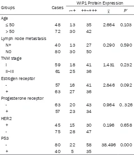
The correlation of WIP1 protein expression with
other clinicopathological characteristics of the breast cancer patients was also investigated and the result is summarized in Table 3. WIP1 expression was not obviously related to age, lymph node metastasis,
estrogen/progester-one receptor levels, HER2, or TNM stage (P > 0.05), but was obviously related to p53 expres-sion (P < 0.001).
Age, tumor size, TNM stage, lymph node metas -tasis, estrogen/progesterone receptor level, and progesterone receptor levels are not relat-ed to WIP1 expression which is compliance with the previous report of Bulavin [16]. However, WIP1 expression is negatively corre-lated with p53 expression, which might indi-cate an inhibitory effect of WIP1 on the p53 gene function of tumor suppressor and/or the
shWIP1-1 was selected in the following research.
After infection, the MCF-7/shWIP-1 group
showed strong fluorescence intensity 48 hr
whereas the infection efficiency in MCF-7/NC was only ~20% and showed weak fluorescence
intensity (Figure 5). The q-PCR and western blot
results indicated that the expression of WIP1 was obviously lower in the MCF-7/shWIP-1 group than in the MCF-7/NC group for both mRNA and protein expression.
Down-regulation of WIP1 inhibits tumor malig-nancy and metastasis potential in vitro
To investigate whether down-regulation of WIP1
in MCF-7 cell affects the proliferation ability of tumor cells, MCF-7/shWIP-1 and MCF-7/NC cells were seeded and cultured for 24, 48, 72
and 96 hr and MTT assay was adopted to evalu-ate cell viability. The results showed that cell
viability in the MCF-7/shWIP-1 group was
obvi-Table 3. The associations between the clinical characteristics
of breast cancer patients and WIP1 protein expression
Groups Cases WIP1 Protein Expression
-~+ ++~+++ _x P*
Age
≤ 50 48 13 35 2.664 0.103
> 50 72 30 42
Lymph node metastasis
N+ 40 13 27 0.290 0.590
N0 80 30 50
TNM stage
I 59 18 41 1.431 0.232
II~III 61 25 36
Estrogen receptor
- 57 16 41 2.846 0.092
+ 63 27 36
Progesterone receptor
- 63 20 43 0.964 0. 326
+ 57 23 34
HER2
+ 45 15 30 0.196 0.658
- 75 28 47
P53
- 80 22 58 38.496 0.000
+ 40 5 35
The Chi-square test was performed for comparisons of patient characteristics between two groups.*P < 0.05.
induction of mutations [17-19]. Overexpression of WIP1 in breast cancer might induce tumor forma-tion via the WIP1/p38MAPK/p53
signaling pathway. The present
results also indicate that overex-pression of WIP1 plays a vital part in the development of breast can-cer by suppressing the function of the tumor suppressor gene p53. Down-regulation of WIP1 expres-sion is achieved by lentivirus infec-tion
Lentivirus infection was used to
pick out breast cancer cells with low WIP1 expression. Three WIP1 -shRNA MCF-7 cell lines that showed
the best effects of WIP1
knock-down were selected and named as MCF-7/shWIP1-1, -2, and -3. MCF-7/ NC (control) and untreated MCF-7
(blank) cell lines were chosen for
comparison. WIP1 mRNA and
pro-tein expression levels in the five
groups are shown in Figure 4. MCF-7/shWIP1-1 showed the low-est expression and therefore
[image:6.612.91.355.98.404.2]ously lower than MCF-7/NC group at each time point (P < 0.05) (Figure 6A). Apoptosis rate of
the MCF-7/shWIP-1 group was also significantly
improved compared to the MCF-7/NC group (17.6 ± 0.9% vs. 5.4 ± 0.06%, P < 0.05; Figure 6B). Furthermore, there was a significantly
higher proportion of MCF-7/shWip1 cells in the G0/G1 phase (72.3 ± 5.2% vs. 53.5 ± 3.6%) and a lower proportion in the S phase (14.6 ± 0.8% vs. 27.3 ± 1.5%) than MCF-7/NC cells (Figure 6C).
To corroborate the effect of WIP1 expression on
tumor metastasis and invasion ability, transwell
invasion assays were carried out. The result
showed that the number of migrating cells in the MCF-7/shWIP-1 group was obviously decreased compared to the MCF-7/NC group (49.0 ± 6.0 vs. 106.0 ± 11.0, P < 0.05; Figure 7). Besides, the number of invading cells in the MCF-7/shWIP-1 group was also reduced as compared to the MCF-7/NC group (42.0 ± 4.0 vs. 96.0 ± 9.0, P < 0.05; Figure 7). The above
findings demonstratethat inhibiting the
expres-sion of WIP1 in MCF-7 cells weakened the cell ability of transwell invasion. This evidence
implied that WIP1 might have endowed MCF-7 cells with higher invasion ability.
Down-regulation of WIP1 promoted the p53 ge- ne expression
Our statistical results of 120 patients proved that WIPl expression is negatively correlated with p53 expression. Previous publications ha- ve proved that tumor genesis and metastasis often accompanied with the p53 tumor sup-pressor inactivation and p53 was an important
regulator in breast cancer. Therefore, we stud -ied the possible mechanism for WIP1-related breast cancer cell invasion and migration by investigating the expression difference of p53 in MCF-7/shWIP-1 group and MCF-7/NC group. Quantitive western blot for p53 indicated that, after WIP1-shRNA infection, the relative expres-sion level of p53 protein was obviously improved compared to the MCF-7/NC group (0.765 ± 0.067 vs. 0.315 ± 0.033, P < 0.05; Figure 8).
Discussion
WIP1 is an oncogene that has been confirmed
in many human cancers. Some previous stud-ies reported that WIP1 hamper the DNA dam-age repair response by inactivating the phos-phorylation removing process of p53 and other tumor suppress gene [20]. Abnormal expres-sion of WIP1 was correlated to cervical cancer, colorectal cancer, salivary adenoid cystic carci-noma, renal cell carcicarci-noma, non-small cell lung
cancer [20-22]. Therefore, the above research
suggesting a close association between WIP1 and its prognosis value. However, little research has been done on the correlation between WIP1 and breast cancer metastasis and tumor-igenicity. In the research, the results indicated that WIP1 expression was increased in breast tumor tissues than normal tissues, and the overall 3-year survival rate was related to WIP1 expression level. Our research proved that WIP1 may involve in the malignancy and metas-tasis of breast cancer.
Breast cancer is a serious hazard that caused one of the most mortality in female cancer. Previous studies proved that down-regulation of WIP1 inhibiting tumor cell proliferation and inducing apoptosis [21]. However, the relation-Figure 4. WIP1 mRNA and protein expression levels
[image:7.612.92.287.73.362.2]ship between WIP1 expression level as well as its clinical
sig-nificance in breast cancer remain unclear. The present
results show that WIP1 mRNA and protein expression in breast cancer tissues was
sig-nificantly higher than the adja -cent tissues. However, WIP1 expression was not obviously correlated with clinicopath- ological characteristics such as age, estrogen/progesterone receptor levels, tumor size,
HER2, TNM stage or lymph
node metastasis. In the future study, it is necessary to further investigate the roles of WIP1 protein expression in breast cancer development.
Anyway, another interesting
finding was that WIP1 expres
-sion was significantly correlat
-ed with p53 expression. The
expression of WIP1 was proved to be related to the wild-type
p53 gene, and exerts a signifi -cant effect on DNA repair pro-cesses [3, 14, 16]. Shreeram reported that WIP1 could sup-press the activity of other
tumor suppressor genes (ATM
e.g.) [14]. Baxter silenced WIP1 expression in medulloblasto-ma D283 cells using RNA inter-ference, which enhanced p53 expression and induced tumor cell apoptosis [23]. Besides, WIP1 over-expression in a vari-ety of tumors acts as a
nega-tive feedback regulator of p53
expression via the p38/MAPK/ p53 signaling pathway and consequently induces p53
mutations [24, 25]. Therefore,
[image:8.612.94.367.65.672.2]in this study the high expres-sion of WIP1 in breast cancer patients may decrease p53 expression, which in turn inhib-it the function of p53 tumor suppresser and lead to the development of breast cancer. It is reported that, inhibition of WIP1 function might enhance
the activity of tumor suppressor genes to pre-vent tumor formation which further support our speculation [26].
We next studied the possible mechanism of WIP1 in the malignancy and metastasis of breast cancer. Our in vitro experiments showed that the constructed lentiviral vector effectively decreased WIP1 protein expression in MCF-7
cells which in turn significantly changed the
MCF-7 cell cycle and inhibited MCF-7 cells growth. Our results demonstrated that WIP1
down-expression dramatically inhibited the invasion and migration of breast cancer cell.
This might be the cause of higher mortality in
WIP1 high expression patients.
Furthermore, we demonstrated in this study
that WIP1 gene silencing significantly inhibited
the migration and invasion of breast cancer
cells, indicating that WIP1 has a significant in-fluence on the metastasis of breast cancer. In
[image:9.612.93.301.72.556.2]Figure 8. A. Western blot analysis for P53 in MCF-7/shWIP-1 group and MCF-7/NC group. B. Quantitative analysis of P53 levels (GAPDH internal control). Independent samples t-test was used to analyze the differences between the MCF-7/shWip1 and MCF-7/NC groups. Data are shown means ± SD (n = 3). (*P < 0.05 MCF-7/shWip1 versus MCF-7/NC).
Figure 7. The invasion and metastasis of MCF-7/shWip1 and MCF-7/NC cells were detected by transwell.
According to the results above, we suggest that the up-expression of WIP1 may induce breast cancer cells metastasis and invasion by inhibit-ing the p53 expression.
In summary, our study indicates that the
expression of WIP1 is significantly increased in
breast cancer tissues suggesting its clinical
significance in breast cancer diagnosis. The
overexpression of WIP1 is closely related to cancer cell viability and invasion in breast
can-cer cell. Down-regulation of WIP1 significantly
ameliorates the malignancy and metastasis of breast cancer cells via regulation of p53 expres-sion. Given that WIP1 expression is strongly associated with the malignancy and metasta-sis of breast cancer cell, WIP1 appears to be a
worthy target for further explo-ration in breast cancer treat- ment.
Acknowledgements
The authors thank all those
who participated in the study for their collaboration. Special
thanks go to Mr. Zheng for his
insightful guidance with writing the manuscript.
Disclosure of conflict of inter-est
None.
Address correspondence to: Jin Zheng, Department of Breast Surgery, Tangshan Gongren Ho-spital, Cultural Road No. 27, Tang-shan 063000, China. Tel: +86133-15577787; Fax: +8603153722- 163; E-mail: jinzhengtsyy@163. com
References
[1] Fan L, Strasser-Weippl K, Li JJ, St LJ, Finkelstein DM, Yu KD, Chen WQ, Shao ZM and Goss PE. Breast cancer in China. Lancet Oncol 2014; 15: 279-89.
[2] Castellino RC, Bortoli MD, Lu X, Moon SH, Nguyen TA, Shepard MA, Rao PH, Done-hower LA and Kim JY. Medul -loblastomas overexpress the p53-inactivating oncogene WIP1/PPM1D. J Neurooncol 2008; 86: 245-56.
[3] Hirasawa A, Saito-Ohara F, Inoue J, Aoki D, Su -sumu N, Yokoyama T, Nozawa S, Inazawa J and Imoto I. Association of 17q21-q24 gain in ovar-ian clear cell adenocarcinomas with poor prog-nosis and identification of PPM1D and APPBP2 as likely amplification targets. Clin Cancer Res 2003; 9: 1995-2004..
[4] Li J, Yang Y, Peng Y, Austin RJ, van Eyndhoven WG, Nguyen KC, Gabriele T, Mccurrach ME, Marks JR and Hoey T. Oncogenic properties of PPM1D located within a breast cancer amplifi -cation epicenter at 17q23. Nat Genet 2002; 31: 133-4.
[image:10.612.88.372.336.434.2]is a potential target for 17q gain in neuroblas-toma. Cancer Res 2003; 63: 1876-83. [6] Bulavin DV, Demidov ON, Saito S, Kauraniemi
P, Phillips C, Amundson SA, Ambrosino C, Sau-ter G, Nebreda AR and Anderson CW. Amplifi -cation of PPM1D in human tumors abrogates p53 tumor-suppressor activity. Nat Genet 2002; 31: 210-5.
[7] Rauta J, Alarmo EL, Kauraniemi P, Karhu R, Kuukasjärvi T and Kallioniemi A. The serine-threonine protein phosphatase PPM1D is fre-quently activated through amplification in ag -gressive primary breast tumours. Breast Can- cer Res Treat 2006; 95: 257-63.
[8] Yu E, Ahn YS, Jang SJ, Kim MJ, Yoon HS, Gong G and Choi J. Overexpression of the wip1 gene abrogates the p38 MAPK/p53/Wip1 pathway and silences p16 expression in human breast cancers. Breast Cancer Res Treat 2007; 101: 269-78.
[9] Pu PY, Liu XW, Liu AX, Wang CY and Wang GX. The effect of antisense epidermal growth fac -tor recep-tor (EGFR) RNA on the proliferation of human glioma cells and induction of cell apop-tosis. Chin J Cancer Res 1999; 11: 164-8. [10] Moon SH, Lin L, Zhang X, Nguyen TA, Darling
-ton Y, Waldman AS, Lu X and Donehower LA. Wild-type p53-induced Phosphatase 1 dephos-phorylates histone variant γ-H2AX and sup -presses DNA double strand break repair. J Biol Chem 2010; 285: 12935-47.
[11] Rossi M, Demidov ON, Anderson CW, Appella E and Mazur SJ. Induction of PPM1D following DNA-damaging treatments through a con-served p53 response element coincides with a shift in the use of transcription initiation sites. Nucleic Acids Res 2008; 36: 7168-80. [12] Tang YL, Liu X, Gao SY, Feng H, Jiang YP, Wang
SS, Yang J, Jiang J, Ma XR and Tang YJ. WIP1 stimulates migration and invasion of salivary adenoid cystic carcinoma by inducing MMP-9 and VEGF-C. Oncotarget 2015; 6: 9031-44. [13] Li ZT, Zhang L, Gao XZ, Jiang XH and Sun LQ.
Expression and significance of the Wip1 proto-oncogene in colorectal cancer. Asian Pac J of Cancer Prev 2013; 14: 1975-9.
[14] Shreeram S, Demidov ON, Hee WK, Yamaguchi H, Onishi N, Kek C, Timofeev ON, Dudgeon C, Fornace AJ and Anderson CW. Wip1 phospha-tase modulates ATM-dependent signaling pathways. Mol Cell 2006; 23: 757-64. [15] Silva J, Silva JG, Garcia JM, Cantos B,
Rodri-guez R, Larrondo FJ, Provencio M, Espana P and Bonilla F. Concomitant expression of p16INK4a and p14ARF in primary breast can-cer and analysis of inactivation mechanisms. J Pathol 2003; 199: 289-97.
[16] Bulavin DV, Phillips C, Nannenga B, Timofeev O, Donehower LA, Anderson CW, Appella E and Fornace AJ. Inactivation of the Wip1 phospha-tase inhibits mammary tumorigenesis through p38 MAPK-mediated activation of the p16Ink4a-p19Arf pathway. Nat Genet 2004; 36: 343-50.
[17] Lu X, Nannenga B and Donehower LA. PPM1D dephosphorylates Chk1 and p53 and abro -gates cell cycle checkpoints. Genes Dev 2005; 19: 1162-74.
[18] Rivlin N, Brosh R, Oren M and Rotter V. Muta -tions in the p53 tumor suppressor gene: im-portant milestones at the various steps of tu-morigenesis. Genes Cancer 2011; 2: 466-74. [19] Jackson JG, Pant V, Li Q, Chang LL, Quintáscar
-dama A, Garza D, Tavana O, Yang P, Manshouri T and Li Y. p53-mediated senescence impairs the apoptotic response to chemotherapy and clinical outcome in breast cancer. Cancer Cell 2012; 21: 793-806.
[20] Freire R. Wip1 regulation: who controls a reset button? Cell Cycle 2013; 12: 390.
[21] Wang HY, Liu ZS, Qiu L, Guo J, Li YF, Zhang J, Wang TJ and Liu XD. Knockdown of Wip1 en -hances sensitivity to radiation in hela cells th- rough activation of p38 MAPK. Oncol Res 2015; 22: 225-33.
[22] Yang S, Dong S, Qu X, Zhong X and Zhang Q. Clinical significance of Wip1 overexpression and its association with the p38MAPK/p53/ p16 pathway in NSCLC. Mol Med Rep 2017; 15: 719-23.
[23] Baxter EW and Milner J. p53 regulates LIF ex-pression in human medulloblastoma cells. J Neurooncol 2010; 97: 373-82.
[24] Chock K, Allison JM and Elshamy WM. BRCA1-IRIS overexpression abrogates UV-induced p3-8MAPK/p53 and promotes proliferation of damaged cells. Oncogene 2010; 29: 5274-85. [25] Batista LF, Roos WP, Christmann M, Menck CF
and Kaina B. Differential sensitivity of malig-nant glioma cells to methylating and chloroeth-ylating anticancer drugs: p53 determines the switch by regulating xpc, ddb2, and DNA dou-ble-strand breaks. Cancer Res 2007; 67: 11886-95.