Case mixing and the right parietal cortex: evidence from rTMS
Braet, W., & Humphreys, G. W. W Braet, G. W. Humphreys
Behavioural Brain Sciences, School of Psychology, University of Birmingham,
Birmingham, B15 2TT, UK e-mail: [email protected]
We investigated the necessary role of the right parietal lobe in visual word recognition using Transcranial Magnetic Stimulation (TMS). TMS was applied to the right posterior parietal lobe and to a control area as participants read aloud words presented either in lower case or in mIxEd-cAsE. The words were presented either with unlimited duration and high contrast (Experiment 1), or with brief presentation and low-contrast (Experiment s 2 and 3). In all 3 experiments TMS over the parietal area disrupted reading, and in Experiments 2 and 3 this effect was most pronounced for mIxEd-cAsE words. This suggests that the right parietal lobe mediates the recognition of words in unfamiliar formats.
Introduction
further showed that attention was spread across the whole of a 5-letter stimulus when words were identified, whereas it could be focused at the letter level when letter discrimination was required.
Neuropsychological evidence supports the argument for a necessary role of attention in visual word recognition. For example, the syndrome of visual neglect is frequently attributed to a deficit in spatial attention (e.g. Heinke & Humphreys, 2003), and can be separated from a simple hemianopic field cut (see Young, Newcombe & Ellis, 1991, for evidence from reading). Patients with neglect can make spatially specific errors in word recognition, and manifest poor report of letters on the contralesional side of words (e.g. Caramazza & Hillis, 1990; Riddoch, Humphreys, Cleton & Fery, 1990). Such errors suggest that, without full attention to some letter positions, there is poor coding of letter features. Unilateral visual neglect is classically associated with damage to the right parietal lobe (e.g. Mort et al., 2003; Vallar, 2001; though see Karnath, Ferber & Himmelbach, 2001). The reading problems associated with the disorder suggest that the right parietal lobe mediates the allocation of attention across words during reading.
In brain imaging studies, however, there have been few attempts to assess this issue. This may be in part because attentional processes are enga ged by words in a relatively habitual fashion, so that contrasts between passive viewing and more explicit reading tasks fail to demonstrate differential attentional activation. An exception to this is when contrasts are made between the reading of words varying in visual format. Mayall et al. (2001) conducted a PET study in which the reading of words in single and mIxEd cAsE was examined. They found that mixed case words were associated with increased activation of the right parietal cortex, when compared with single case baselines. They interpreted this result in terms of the higher attentional demands placed on the reading of mixed relative to single case words.
In the present study we sought to investigate the functional necessity of the right posterior parietal cortex, linked to the allocation of visual attention, in word recognition. To do this we used repetitive Transcranial Magnetic Stimulation (rTMS) to disrupt activity at the same location that showed an increase of activation in the PET study of Mayall et al. (2001). If this region in the brain is critically involved in word processing, reading should be selectively disrupted by applying TMS to this area, compared with when TMS is applied to a ‘control’ site unlikely to be involved in word recognition. Furthermore, if attentional processes supported by the right posterior parietal cortex are particularly involved in the recognition of mixed case words, then TMS applied to this location should be most detrimental to the reading of these stimuli. In contrast, peripheral effects of TMS (e.g. due to increased arousal or to distracting effects of peripheral stimulation) should be apparent on single as well as mixed-case stimuli).
Materials and methods
Three experiments are reported, contrasting the effects of TMS applied to right posterior parietal cortex with the effects of TMS applied to a control site (over electrode site Pz, in the midline between the parietal lobes, in Experiments 1 and 2, or over area V1 in Experiment 3). Participants were required to name single and mixed case words as fast and accurately as they could. In Experiment 1, the stimulus words were presented for an unlimited duration. In Experiments 2 and 3 the difficulty of the task was increased, with the stimuli being presented at lower contrast and for shorter duration. The data for Experiment 3 were taken from another study (Braet & Humphreys, in preparation), and are explored further there.
Participants
too much variability over trials. Seven participants took part in both experiments 1 and 2. Data-collection for Experiment 2 occurred on average 3 months after participants completed the first experiment, which makes it unlikely that there were large effects of learning,
All participants gave written consent to participate, were native speakers of English, had normal or corrected-to-normal vision, and reported an absence of epilepsy or other neurological disorders in themselves and immediate members of their family (first degree relatives). The study had approval of the local ethics committee, and conformed to the Declaration of Helsinki (1964) and safety-procedures for rTMS as outlined in Wasserman (1998).
Stimulus presentation
For all 3 experiments, stimuli were presented on a 17” Gateway VX720–monitor, at an approximate viewing distance of 100 cm, using E-Prime software (Psychology Software Tools, Pittsburg) run on a Pentium 4 (1.8 GHz). For Experiment 1, the stimuli were presented in Arial font, white lettering on a black background, font size 18 (2.2° visual angle). The low-contrast stimuli used in Experiment s 2 and 3 were generated using the same software used in Mayall et al. (2001), and were light- grey on a dark-grey background (3.1° visual angle). All stimuli were presented in the centre of the screen.
Stimulus words
Experiments 1 and 2 each used 4 lists of 50 6- letter words, with a mean frequency of respectively 157.42, 153.8, 144.08, and 146.5 occurrences per million (Kucera & Francis, 1967). Experiment 3 used 6 lists of 50 6- letter words, with a mean frequency of respectively 54.92, 49, 46.68, 51.7, 47.76, and 53.3 occurrences per million. These lists were assigned to the different conditions within an experiment, which were counterbalanced over participants, so that for any participant, each individual word would be presented twice, once in single case and once in mixed case.
The stimulator used was a Magstim Rapid with 2 external boosters, in conjunction with a Double Circular 70 mm coil which produces a maximum output of 2.2 Tesla. With this coil-configuration the magnetic fields generated by both halves of the coil will add up, ensuring that the induced current is strongest in the region directly beneath the centre of the coil (Jalinous, 1998).
Prior to the first experimental block, each participant’s individual Motor Threshold (MT) was established, by finding the lowest stimulation- intensity at which finger movements could be elicited reliably to visual observation, with single-pulse stimulation of the motor cortex. The stimulation during both experiments involved a train of 20Hz 3-pulses at 10% below an individual’s MT, with the first pulse occurring 50ms prior to stimulus onset. The total number of TMS-pulses in an experiment was 600, with an intertrial interval of 1s + the RT of the participant.
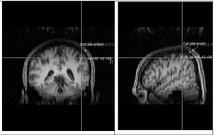
--- figure 1 here
---
The experimental site was subsequently assessed by testing 7 participants (2 of whom had participated in the experiments), using their individual MRI. Talairach coordinates were transformed into individual coordinates using FSL (FMRIB's Software Library - www.fmrib.ox.ac.uk/fsl). Brainsight frameless stereotaxy was then used to verify the distance on the scalp between the intended point of stimulation and the point were stimulation was applied. Coil placement was found to be over the bank of the inferior parietal lobule in all 7 participants, but on average 1.6 cm posterior to the +48, -42, +56 site (sd=.74, min=0.8, max=3; see figure 1). Given the consistency of this result, it is likely that this site was stimulated for all participants in the main study.
Procedure
Both experiments 1 and 2 consisted of two blocks (parietal stimulation and a Pz-control block), each of 200 trials, the order of which was counterbalanced across participants. The two blocks were always completed within a single session of testing. Within a block, half of the trials contained words in single case, and TMS was administered on half of the trials. There were thus 50 trials in each of the following conditions: single case no TMS, single case TMS, case no TMS and mixed-case TMS. Trials were presented in a random order for Experiment 1 and in a semi-random order (semi-randomised prior to the experiment) for Experiments 2 and 3(the latter was done to facilitate scoring for accuracy).
Experiment 3 consisted of three blocks (only 2 of which are reported here), each of 100 trials. In each block, half of the trials contained words in single case, and TMS was administered on every trial. The two blocks reported here are with stimulation over the parietal site or with stimulation over the occipital site.
Every trial started with a 1s fixation cross in the centre of the screen, which was then replaced with the stimulus word, which participants were asked to name as quickly and accurately as possible. Response latencies were measured with a voice-key, and accuracy was scored manually.
Results
Experiment 1 – RTs
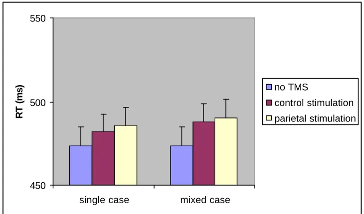
Experiment 1 was analysed using a repeated- measures ANOVA with 2 factors: TMS (no TMS, TMS at the control site, and TMS at the parietal site), and case (single or mIxEd). The no-TMS baseline consisted of no-TMS trials averaged across both blocks (control and parietal stimulation), which did not differ significantly (477 and 471 ms respectively, t(1,10)=.815, p=.434). Order of the blocks did not interact with any of the other variables, and was not included in further analyses.
There was no effect for case (F(1,10)=2.032, p=.184), and the interaction between TMS and case was also not significant (F(2,20)=2.136, p=.144). There was only a trend for a TMS main effect, with higher naming latencies on trials in the TMS conditions (F(2,20)=3.207, p=.062). Reaction times in the parietal- TMS condition were higher than in the no-TMS condition (F(1,10)=7.869, p=.019), while there were no significant differences in RTs between the no-TMS condition and the control-site (F(1,10)=2.519, p=.144). The difference between the two TMS-sites was also not significant (F(1,10)=.336, p=.575).
--- Figure 2 here ---
Experiment 2 – Efficiency of recognition
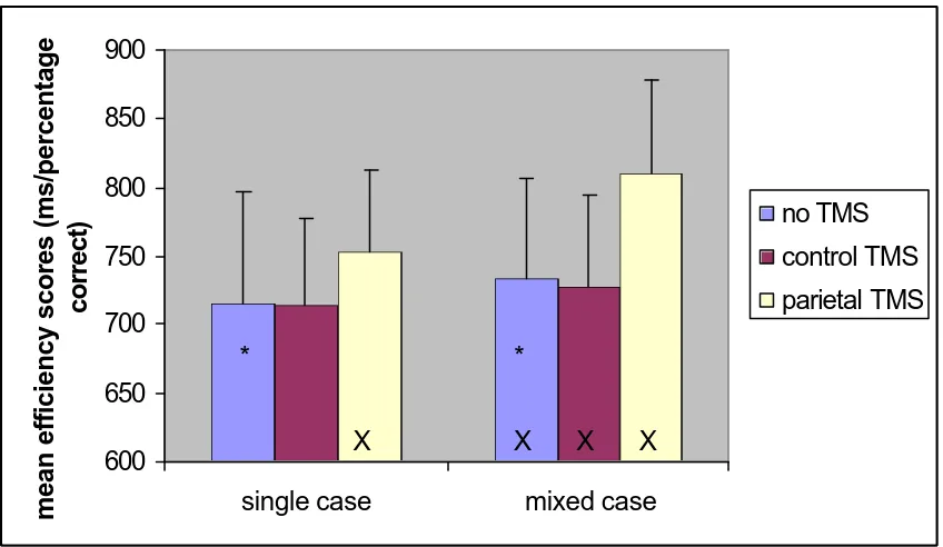
RT- and error-data were combined in a single measure, efficiency, by dividing the participant’s mean RT by the proportion correct of their responses in that condition (see Townsend & Ashby, 1983). The data were then analysed as a two- factor repeated measures ANOVA, with TMS (no TMS, control TMS, and parietal TMS) and case (single or mixed) as factors. Like in Experiment 1, no TMS trials from both blocks were combined in a single no- TMS baseline (mean efficiency scores 736 and 731 ms/percentage correct respectively, t(1,10)=.840, p=.421). The order of the blocks did not interact with any of the other variables, and was not included in further analyses.
There were main effects of TMS (F(2,20)=5.27, p=.014), and case (F(1,10)=40.942, p<.001). Participants found it more difficult to read words presented in mixed case. There was also a reliable interaction between TMS and case (F(2,20)=9.839, p=.001). This interaction was decomposed by comparing the 3 TMS conditions separately.
--- Figure 3 here ---
1. No TMS vs control stimulation (Pz)
Participants were slower to read words presented in mixed-case (F(1,10)=14.753, p=.003). There was no main effect of TMS (F(1,10)=.087, p=.774), nor did TMS interact with case (F(1,10)=.208, p=.658).
There was a main effect of case (F(1,10)=47.133, p<.001) and a trend for a main effect of TMS (F(1,10)=4.427, p=.062). The interaction between case and TMS was significant (F(1,10)=11.76, p=.006). Participants found it more difficult to read words in mixed case compared to single case, and this held particularly when stimulation was applied to the right posterior parietal site, compared to in the no-TMS baseline.
3. Control stimulation vs right parietal stimulation
There were main effects of case (F(1,10)=26.292, p<.001) and TMS (F(1,10)=8.698, p=.015). The interaction between them was also significant (F(1,10)=23.213, p=.001). Participants were worse with words presented in mixed case, and when TMS was applied to the parietal compared to the control site. In addition, there was a stronger case effect when stimulation was applied to the right posterior parietal site, compared to when stimulation was applied to the control site.
Experiment 3 – Efficiency of recognition
As in Experiment 2, RT- and error-data were combined in a single measure, efficiency, by dividing the participant’s mean RT by the proportion correct of their responses in that condition. The data were then analysed as a two- factor repeated measures ANOVA, with TMS (control TMS, and parietal TMS) and case (single or mixed) as factors.
There was a main effect of case (F(1,9)=16.82, p=.003), but not of TMS (F<1). The interaction between case and TMS was also significant (F(1,9)=7.80, p=.021). Participants found it more difficult to read words presented in mixed case, especially when stimulation was applied over the parietal site.
In both experiments 2 and 3, stimulation of the parietal site, but not the control site, had a detrimental effect on reading efficiency, particularly for words presented in mixed case.
Discussion
TMS to the right posterior parietal lobe disrupted word recognition compared to a no TMS baseline and compared to when stimulation was given to a control site. This is interesting as the right posterior parietal lobe is not an area classically associated with reading, and it is not an area that is typically highlighted in studies of reading using functional imaging, though the area is associated with neglect dyslexia in patients with brain lesions. The present data suggest that the area is necessarily involved in visual word recognition. We suggest that its involvement reflects the recruitment of attentional processes when words are read. The effect was strongest on mixed case words. It is unlikely that the effect can be attributed to non-specific effects of TMS, such as increased arousal, as this pattern was not found when stimulation was applied to control site Pz, a site used as a control in other studies of attention (e.g. Cooper et al., 2004), or to an occipital control site where stimulation has been found to ha ve detrimental effects on reading when words are varied over contrast, but not here when they were varied over case (Braet & Humphreys, in preparation).
The finding that TMS to the right posterior parietal cortex impaired reading especially for mixed case words is in line with prior data from functional brain imaging, where there was greater right parietal involvement when stimulus words were presented in mixed case (Mayall et al., 2001). This may be for any of several reasons. The unfamiliar format of mixed case words may mean that more attention must be recruited to read the letters in parallel, or to help recover the features of lower case letters that are masked by upper case letter forms (cf. Besner & Johnston, 1989). Mixed case stimuli may also be tend to read sequentially, based on serial scanning of attention across the string (cf. Mayall et al., 2001). Stimulation of the right parietal cortex may have disrupted this serial reading-strategy, needed for mixed case words, while single case words, which can be read in a parallel fashion, remained largely unaffected.
become ‘case-normalised’ before being read. Both the superior and inferior parietal lobes have been implicated in mirror-reading (e.g. Kassubek et al., 2001), and they form the ‘dorsal pathway’ which is involved in spatial processing and spatial working memory (Ungerleider, Courtney & Haxby, 1998). These two accounts are not necessarily mutually exclusive. When reading words in mixed case or words that require some spatial transformation first, word recognition is unlikely to occur in a parallel fashion, with direct lexical access, as the familiar word and co-occurring letter shapes are not available. Rather, a slower (more serial) strategy is used, where words presented in an unusual format require some form of normalisation before reading can occur. If these processes do rely on a common mechanism, we would expect stimulation of the same site to also disrupt mirror reading, and other similar tasks.
The results support neurological data on attentional dyslexia, where performance is worse with mixed case than single case strings (Hall et al., 2001). Such patients can also have severe problems in the serial scanning of attention (e.g. Friedman-Hill, Robertson & Treisman, 1995), and in detecting visual targets in a spatially parallel manner as feature discriminability decreases (Humphreys & Price, 1994). Their problems with mixed case words, then, are consistent with the greater demand of these stimuli on attentional processes that are impaired in these patients.
information processing, such as visual and linguistic processing, or attentional processes.
In our case, the effects of right posterior parietal stimulation emerged most strongly when the words were briefly presented under low-contrast conditions (in Experiments 2 and 3 relative to Experiment 1). When considered along with the prior neuropsychological and functional imaging evidence indicating the right parietal lobe in visual attentional processing (Corbetta et al., 2002), the data suggest that visual attention needs to be recruited particularly when the visual reading conditions become more difficult (with linguistic processes held constant over the experiments)
Acknowledgements:
This work was supported by grants from the BBSRC and the MRC to G.W.H., and a University of
Birmingham studentship to W.B. We would like to thank Peter Praamstra, as well as the reviewers, for
References:
Baylis GC, Driver J, Baylis LL, & Rafal RD (1994) Reading of letters and words in a patient with Balint’s syndrome. Neuropsychologia, 32: 1273-1286
Besner D, & Johnston JC (1989) Reading the mental lexicon: On the uptake of visual information. In: Marslen-Wilson W (Ed.), Lexical representation and process. Cambridge, MA: MIT Press, pp 291-316
Besner D, & Stolz JA (1999) What kind of attention modulates the Stroop effect? Psychon bull rev, 6: 99-104
Caramazza A, & Hillis AE (1990) Levels of representation, co-ordinate frames, and unilateral neglect. Cognitive Neuropsychology, 7: 391-445
Corbetta M, Kincade JM, & Shulman GL (2002). Neural systems for visual orienting and their relationships to spatial working memory. J Cogn Neurosci, 14: 508-523
Cooper AC, Humphreys GW, Hulleman J, Praamstra P, &Georgeson M (2004) Transcranial magnetic stimulation to right parietal cortex modifies the attentional blink. Exp Brain Res, 155: 24-29
Evett LJ, & Humphreys G W (1981). The use of abstract graphemic information in lexical access. Q J Exp Psychol, 33: 225-250
Ferrand L, & New B (2003) Syllabic length effects in visual word recognition and naming. Acta Psychologica, 113:167-183
Flitman SS, Grafman J, Wasserman EM, Cooper BA., Pascual- Leone A, Hallet M (1998) Linguistic processing during repetitive transcranial magnetic stimulation. Neurology, 50: 175-181
Forster KI, & Davis C (1984) Repetition priming and frequency attenuation in lexical access. J Exp Psychol Learn Mem Cogn, 10: 680-698
Frederiksen JR & Kroll JF (1976) Spelling and sound: approaches to the internal lexicon. J Exp Psychol Hum Percept Perform, 2: 361-379
Friedman-Hill SR, Robertson LC, & Treisman A (1995) Parietal contributions to visual feature binding – evidence from a patient with bilateral lesions. Science, 269: 853-855
Heinke D, & Humphreys GW (2003) Attention, spatial representation, and visual neglect: simulating emergent attention and spatial memory in the selective attention for identification model (SAIM). Psychol Rev, 110: 29-87
Humphreys GW, & Price CJ (1994) Visual feature discrimination in simultanagnosia – a study of 2 cases. Cognitive Neuropsychology, 11: 393- 434
Jalinous R (1998) Guide to Magnetic Stimulation, The MagStim Company, Whitland, Wales.
Johnston JC, & McClelland JL (1974) Perception of Letters in Words: Seek Not and Ye Shall Find. Science, 184: 1192-1194
Karnath HO, Ferber S, Himmelbach M. (2001) Spatial awareness is a function of the temporal not the posterior parietal lobe. Nature, 411: 950-953
Kassubek J, Schmidtke K, Kimmig H, Lücking CH, &Greenlee MW (2001) Changes in cortical activation during mirror reading before and after training: an fMRI study of procedural learning. Cogn Brain Res, 10: 207-217
Kucera H., & Francis WN (1967) Computational analysis of present-day American English. Brown University Press, Providence, Rhode Island
Laberge D (1983) Spatial extent of attention to letters and words J Exp Psychol Hum Percept Perform, 9: 371-379
Lavidor M, & Walsh V (2003) Magnetic stimulation studies of foveal representation. Brain Lang, 88: 331-338
Mari-beffe P, Estevez AF, & Danziger, S (2000). Stroop interference and negative priming: problems with inferences from null effects. Psychon Bull Rev, 7: 499-503
Mayall KA, Humphreys GW, Mechelli A., Olson A., & Price CJ (2001) The effects of case mixing on word recognition: evidence from a PET study. J Cogn
Neurosci,13: 844-853
Mort DJ, Malhotra P, Mannan SK, Rorden C, Pambakian A, Kennard C, & Husain M (2003) The anatomy of visual neglect. Brain, 126: 1986-1997
Pascual- Leone A, Gates JR, & Dhuna A (1991) Induction of speech arrest and counting errors with rapid-rate transcranial magnetic stimulation. Neurology, 41: 697-702
Stolz JA, & Besner D (1996) Role of set in visual word recognition: Activation and activation blocking as nonautomatic processes. J Exp Psychol Hum Percept Perform, 22: 1166-1177
Stolz JA, & Besner D (1998) Levels of representation in visual word recognition: a dissociation between morphological and semantic processing. J Exp Psychol Hum Percept Perform, 24:1642-55
Steinsträter O, Sommer J, Deppe M, Knecht S (2002) The Münster T2T-converter. Available at: http://neurologie.uni- muenster.de/ger/t2tconv/
Stewart L, Walsh V, Frith U, & Rothwell JC (2001) TMS produces two types of speech disruption. Neuroimage, 13: 472-478
Talairach J, & Tournoux P (1988) Co-planar stereotaxic atlas of the human brain, Thieme medical publishers, New York
Townsend JT, & Asby FG (1983). Stochastic modelling of elementary psychological processes. Cambridge University Press
Ungerleider LG, Courtney SM, & Haxby JV (1998) A neural system for human visual working memory. Proc Natl Acad Sci, 95 : 883–889
Vallar G (2001) Extrapersonal visual unilateral spatial neglect and its neuroanatomy. Neuroimage, 14: 52-58
Walsh V, & Cowey A (2000) Transcranial magnetic stimulation and cognitive neuroscience. Nat Rev Neurosci, 1: 73-79
Walsh V, & Pascual-Leone A (2003) Transcranial Magnetic Stimulation: A Neurochronometrics of Mind: MIT Press
Wasserman EM (1998) Risk and safety of repetitive transcranial magnetic stimulation: report and suggested guidelines from the International Workshop on the Safety of Transcranial Magnetic Stimulation, June 5-7, 1996. Electroencephal Clin Neurophysiol, 107: 1-16
Weekes BS (1997) Differential effects of number of letters on word and nonword naming latency. Q J Exp Psychol, 50: 439-456
X
Coil placement
X tal +48 -42 +56 X
Coil placement
X tal +48 -42 +56
x Coil placement
X àtal +48 -42 +56 x
Coil placement
[image:16.596.86.513.70.340.2]X àtal +48 -42 +56
450 500 550
single case mixed case
RT (ms)
no TMS
control stimulation
[image:17.596.114.479.101.316.2]parietal stimulation
Figure 3: Experiment 2 efficiency scores (ms/proportion correct). Error bars represent 95% confidence intervals, calculated as in Masson & Loftus (2003). No-TMS mixed case differs significantly from no-TMS single-case (*); Mixed case (parietal TMS) differs from mixed case (no-TMS and control TMS), as well
as single case (parietal TMS) (X)
600 650 700 750 800 850 900
single case mixed case
mean efficiency scores (ms/percentage
correct)
no TMS
control TMS
parietal TMS
* *
X X X X 600
650 700 750 800 850 900
single case mixed case
mean efficiency scores (ms/percentage
correct)
no TMS
control TMS
parietal TMS
* *
550 600 650 700 750 800 850
single case mixed case
mean efficiency scores (ms/percentage
correct)
control stimulation
parietal stimulation