Original Article
Evaluation of serum high sensitive C-reactive protein,
procalcitonin, neopterin and leukocyte on different
respiratory infectious disease in Chinese children
Han-Rong Cheng1, Dong-Cai Li2, Ben-Qing Wu1, Jian-Liang Yang1, Li Chen1
1Division of Pediatrics, Shenzhen People’s Hospital, Second Clinical Medical College of Jinan University, Shenzhen 518020, Guangdong Province, China; 2Division of Otolaryngology, The Central Hospital of Longgang, Shenzhen 518116, Guangdong Province, China
Received January 9, 2016; Accepted May 5, 2016; Epub July 15, 2016; Published July 30, 2016
Abstract: Objective: To investigate serum high sensitive C-reactive protein (hs-CRP), procalcitonin (PCT), neopterin (NP) and leukocyte (WBC) on different pathogens of respiratory infectious disease, and to provide antibiotic therapy with favorable evidences in Chinese children in the region of guangdong. Methods: 438 infants and children were diagnosed and divided into normal bacterial infection (group A1), suppurative tonsillitis (group A2), mycoplasma (Mp) infection (group B) and virus infection (group C). 50 healthy children were taken as control. Serum hs-CRP, PCT, NP, WBC and blood routine were detected and compared in all groups before and after antibiotic therapy. Results: Serum hs-CRP (mg/L) was higher in A1 (11.55±9.31), A2 (46.38±40.17) and B (6.25±2.64) groups than in
con-trol (1.39±1.76) group, significantly (P<0.05). Serum PCT (ng/mL) was higher in A1 (1.33±6.90), A2 (1.41±4.31), B (0.26±4.98) and C (0.18±7.10) groups than in control (0.05±5.75) group, significantly (P<0.05). Serum NP
(nmol/L) in A1 (19.05±8.94), A2 (35.86±12.76), B (33.75±10.44) and C (43.51±15.90) groups was higher than in
control (6.67±3.32) group, significantly (P<0.05). Serum WBC (1×10⁹/L) was higher in A1 (13.82±10.81) and A2
(13.64±10.55) groups than in control (7.45±3.30) group, significantly (P<0.05). The positive rate of hs-CRP in A1
(71.33%) and A2 (77.78%) groups were higher than in B (30.86%) and C (14.80%) group. The positive rate of PCT in A2 (100%) group was the highest; while in A1 (40.56%) group was higher than in B (32.10%) and C (17.35%) groups. The positive rate of NP in A1 (68.51%), A2 (72.22%), B (80.25%) and C (96.94%) increased gradually. The positive rate of WBC in A1 (51.75%) group was higher than in A2 (33.33%), B (34.57%) and C (15.31%) groups. The
sensitivi-ties and specificisensitivi-ties of hs-CRP, PCT and WBC for bacterial respiratory infection were 72.05% and 100%, 47.20%
and 98.00%, 49.69% and 96.00%, separately, while NP for viral were 96.94% and 100%. Conclusions: Detection
of serum hs-CRP, PCT, NP and WBC was helpful for identification of bacterial, Mp pneumonia and virus infection.
Using serum hs-CRP to evaluate bacterial respiratory infection was more sensitive than using serum PCT or WBC, independently, while using serum NP to evaluate viral respiratory infection was more sensitive. Combining serum hs-CRP, PCR, NP and WBC to evaluate different kinds of respiratory infection was more suitable clinically.
Keywords: C-reactive protein, procalcitonin, neopterin, leukocyte, respiratory infectious disease, children
Introductions
Respiratory infection was one of the large glob-al burden of diseases. In 2010, lower respira-tory tract infections accounted for 115,227,000 disability-adjusted life years (DALYs) [1]. Acc- ording to Walker [2] et al., about 120 million were infected with pneumonia episodes and 1.2 million died for it, and 72% of the deaths were children with age under 2 years. The majority of the patients and the mortality occurred in Southeast Asia and Africa.
and nuclear antigens. Different from the spe-cific recognition of IgG to antigen, hs-CRP rec -ognized the changed auto-antigens and exotic molecules according to pattern recognition. According to the phenomenon mentioned above, Du Clos [6] et al. proposed the basic theories including early-stage defense, signal-ing and activation of proinflammatory cells, hu-moral immunity and the activation of acquired immunity.
In previous studies, hs-CRP combined with dif-ferent indexes was used for evaluating acute coronary syndrome (ACS) [7], carotid athero-sclerosis (CAS) [8] and so on. Xie [9] et al. exam-ined the serum PCT, CRP and soluble triggering receptor expressed on myeloid cells-1 (sTREM-1) for predicting the survival of the patients with early-onset stroke associated pneumonia (EOP). Zhao [10] and Pang [11] et al. investigat-ed the serum hs-CRP in prehypertension com-bined with cardiovascular risk and hyperten-sive heart diseases accompanied with coronary artery disease. PCT, NP and CRP were both evaluated on differentiating bacterial from viral etiologies in patients with lower respiratory tract infections [12]. However, there was no relative report about serum hs-CRP combines with PCT, NP, and leukocyte (WBC) evaluated the different respiratory infectious disease in children.
PCT [13], NP [14] and leukocyte (WBC) [15] were also used for diagnosis of whether there was bacterial or virus infection, clinically. In our study, we focused on the detection of serum hs-CRP combined with PCT, NP and WBC in the infants and children, who were diagnosed as with respiratory infectious diseases, aiming at Chinese children in the region of guangdong, in order to investigate the evaluation of hs-CRP combined with PCT, neopterin and WBC in the early diagnoses of respiratory infectious dis-ease, and to reduce the abuse of antibiotics in infants and children.
Methods and material
Patients
438 children was diagnosed as respiratory infection according to the diagnostic criteria [16], and hospitalized in Second Clinical Medi- cal College of Jinan University from January 1st,
2009 to December 31st, 2014. There were 64 newborns and 374 children, including 274 male and 164 female, age from 2 months to 11 years old. In the 438 patients, 161 patients were diagnosed as bacterial infection (group A) by sputum culture [17] and pharyngeal swab cul-ture [18], including 143 cases with normal bac-terial infection (group A1) and 18 cases with suppurative tonsillitis (group A2). 81 patients were diagnosed as mycoplasma (Mp) infection (group B) and 196 patients were diagnosed as virus infection (group C) by serologic confirm -ing [19]. 50 healthy children were taken as control.
The results of bacterial culture showed that there were 65 cases with escherichia coli (40.37%, 65/161), 40 cases with klebsiella pneumonia (24.84%, 40/161), 31 cases with staphylococcus aureus (19.25%, 31/161), 9 cases with pseudomonas aeruginosa (5.59%, 9/161), and 16 cases with others (9.94%, 16/161).
Informed consent was obtained from all the parents of the patients. This investigation was approved by the medical ethics committee of Second Clinical Medical College of Jinan University.
Blood samples collection
After hospitalization, 2 ml of the venous blood sample was collected in ethylenediaminetet-raacetic acid (EDTA)-K2 anticoagulative tube (Becton, Dickinson and Company, American) from each patient in 24 h. 3 ml of the venous blood sample was collected and the serum was separated for detections. After antibiotic thera-py, the venous blood was collected according to previous methods
Detections
China). Blood routine examination was carried on with BC-5500 and relative reagents (Mindr- ay Medical International Limited. Shenzhen, China).
hs-CRP assay
hs-CRP was detected with enzyme linked immu-noassay (ELISA) kit (Kehua Bioengineering Co., Ltd. Shanghai, China) according to the specifi -cations. The absorbance at 450 nm was mea-sured using immune turbidimetry with AU400 automatic biochemistry analyzer (Olympus im- aging China, Beijing). The calibration curve was linear between 0 and 10 mg/L with the detect-ing limit of 0.2 mg/L. The inter- and intra-assay variance was <10%. The healthy control was with the reference level of 10 mg/L.
PCT assay
PCT was detected with PCT ELISA kit (Thermo) according to the specifications. The functional assay sensitivity of PCT was 0.06 μg/L with the critical values for pneumonia of bacterial etiol-ogy was ≥0.15 μg/L [20]. The healthy control was with the reference level less than 0.5 ng/ mL.
NP assay
NP was detected with ELItest® Neopterin-Screening kit (BRAHMS, Thermo) according to the specification. The absorbance at 405 nm
was measured using immune turbidimetry with AU400 automatic biochemistry analyzer (Oly- mpus) with the detecting limit of 2 nmol/L. The inter- and intra-assay variance was <10%. The healthy control was with the reference level of 10 nmol/L [21].
WBC assay
WBC counting, as one of the blood routine examinations, was carried on with BC-5500 blood-counter system (Mindray). The healthy control was with the reference level of (3.5~ 9.5)×109/L [22].
Statistical analysis
All the data were analyzed by SPSS 13.0 soft-ware. The measuring data were presented as mean ± standard deviations (X±SDs). Compa- risons between groups were used t-test. A value of P<0.05 considered significant difference. Results
hs-CRP, PCT, NP and WBC comparisons before antibiotic therapy
[image:3.612.89.523.85.164.2]According to the results showed in Table 1, serum hs-CRP (mg/L) was higher in A1 (11.55 ±9.31), A2 (46.38±40.17) and B (6.25±2.64) groups than in control (1.39±1.76) group, sig-nificantly (P<0.05). hs-CRP in A2 group incr-eased more obvious than in A1 and B groups.
Table 1. hs-CRP comparisons among groups before antibiotic therapy (X±SDs)
Groups Cases (n=438) hs-CRP (mg/L) PCT (ng/mL) NP (nmol/L) WBC (1×10⁹/L)
A1 143 11.55±9.31* 1.33±6.90* 19.05±8.94* 13.82±10.81*
A2 18 46.38±40.17* 1.41±4.31* 35.86±12.76* 13.64±10.55*
B 81 6.25±2.64* 0.26±4.98* 33.75±10.44* 8.39±4.61
C 196 1.84±2.03 0.18±7.10* 43.51±15.90* 7.58±6.02
Control 50 1.39±1.76 0.05±5.75 6.67±3.32 7.45±3.30
Note: *comparing to control group, P<0.05.
Table 2. Positive rate of hs-CRP, PCT and WBC among groups before antibiotic therapy
Groups (n=438)Cases hs-CRP PCT NP WBC
Cases (n) Rate (%) Cases (n) Rate (%) Cases (n) Rate (%) Cases (n) Rate (%)
A1 143 102 71.33 58 40.56 98 68.51 74 51.75
A2 18 14 77.78 18 100 13 72.22 6 33.33
B 81 25 30.86 26 32.10 65 80.25 28 34.57
C 196 29 14.80 34 17.35 190 96.94 30 15.31
[image:3.612.89.523.211.303.2]There was no significant difference of hs-CRP (mg/L) between in group C (1.84±2.03) and control group.
Serum PCT (ng/mL) was higher in A1 (1.33 ±6.90), A2 (1.41±4.31), B (0.26±4.98) and C (0.18±7.10) groups than in control (0.05±5.75) group, significantly (P<0.05). PCT in A1 and A2 groups increased more obvious than in B and C groups.
Serum NP (nmol/L) in A1 (19.05±8.94), A2 (35.86±12.76), B (33.75±10.44) and C (43.51 ±15.90) groups was higher than in control (6.67±3.32) group, significantly (P<0.05). NP in C group increased more obvious than in A1, A2 and B groups, while in A2 and B groups in- creased more obvious than in A1 group. Serum WBC (1×10⁹/L) was higher in A1 (13.82 ±10.81) and A2 (13.64±10.55) groups than in control (7.45±3.30) group, significantly (P< 0.05). There were no significant difference of WBC (1×10⁹/L) between in B (8.39±4.61), C (7.58±6.02) groups and control group.
Positive rate of hs-CRP and WBC comparisons before antibiotic therapy
The value of hs-CRP >10 mg/L, PCT >0.5 ng/ mL, NP >10 nmol/L and WBC >10×109/L con-sidered as positive. According to Table 2, the positive rate of hs-CRP in A1 (71.33%) and A2 (77.78%) groups were higher than in B (30.86%) and C (14.80%) group.
The positive rate of PCT in A2 (100%) group was the highest, while in A1 (40.56%) group was
ences in A1 (11.55±9.31 v.s. 5.49±6.31) and A2 (46.38±40.17 v.s. 5.88±9.16) groups before and after antibiotic therapy, the same was to serum WBC (1×10⁹/L) in A1 (13.82±10.81 v.s. 10.78±8.44) and A2 (13.64±10.55 v.s. 10.86±12.38) groups, significantly (P<0.05). However, comparing to control group, serum hs-CRP and WBC were both higher after antibi-otic therapy. There was difference of serum NP (nmol/L) in C (43.51±15.90 v.s. 10.50±4.83) group before and after treatments, significant (P<0.05), but still higher than in control group (6.67±3.32).
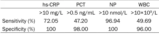
Sensitivities and specificities for hs-CRP, PCT, NP and WBC
According to Table 4, the sensitivities and spec-ificities of hs-CRP, PCT and WBC for bacterial respiratory infection were 72.05% and 100%, 47.20% and 98.00%, 49.69% and 96.00%, se- parately, while NP for viral were 96.94% and 100%. Using serum hs-CRP to evaluate bacte-rial respiratory infection was more sensitive than using serum PCT or WBC, independently.
Discussions
[image:4.612.90.342.86.163.2]hs-CRP was synthesized in liver and transport-ed to the blood, cerebrospinal fluid (CSF), hydrothorax and ascites and so on. hs-CRP was with the regulatory and agglutinating functions similar to IgG and its complement, which could promote the phagocytosis of macrophage and stimulated the expression of the tissue factors on the surface of monocyte [23]. Recently, hs-CRP was found to participate in the cytotoxicity
Table 3. hs-CRP and WBC after antibiotic therapy (X±SDs)
Groups (n=211)Cases hs-CRP (mg/L) NP (nmol/L) WBC (1×10⁹/L)
A1 143 5.49±6.31 - 10.78±8.44
A2 18 5.88±9.16 - 10.86±12.38
C 196 - 10.50±4.83
-Control 50 1.39±1.76 6.67±3.32 7.45±3.30
higher than in B (32.10%) and C (17.35%) groups.
The positive rate of NP in A1 (68.51%), A2 (72.22%), B (80.25%) and C (96.94%) increased gradually.
The positive rate of WBC in A1 (51.75%) group was the highest, while in A2 (33.33%) and B (34.57%) groups were higher than in C (15.31%) group. hs-CRP and WBC comparisons after antibiotic therapy
According to Table 3, comparing the serum hs-CRP (mg/L) before and after antibiotic therapy, there were
differ-Table 4. Sensitivities and specificities of hs-CRP, PCT and WBC for bacterial and NP for viral respiratory infection
hs-CRP PCT NP WBC
>10 mg/L >0.5 ng/mL >10 nmol/L >10×109/L
Sensitivity (%) 72.05 47.20 96.94 49.69
[image:4.612.89.343.209.264.2]mediated by platelet [24]. On one hand, hs-CRP could oxide and activate platelets by itself, on the other hand, hs-CRP could combined with platelet activated factor (PAF) to inhibit the release of arachidonic acid, which would inhibit the neutrophil induced by PAF and the produc -tion of peroxide.
Oh [25] et al. found that when with bacterial infection, inflammatory cell infiltrated and released endogenous transmitters to stimulate hepatocyte to promote the synthesis of hs-CRP, and the serum PCT, as a marker of bacterial infection [26], would also change. At the early stage of bacterial infection, serum hs-CRP would increase rapidly, with tendency positive to the severity of infection. However, the am- ount of WBC was normal or just increased slightly. In our study, at the early stage of bacte-rial infection, the positive rate of hs-CRP in A1 and A2 groups were 71.33% and 77.78%, sepa-rately, the PCT were 40.56% and 100%, and the WBC were 51.75% and 33.33%. Comparing the serum hs-CRP, PCT and WBC in bacterial respiratory infection, the sensitivities and spec-ificities of hs-CRP, PCT and WBC for bacterial respiratory infection were 72.05% and 100%, 47.20% and 98.00%, 49.69% and 96.00%, separately, which indicating that serum hs-CRP was more sensitive to evaluate bacterial respi-ratory infection.
However, there were large individual differenc-es between each child, differenc-especially the emotion of the infants and children with infectious dis-ease would induced large fluctuation of serum WBC, which would result in the false positive of WBC. hs-CRP was not influenced by emotion easily, as well as PCT. Therefore, combining serum hs-CRP with PCT and WBC was more suitable for early diagnosis of bacterial infection.
In healthy people, the content of serum hs-CRP was very low (<3 mg/L). But in those with inflammation or acute injury, hs-CRP increased in 4 to 6 hours and reached the peak in 36 to 50 h, approximately 100 to 1,000 times of healthy people. hs-CRP was not easily affected by sex, age, anemia, hyperglobulinemia, even the temperature, and would return to normal with remission [27, 28]. Although WBC was the indicator of bacterial infection, WBC failed to support with effective information in infants or children for the not significant changes.
The main pathogens in respiratory infection in pediatrics department included bacteria, Mp pneumonia and virus. NP was the predictive marker of virus infection [29], which was sensi-tive for early assessment of acute respiratory syndrome both in adults and children. When with virus infection, the serum NP would increase more than 10 nmol/L [21]. In our study, the sensitivity of NP for viral respiratory infection was 96.94%, which was accorded to and indicated the fact that NP played an impor-tant role in diagnosis of virus infection. With the limitations including not the separation of virus or detection of calcitonin zymogen, and the long time of bacteria culture, hs-CRP combined with PCT NP could be the diagnostic and treat-ing indicator at the early infectious stage. From the results mentioned in Table 1, hs-CRP in the infants and children with bacterial or Mp infection were both higher than in healthy infants and children (control group), significant -ly, while in those with virus infection was not significantly different from the control. There were significant differences of the positive rate of hs-CRP among the bacterial (A1, 71.33%; A2, 77.78%), Mp (30.86%) and virus (14.80%) infection, which was similar to PCT (A1, 40.56%; A2, 100%; B, 32.10%; C, 17.35%) and NP (A1, 68.51%, A2, 72.22%; B, 80.25%; C, 96.94%) among each group. These phenomenon indi-cated that hs-CRP combined with PCT could be the index to identify and distinguish different infections induced by different kinds of patho-gen, while hs-CRP combined with NP could identify and distinguish infections induced by bacteria or virus, accorded to previous rese- arches [30, 31].
Additionally, before antibiotic therapy on bacte-rial infection, serum hs-CRP (mg/L) was higher in A1 (11.55±9.31) and A2 (46.38±40.17) groups comparing to the control (1.39±1.76) group, significantly (P<0.05). After therapy, serum hs-CRP (mg/L) both decreased in A1 (5.49±6.31) and A2 (5.88±9.16) groups, which indicated that hs-CRP could be the index for infection control, accorded to previous researches [32, 33].
evidences. However, our study only focused on the infants and children in the region of Shenzhen guangdong with limited quantity of samples and large individual and regional dif-ferences. There were differences of the sensi-tivities and specificities of serum hs-CRP, PCT, NP and WBC for diagnosing different kinds of respiratory infection. Therefore, identification of different kinds of respiratory infectious dis-ease still needed to accord to the clinically practical situations and analyzed comprehe- nsively.
Disclosure of conflict of interest
None.
Address correspondence to: Dong-Cai Li, Division of Otolaryngology, The Central Hospital of Longgang,
Shenzhen 518116, Guangdong Province, China. Tel: +86-13510099301; Fax: +86-13510099301;
E-mail: dongcai517@hotmail.com
References
[1] Murthy NS, Nandakumar BS, Pruthvish S,
George PS and Mathew A. Disability adjusted
life years for cancer patients in India. Asian Pac J Cancer Prev 2010; 11: 633-640. [2] Walker CL, Rudan I, Liu L, Nair H, Theodoratou
E, Bhutta ZA, O’Brien KL, Campbell H and
Black RE. Global burden of childhood pneumo -nia and diarrhoea. Lancet 2013; 381: 1405-1416
[3] Wang SM. Use of serum C-reaction protein es-timation in diseases of respiratory tract. Aca-demic Journal of Plan Postgraduate Medical School. 1991.
[4] Hu XJ, Zhou F, Qiu YR and Li Q. [Diagnostic
value of serum procalcitonin and C-reaction protein in acute exacerbation of chronic
bron-chitis]. Nan Fang Yi Ke Da Xue Xue Bao 2010;
30: 1618-1620.
[5] Rasmussen F, Mikkelsen D, Rj, Lambrechtsen
J, Nybo M, Hansen H and Siersted H. High-sen-sitive C-reactive protein is associated with re-duced lung function in young adults. Eur Respir J 2009; 33: 382-388.
[6] Du Clos TW. Function of C-reactive protein. Ann
Med 2000; 32: 274-278.
[7] Wang XH, Liu SQ, Wang YL and Jin Y. Correla-tion of serum high-sensitivity C-reactive protein and interleukin-6 in patients with acute
coro-nary syndrome. Genet Mol Res 2014; 13:
4260-4266.
[8] Zhang Q, Qian G and Ding Z. Xuemaitong gran -ules attenuate carotid atherosclerosis by
de-creasing the expression of CD14+CD16+ mo-
nocytes, IL-6, TNF-alpha, and hsCRP. Genet
Mol Res 2014; 13: 7519-7527.
[9] Xie J, Zhang XH and Zhu WY. Values for serum procalcitonin, C-reactive protein, and soluble triggering receptor expressed on myeloid ce- lls-1 in predicting survival of patients with
ear-ly-onset stroke-associated pneumonia. Genet
Mol Res 2015; 14: 4716-4723.
[10] Zhao XS, Wang R, Bin LR and Wa SQ. Interven-tion for prehypertension and its cardiovascular
risk factors in Inner Mongolia. Genet Mol Res
2014; 13: 4867-4882.
[11] Pang H, Han B, Li ZY and Fu Q. Identification of
molecular markers in patients with hyperten-sive heart disease accompanied with coronary
artery disease. Genet Mol Res 2015; 14:
93-100.
[12] Ip M, Rainer TH, Lee N, Chan C, Chau SS,
Leung W, Leung MF, Tam TK, Antonio GE, Lui G, Lau TK, Hui DS, Fuchs D, Renneberg R and
Chan PK. Value of serum procalcitonin, neop-terin, and C-reactive protein in differentiating bacterial from viral etiologies in patients pre-senting with lower respiratory tract infections. Diagn Microbiol Infect Dis 2007; 59: 131-136. [13] Certain L and Schuetz P. The role of procalcito-nin in respiratory infections. Curr Infect Dis Rep 2012; 14: 308-316.
[14] Luczak G, Plata-Nazar K, Kozielska E, Delins
-ka-Galinska A, Borkowska A, Kotlowska-Kmied
A, Wos-Wasilewska E and Kaminska B. [Serum neopterin level in children with lower respira-tory tract infections]. Med Wieku Rozwoj 2007; 11: 423-427.
[15] Schwartz J and Weiss ST. Peripheral blood leu-kocyte count and respiratory symptoms. Ann Epidemiol 1993; 3: 57-63.
[16] Li Y. Pediatric disease diagnostic criteria Sci-ence Press 2001.
[17] Verenkar MP, Pinto MJ, Savio R, Virginkar N and Singh I. Bacterial pneumonias--evaluation of various sputum culture methods. J Postgrad Med 39: 1993; 60-62.
[18] Bruckova M, Grandien M, Pettersson CA and
Kunzova L. Use of nasal and pharyngeal swabs for rapid detection of respiratory syncytial virus and adenovirus antigens by enzyme-linked im-munosorbent assay. J Clin Microbiol 1989; 27: 1867-1869.
[19] Falsey AR, Walsh EE and Betts RF.Serologic
evidence of respiratory syncytial virus infection in nursing home patients. J Infect Dis 1990; 162: 568-569.
[20] Christ-Crain M, Jaccard-Stolz D, Bingisser R,
Gencay MM, Huber PR, Tamm M and Muller B.
single-blinded intervention trial. Lancet 2004; 363: 600-6007.
[21] Zheng B, Cao KY, Chan CP, Choi JW, Leung W,
Leung M, Duan ZH, Gao Y, Wang M, Di B, Hol -lidt JM, Bergmann A, Lehmann M, Renneberg I,
Tam JS, Chan PK, Cautherley GW, Fuchs D and
Renneberg R. Serum neopterin for early as-sessment of severity of severe acute respira-tory syndrome. Clin Immunol 2005; 116: 18-26.
[22] Perez IA, Santana SP, Argudin TD and Gardon
DO. Analysis of blood processing conditions to obtain high-quality total RNA from human
leu-kocyte concentrate. Genet Mol Res 2007; 6:
298-307.
[23] Chen YX, Wang JF, Yao YJ, Yuan WL, Kong MY, Lin YQ, Geng DF and Nie RQ. CRP regulates the
expression and activity of tissue factor as well
as tissue factor pathway inhibitor via NF-κB and ERK 1/2 MAPK pathway. FEBS Lett 2009;
583: 2811-2818.
[24] Volanakis JE. Human C-reactive protein: ex-pression, structure, and function. Mol Immu-nol 2001; 38: 189-197.
[25] Oh KJ, Park KH, Kim SN, Jeong EH, Lee SY and Yoon HY. Predictive value of intra-amniotic and
serum markers for inflammatory lesions of pre -term placenta. Placenta 2011; 32: 732-736. [26] Simon L, Gauvin F, Amre DK, Saint-Louis P and
Lacroix J. Serum procalcitonin and C-reactive protein levels as markers of bacterial infection: a systematic review and meta-analysis. Clin In-fect Dis 2004; 39: 206-217.
[27] Wener MH, Daum PR and McQuillan GM. The influence of age, sex, and race on the upper
reference limit of serum C-reactive protein con-centration. J Rheumatol 2000; 27: 2351-2359.
[28] Breiterman-White R. C-reactive protein and anemia: implications for patients on dialysis. Nephrol Nurs J 2006; 33: 555-558.
[29] Kandelaki ET, Nemsadze KP, Chkhaidze IG, Ka
-vlashvili NI and Kamkamidze GK.[Neopterine
and ige during respiratory-syncytial virus
infec-tion in infants]. Georgian Med News; 2006. pp.
76-80.
[30] Wang SM, Lei HY, Huang KJ, Wu JM, Wang JR, Yu CK, Su IJ and Liu CC. Pathogenesis of en-terovirus 71 brainstem encephalitis in pediat-ric patients: roles of cytokines and cellular im-mune activation in patients with pulmonary edema. J Infect Dis 2003; 188: 564-570. [31] Meloni GF, Tomasi PA, Spanu P, Piga S and
Porcu A. C-reactive protein levels for diagnosis of Salmonella gastroenteritis. Pediatr Infect Dis J 1999; 18: 471-473.
[32] Ehl S, Gering B, Bartmann P, Hogel J and Poh
-landt F. C-reactive protein is a useful marker
for guiding duration of antibiotic therapy in suspected neonatal bacterial infection. Pediat-rics 1997; 99: 216-221.