Original Article
Long-term evaluation of motor function and
learning-memory ability in a 3-day-old
ischemic brain injury rat model
Lan Hu, Mili Xiao, Zhihua Li, Chao Chen
Department of Neonatology, Children’s Hospital of Fudan University, Shanghai, China
Received March 30, 2016; Accepted September 7, 2016; Epub November 15, 2016; Published November 30, 2016
Abstract: To evaluate the histopathological changes, physical development and long term motor and learning-mem-ory ability of a 3-day-old rat brain injury model. Postnatal day-3 male Sprague-Dawley rats were randomly assigned to experimental group induced by ligation of bilateral carotid arteries, and sham-operated control group (48 rats/ group). At 1 day after the procedure and 3 weeks old, we used hematoxylin-eosin stain, myelin related protein
im-munohistochemistry, pre-oligodendrocyte marker O4 and TUNEL double immunofluorescence stain to investigate
the pathological changes. Inspections of physical development were made after operation until 3 weeks old. Motor activity and coordination of the rats were evaluated by four sensorimotor tests between 3 to 6 weeks old. Learning and memory ability were tested by Morris water maze between 7 to 13 weeks old. The survival rate was 100% (48/48) in the control group and 68.8% (33/48) in the experimental group 24 hours after operation. Pathological changes including periventricular leukoaraiosis, dilated ventricles, and pre oligodendrocyte apoptosis. The expres-sion of myelin basic protein was reduced at 3 weeks old. Rats in the experimental group showed decreased weight gain (P<0.05), poor motor ability and decreased maintenance of balance (P<0.05) and learning-memory defects compared with rats in the control group (P<0.05). Our rat model revealed a preferential occurrence of brain
dam-age which is similar to preterm infant, and confirmed the early outcome is delayed growth development during
childhood. The long term outcome including motor and learning-memory ability defect will persist until adulthood.
Keywords: White matter injury, preterm birth, oligodendrocyte, neurobehavioral manifestations
Introduction
Preterm brain injury has become a significant cause of neonatal brain injury and often leads to severe neurological sequelae that mostly involve motor and visual abnormalities [1], cerebral palsy accounts for up to 20% [2-4]. Preterm brain injury can also widely affect life quality through speech and language problems [5] behavioral, social, attention [6], motor skill [7] and cognitive deficits [8].
Recent studies suggest that approximately 1% of live-born infants are very low birth weight (VLBW), among which the incidence of white matter damage is 20% [9-11]. Over the past 10-15 years, cystic PVL has declined in inci-dence and currently occurs in less than 5% of VLBW infants [12]. However, the increasing application of MRI to the clinical assessment of
brain injury in the preterm newborn has now revealed that diffuse noncystic white matter injury is the dominant pattern of white matter injury [13, 14], accounting for more than 90% of PVL and occurring in up to half of premature VLBW newborns [12, 15, 16]. Diffuse noncystic white matter injury (WMI) is increasingly being recognized as a risk factor for autism-spectrum disorders, ADHD, and other psychological dis-turbances [17]. Thus, proper selection of an ani-mal model for studying the pathogenesis in- volved in preterm brain injury allowing observa-tion of neuromotor and cognitive development is important for studying the mechanism of neuroprotection from brain injury.
mecha-nism of brain injury in preterm infants that of inappropriate vasoconstriction resulting from the immature vascular system [19]. The most common rodent models are based upon the Vannucci method [20, 21], generally estab-lished at postnatal day 7 and 10 with adapta-tions for younger rats at postnatal day 3 to 6 [22] and postnatal day 6 to 7 mice [23, 24] have also been developed. Carotid artery liga-tion followed by exposure to low oxygen has recently been successfully applied to study the mechanism of hypoxic-ischemic injury related to immature oligodendrocytes. Lin et al. [25] established a hypoxic-ischemic model in post-natal day 4rats by bilateral carotid artery liga-tion followed by 8% O2 inhalation for 20 min and performed immunohistochemical analy-sis. The results suggested that pre-oligoden-drocytes positively labelled with O4+ and O1+
and staining for myelin basic protein decreased and amyloid precursor protein (APP) immuno-reaction increased in postnatal day 6 and post-natal day 9 experimental group rats compared to the control group, confirming the occurrence of white matter damage to rats in the experi-mental group. A previous report [26] by Uehara demonstrated that the decrease of cerebral blood flow was up to about 25% of baseline in the subcortical white matter in neonatal rats with bilateral carotid artery occlusion, which is higher than that in rats subjected to unilateral common carotid artery ligation combined with systemic hypoxia (15%-17%) and which prefer-entially caused white matter injury and reduced gray matter injury. Back [27, 28] described that cerebral white matter damage is most com-mon at 24-32 weeks gestation in humans, a time at which pre-oligodendrocytes are the predominating oligodendroglial cell [29], whereas in rats it occurs much more frequently on postnatal days 2-4. Thus, it is proper to use postnatal day 2 to day 4 immature rats for study of cerebral white matter injury in preterm infants. Sizonenko [30] has shown that by right carotid ligation followed by 6% hypoxia for 30 min in postnatal d3 rat compromised brain cor-tical growth and led to a selective alteration of cortical myelinated axons with persistent glio-sis. These alterations share neuropathological similarities with the diffuse white matter lesions found in VLBW infants.
So far several studies have evaluated a brain injury model established by bilateral carotid
31], but no one has follow up longer enough to evaluate the long-term motor and cognitive development which may provide important information for preterm brain injury. In this study, we established the hypoxic-ischemic brain injury model by ligation of bilateral carotid arteries in postnatal day 3 rats with the aim of investigating the histopathological changes, short-term physical development and long-term motor and learning-memory ability.
Materials and methods
Animals and experimental groups
96 clean male Sprague-Dawley (SD) rats aged 3 days were obtained from the Laboratory Animal Center of Shanghai Medical College, Fudan University, weighing 7.5-12 g, which were divided into 2 groups: sham operation group (normal control) and bilateral carotid artery li- gation group (experimental group). This experi-ment was undertaken with approval from Fudan University Affiliated Children Hospital animal use and care committee.
Model establishment
The rats were anesthetized with diethyl ether. In a supine position, the skin of the neck was sterilized with disinfectant, and a 0.5 cm inci-sion was made at the cervical ventral midline with an aseptic scalpel under sterile conditions. The subcutaneous tissue was separated layer by layer until the euro vascular bundle was found and bilateral common carotid arteries were separated. The skin was then sutured for the control group and the bilateral carotid arter-ies were ligated with 9-0 sutures for the experi-mental group followed by skin suturing. The rats were allowed to recover to their normal body temperature and activity using a water bath at 37°C after the operation before being placed back to the cage. 31.2% (15/48) of rats in the experimental group died during or right after surgery resulting from intolerance to the surgical treatments.
Evaluation of growth and development
We randomly selected 8 live rats from each group after operation until 3 weeks old and recorded their weight, eye opening, ear erect-ing, and incisor eruption by daily video footage. Motor development assessment
sorimotor tests including the vertical screen test, foot fault test, beam traversal test and rotarod test were performed twice per week and the whole process for each test was record-ed by video. A neurologist from the Children’s Hospital of Fudan University evaluated the video footage. Selected reflexes and evaluation criteria were based on Wang [32]. Results were taken as means of the two experiments in a week. The apparatus and software were from Jing Mei Company, Shanghai.
Learning and memory assessment
We randomly selected 8 live rats from each group at 7, 9, 11 and 13 weeks of age to assess the learning and memory ability by water maze. During training period, 8 live rats from each
group were allowed to swim in the water maze three times a day and two minutes for each time from each quadrant. If some of the rats can’t find the platform in the water within two minutes, then help them to. After finishing the training period, the Morris water maze test [33] was carried out once in two weeks to determine the cognition and memory of rats. The water was stained by ink so that the platform can’t be seen above the water. The tracking software automatically recorded original data and ana-lyzed swim speed, swimming time around the platform, swimming curve and success rate on the platform. The water maze and software were from Jing Mei Company, Shanghai.
Histology
At 24 hours after the procedure and at postna-tal 3 weeks, we selected 8 live rats from each group for histological study. After being anes-thetized by ether, the rats were perfused with normal saline and 4% PFA successively, by left ventricular catheterization. Next, we carefully stripped the brain tissue and subsequently fixed the tissue in 4% PFA for 48 hours. Brain tissue was dehydrated by ethanol and then par-affin-embedded. Continuous coronal sections of 6-μm thickness were taken and analyzed in the anterior commissure lateral ventricle plane. Changes in the structure of brain tissue were observed after hematoxylin-eosin staining of the brain tissue section including the histologi-cal profile of the cortex, subcortex, periventricu -lar white matter, and ventricles under a light microscope.
Myelin basic protein (MBP) immunohistochemi-cal staining was conducted to compare the dif-ference in staining of the subcortical white mat-ter versus the periventricular whimat-ter matmat-ter. MBP antibody was from ABCAM Company, British. We used the TUNEL assay to compare the number of apoptotic cells in the subcortical and periventricular areas of the two groups under a fluorescence microscope. The nuclei of apoptotic cells emitted yellow-green fluores -cence under a fluores-cence microscope (TRITIC with an emission wavelength of 620 nm and an absorption wavelength of 550 nm). Pre-OL marker O4 immunohistochemical staining was conducted to compare the number of pre-OL cells in the subcortical and periventricular area of the two groups under the fluorescence micro -scope. TRITIC emission was used to detect pre-Figure 1. Weights of the experimental group and
con-trol group. A significant difference in weight between
[image:3.612.92.287.74.210.2]the two groups was seen every day until 3 weeks af-ter the procedure (*P<0.05).
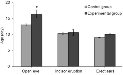
Figure 2. Physical development of experimental group and control group. Eye lid opening for rats in the experimental group happened obviously later than that for rats in the control group. The difference
was statistically significant (*P<0.05). Other physi -ological phenomena such as incisor eruption and
[image:3.612.91.289.288.409.2]OL cells, and a laser confocal scanning micro-scope to observe O4 and TUNEL double posi-tive cells. The O4 antibody was from R&D sys-tems Company. Fluorescence microscope was Leica Fluorescence Microscope, USA. The laser confocal scanning microscope was Leica TCS SP2, USA.
Statistics
Normally distributed continuous variables are presented as mean ± standard deviation (mean ± SD) and were compared using the indepen-dent sample t-test. The differences were con-sidered statistically significant when the P value was less than 0.05. Statistical analysis was performed by SPSS 10.0 (SPSS Inc., Chicago, IL, USA). Water maze tracking soft-ware was used to analyze learning and memo-ryability in rats.
duced diet. 3% (1/33) of rats in the experimen-tal group suffered from convulsions after the operation and died 48 hours after operation. Among the experimental group, within one week after operation, 28.1% (9/32) of rats suf-fered from monoplegia and circled to the healthy limb side while crawling, and 12.5% (4/32) of rats had paralysis of the lower limbs, leading to falling down when crawling.
[image:4.612.91.522.83.298.2]Before the procedure, there was no significant difference in body weight of rats between the control group and the experimental group. At the first day after the procedure, all rats in the experimental group experienced a decrease in body weight. The average body weight before the procedure was 7.11±0.16 g in the experi-mental group and 7.28±0.25 g in the control group, and the average body weight after the procedure was 6.92±0.16 g and 8.71±0.17 g, Table 1. Tests for 3-day-old neonatal rats after bilateral carotid artery ligation at different ages
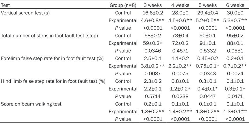
Test Group (n=8) 3 weeks 4 weeks 5 weeks 6 weeks
Vertical screen test (s) Control 16.6±0.2 28.0±0 29.4±0.4 30.0±0 Experimental 4.6±0.8** 4.5±0.6** 5.2±0.5** 5.3±0.7**
P value <0.0001 <0.0001 <0.0001 <0.0001 Total number of steps in foot fault test (step) Control 68±0.2 73±0.4 90±0.1 95±0.2
Experimental 59±0.2* 72±0.2 91±0.1 88±0.1
P value 0.0346 0.4571 0.5332 0.0551 Forelimb false step rate for in foot fault test (%) Control 2.5±0.1 1.1±0.2 0.45±0.2 0.2±0.1 Experimental 3.8±0.2** 2.2±0.2** 0.75±0.1* 0.7±0.2**
P value 0.0087 0.0075 0.0343 0.0024 Hind limb false step rate for in foot fault test (%) Control 2.3±0.2 0.8±0.1 0.3±0.1 0.1±0.1 Experimental 2.2±0.1 1.2±0.2* 0.4±0.1* 0.3±0.1*
P value 0.5714 0.0238 0.0447 0.0171 Score on beam walking test Control 0.2±0.1 0.1±0.1 0.1±0.1 0.1±0.1
Experimental 1.8±0.2** 1.4±0.2** 1.3±0.2** 1.3±0.1**
P value <0.0001 <0.0001 <0.0001 <0.0001
*P<0.05 vs control, **P<0.01 vs control.
Table 2. Rotarod test for 3-day-old neonatal rats after bilateral carotid artery ligation at different ages
Group (n=8) 4 weeks 5 weeks 6 weeks Rotarod distance (cm) Control 682.9±0.3 850.6±0.2 1027.3±0.4
Experimental 285.8±0.7* 339.8±0.6* 559.5±0.7*
P value 0.0353 0.0327 0.0191 Rotarod speed (cm/s) Control 8.3±0.6 11.6±0.5 13.0±0.6
Experimental 6.0±0.7* 8.4±0.3* 9.3±0.1*
P value 0.0314 0.0256 0.0212
*P<0.05 vs control.
Results
[image:4.612.91.394.356.448.2]respectively. Subsequently, the rats in the ex- perimental group slowly gained weight, with an average weight of 7.55±0.24 g at day 2 after the procedure compared with 10.55±0.23 g in the control group. A significant difference in weight between the two groups was seen every day until 3 weeks after the procedure (P<0.05) (Figure 1). Eye lid opening for rats in the experi-mental group happened obviously later than that for rats in the control group at 16.4±1.2
and 13.0±0.3 days, respectively (Figure 2). The difference was statistically significant (P<0.05). Other physiological phenomena such as incisor eruption which appeared at 10.3±0.4 days for the control group and 10.6±0.9 days for the experimental group, and ear erecting that app- eared at 9.0±0.1 days for the control group and 10.0±0.2 days for the experimental group were not statistically significant (Figure 2).
Vertical screen test
The vertical screen test was performed twice a week in rats 3 weeks after birth to assess fore-limb and hind fore-limb strength. Results for a week were taken as means of the two experiments within that week. Time of seizing the screen for the control group gradually became longer as their age increased. The ability to grasp for the experimental group developed slowly and time of seizing the screen was obviously decreased compared to the control group. The difference between the two groups was statistically signifi -cant (P<0.01) (Table 1).
Foot fault test
The foot fault test was used twice a week in rats 3 weeks after birth to evaluate their motor coordination. Results for one week were shown as means of the two experiments within that week. Compared with the control group, the total number of steps for the rats in the experi-mental group was less at 3 weeks’ postnatal age (P<0.05), (Table 1). In contrast, no statisti-cally significant differences were observed when testing at 4, 5 and 6 weeks’ postnatal age. Rats in the experimental group made more false steps than that in the control group. False steps rates for forelimb and hind limb were cal-culated. Higher false steps rates were seen in the experimental group compared to the con-trol group and the difference was statistically significant (P<0.05) (Table 1).
Beam traversal test
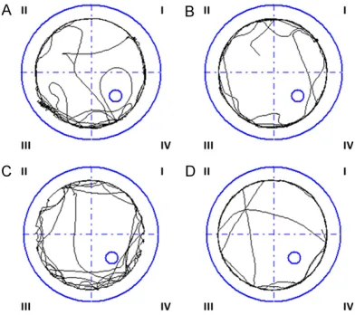
[image:5.612.90.289.71.254.2]The balancing ability of rats in the experimental group obviously decreased compared to the control group. The beam traversal test was used for rats 3, 4, 5 and 6 weeks old. The score in the experimental group was much higher than in the control group and the difference was statistically significant (P<0.05) (Table 1). Most of the rats in experimental group grasped Figure 3. The swimming route of control rats in the
Morris water maze. A. Rats go into the water at the
first quadrant. B. Rats go into the water at the second
quadrant. C. Rats go into the water at the third quad-rant. D. Rats go into the water at the fourth quadquad-rant.
Figure 4. The swimming route of experimental rats in the Morris water maze. A. Rats go into the water
at the first quadrant. B. Rats go into the water at the
[image:5.612.91.287.336.510.2]one side of the beam or rotated more than 90 degrees when traversing the beam. 12.5% of the experimental rats (1/8) fell down from the beam. In contrast, rats in control group revealed normal motor action and 100% (8/8) of them could reach the platform located at the end of the beam.
Rotarod test
The rotarod distance and rotarod speed for control rats increased with age. Rats in the experimental group showed shorter rotarod distance and lower rotarod speed in compari-son of control group (P<0.05). In the experi-mental group, the ability to remain on the rotat-ing rod increased slowly and shorter rotarod distance and lower rotarod speed were also required in comparison with the control group (Table 2) (P<0.05).
Water maze test
Rats without memory of the platform will swim along the pool in a circular motion while rats with spatial cognition and memory of the plat-form change their swimming pattern and look for the location of the platform (Figures 3 and 4). The success rate of getting to the platform was 93.8% (8/8) for control group rats and 25% (2/8) for experimental group rats, sug-gesting a lower cognition ability of rats with brain damage. The longer time the rats spend in the quadrant of the platform and the higher the percentage of time around the platform, the stronger the cognition function of the rats.
The experimental group spent shorter time on the platform compared to control group and the difference was statistically significant (P<0.05) (Table 3). The parameter of movement velocity was used to assess motor function. Movement velocity of the experimental group was slower than that of the control group and the differ-ence was statistically significant (P<0.05) (Table 4).
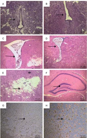
Comparison of histology
[image:6.612.89.525.100.150.2]HE staining showed that brain lesions altered with age. 24 hours after operation, subcortical and periventricular leukoaraiosis occurred in the experimental group. Damage to the cortex and ventricular dilatation was not obviously observed in the experimental group compared to control group. Ventricular dilatation and leu-komalacia appeared in rats in the experimental group at 3 weeks postnatal age. All experimen-tal group rats suffered from gliocyte hyperpla-sia to varying degrees (Figure 5A-F). MBP imm- unohistochemical staining results did not show any difference between control and experimen-tal groups 24 hours after operation due to the immature myelination. As the myelination matures with age, the difference between con-trol and experimental groups became more and more evident. Gray matter myelin was evidently seen in the corpus callosum and internal cap-sule of the rats from the experimental group, but not the control group rats which showed a sparse pattern of gray matter myelin (Figure 5G, 5H).
Table 3. The percentage of time around the platform for 3-day-old neonatal rats after bilateral carotid artery ligation at different ages in water maze test
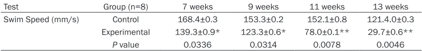
Test Group (n=8) 7 weeks 9 weeks 11 weeks 13 weeks
Percentage of time around the platform (s) Control 62%±0.7% 63%±0.4% 61%±0.6% 72%±0.3%
Experimental 7%±0.7%** 19%±0.6%** 19%±0.8** 12%±0.2%**
P value <0.0001 <0.0001 <0.0001 <0.0001
**P<0.01 vs control.
Table 4. Swim speed of 3-day-old neonatal rats after bilateral carotid artery ligation at different ages in water maze test
Test Group (n=8) 7 weeks 9 weeks 11 weeks 13 weeks
Swim Speed (mm/s) Control 168.4±0.3 153.3±0.2 152.1±0.8 121.4.0±0.3 Experimental 139.3±0.9* 123.3±0.6* 78.0±0.1** 29.7±0.6**
P value 0.0336 0.0314 0.0078 0.0046
[image:6.612.89.522.207.260.2]The experimental group showed higher num-bers of apoptotic cells than the control group 24 hours after operation. Oligodendrocytes labeled with anti-O4 were detected by TRITC excitation. Observation of O4 and TUNEL-double-positive cells were processed using confocal laser microscopy. Our results
[image:7.612.89.375.69.521.2]suggest-opening, primitive reflexes inhibition, limb movement disorder and declined motor coordi-nation. Our results also showed declined growth and development of rats in the experi-mental group. Weight loss is associated with water deprivation caused by reduced food intake and consciousness depression after Figure 5. Pathology in rat brain tissue. A. Periventricular leukoaraiosis is
seen in the experimental group at 24 h after surgery. B. No periventricular abnormalities in the control group at 24 h after surgery. C. Dilated brain ventricles in the experimental group. D. Brain ventricles are normal in the control group. E. Damaged intracranial structure. F. Intact intracranial struc-ture in the control group. G. Positive staining of myelin basic protein (MBP) was occasionally seen in the control group. H. Positive staining of MBP was obviously seen in the control group.
ed that the apoptotic cells were mainly oligodendrocytes (Figure 6).
Discussion
brain injury, and may also relate to a decreased appetite from ischemic damage to feeding cen-ter in hippocampus. In the current study we found one rat in the experimental group suf-fered from convulsions. Because all of the experimental group rats must be returned to their cages for maternal feeding, which was unfavorable for observation of convulsions, it is difficult to calculate the incidence of convul -sions. Eyelid opening and eyelid reflex were obviously delayed in rats in the experimental group compared to the control group. Whereas differences in time spent on pricking ear and auricle reflex were not statistically significant. Poggi et al. [35] developed a white matter dam-age model by intracranial injection of lipopoly-saccharide (LPS) in rats and evaluated behav-ioral and motor development. Rats were intra-cervically injected with LPS or saline and evalu-ation for motor developmental milestones was performed in neonatal rats on days 1 to 21 after delivery. On postnatal day 21, animals we- re sacrificed for immunohistochemistry detec -tion. The results suggested that there was no significant difference in neonatal weight, bal
-ance function, negative geotaxis, forelimb gra- sp, audio startle, eye opening and activity. Sur- prisingly, neonatal rats subjected to LPS injec-tion revealed better ability of forelimb place-ment and balance. Although the immunohisto-chemical findings revealed cerebral white mat -ter damage, there was no obvious difference in the motor outcome of neonatal animals, relat-ing to manifestations of cerebral palsy in hu- mans. This could be due to the strong compen-satory adjustment of neonatal rats after brain injury induced by injection of LPS.
[image:8.612.91.524.70.354.2]brain injury with long-term follow-up, and de- tected motor and cognitive deficit in the experi -mental group rats.
The Morris water maze test was mainly for study of cognition and memory. We combined the Morris water maze test with motor tests, which could comprehensively evaluate neuro-functional changes of neonatal rats after brain injury. In this study, for the first time we applied brain injury model by bilateral carotid artery ligation in 3-day-old newborn rats and evaluat-ed long-term motor outcomes and cognition development of model animals, which would provide a basis for intervention study on hypox-ic-ischemic brain injury in preterm infants using immature rat model. Ikeda et al. [38] suggest-ed that neonatal rats with cerebral white mat-ter injury revealed a sustained cognitive impair-ment during infancy, adolescence and maturity. Mishima et al. [39] set up the hypoxia-ischemic brain injury model by unilateral common carot-id artery ligation followed by exposure to sys-temic hypoxia in 7-day-old rats and evaluated the cognition development by 8-arm radial maze task between 2 and 17 weeks after oper-ation. The results suggested that the cognitive level 16 weeks after surgery was more severe than that for 3 weeks in experimental group rats, confirming that cognitive impairment slow -ly progressed with time. In our study, the Morris water maze test was carried out in rats after birth for 7, 9, 11 and 13 weeks, equivalent to adolescence and maturity in humans, and the results showed decreased swim speed, shorter residence time on the platform, and lower suc-cess rate on the platform in rats subjected to bilateral carotid artery ligation compared to sham-operated rats, indicating sustained spa-tial cognition function and long-term memory defects.
In summary, our present study established a brain injury model by bilateral carotid artery ligation in 3-day-old premature rats and the his-tological findings revealed a preferential occur -rence of cerebral white matter damage. Results for assessment of motor function and cognition revealed retarded growth and development, inhibition of primitive reflexes, decreased physi -cal activity, even convulsion and paralysis in the early stage, and cognition dysfunction in the later stage in the experimental rats. These results suggest that the model demonstrated a similar condition of brain injury as that found in preterm brain injury in human.
Disclosure of conflict of interest None.
Address correspondence to: Dr. Chao Chen, Depart- ment of Neonatology, Children’s Hospital of Fudan University, 399 Wanyuan Road, Shanghai 201102, China. Tel: 86 -21-64931186; Fax: 86 -21-6493- 1146; E-mail: [email protected]
References
[1] Kidokoro H, Anderson PJ, Doyle LW, Woodward LJ, Neil JJ, Inder TE. Brain injury and altered brain growth in preterm infants: predictors and prognosis. Pediatrics 2014; 134: 444-453. [2] Payne AH, Hintz SR, Hibbs AM, Walsh MC, Vohr
BR, Bann CM, Wilson-Costello DE. Neurodevelo- pmental outcomes of extremely low-gestation-al-age neonates with low-grade periventricul- ar-intraventricular hemorrhage. JAMA Pediatr 2013; 167: 451-459.
[3] Elitt CM, Rosenberg PA. The challenge of un-derstanding cerebral white matter injury in the premature infant. Neuroscience 2014; 276: 216-238.
[4] Volpe JJ. Brain injury in premature infants: a complex amalgam of destructive and develop-mental disturbances. Lancet Neurol 2009; 8: 110-124.
[5] Vohr B. Speech and language outcomes of very preterm infants. Semin Fetal Neonatal Med 2014; 19: 78-83.
[6] Fan RG, Portuguez MW, Nunes ML. Cognition, behavior and social competence of preterm low birth weight children at school age. Clinics (Sao Paulo) 2013; 68: 915-921.
[7] Bos AF, Van Braeckel KN, Hitzert MM, Tanis JC,
Roze E. Development of fine motor skills in pre -term infants. Dev Med Child Neurol 2013; 55: 1-4.
[8] Marret S, Marchand-Martin L, Picaud JC, Has- coët JM, Arnaud C, Rozé JC, Truffert P, Larroque B, Kaminski M, Ancel PY. Brain injury in very preterm children and neurosensory and cogni-tive disabilities during childhood: the EPIPAGE cohort study. PLoS One 2013; 8: 626-683. [9] Blumenthal I. Periventricular leucomalacia: a
review. Eur J Pediatr 2004; 163: 435-42. [10] Izbudak I, Grant PE. MR imaging of the term
and preterm neonate with diffuse brain injury. Magn Reson Imaging Clin N Am 2011; 19: 709-731.
[11] Volpe JJ, Kinney HC, Jensen FE, Rosenberg PA. Reprint of The developing oligodendrocyte: key cellular target in brain injury in the premature infant. Int J Dev Neurosci 2011; 29: 565-582. [12] Volpe JJ. Neurology of the Newborn, WB Saun-
[13] Counsell SJ, Ball G, Edwards AD. New imaging approaches to evaluate newborn brain injury and their role in predicting developmental dis-orders. Curr Opin Neurol 2014; 27: 168-175. [14] de Vries LS, Benders MJ, Groenendaal F. Pro-
gress in Neonatal Neurology with a Focus on Neuroimaging in the Preterm Infant. Neurope- diatrics 2015; 46: 234-241.
[15] Miller SP, Ferriero DM, Leonard C, Piecuch R, Glidden DV, Partridge JC, Perez M, Mukherjee P, Vigneron DB, Barkovich AJ. Early brain injury in premature newborns detected with magnet-ic resonance imaging is associated with ad-verse early neurodevelopmental outcome. J Pediatr 2005; 147: 609-616.
[16] Miller SP, Cozzio CC, Goldstein RB, Ferriero DM, Partridge JC, Vigneron DB, Barkovich AJ. Comparing the diagnosis of white matter injury in premature newborns with serial MR imaging
and transfontanel ultrasonography findings.
AJNR Am J Neuroradiol 2003; 24: 1661-1669. [17] Tilborga EV, Heijnenb CJ, Bendersc MJ, Belc
FV, Fleiss B, Gressens P, Nijboer CH. Impaired oligodendrocyte maturation in preterm infants: Potential therapeutic targets. Prog Neurobiol 2016; 136: 28-49.
[18] Scafidi J, Fagel DM, Ment LR, Vaccarino FM.
Modeling premature brain injury and recovery. Int J Dev Neurosci 2009; 27: 863-871. [19] Brew N, Walker D, Wong FY. Cerebral vascular
regulation and brain injury in preterm infants. Am J Physiol Regul Integr Comp Physiol 2014; 306: 773-786.
[20] Rice JE 3rd, Vannucci RC, Brierley JB. The influ -ence of immaturity on hypoxic-ischemic brain damage in the rat. Ann Neurol 1981; 9: 131-141.
[21] Vannucci RC, Vannucci SJ. Perinatal hypoxic-ischemic brain damage: evolution of an animal model. Dev Neurosci 2005; 27: 81-86. [22] Sizonenko SV, Sirimanne E, Mayall Y, Gluckman
PD, Inder T, Williams C. Selective cortical al-teration after hypoxic-ischemic injury in the very immature rat brain. Pediatr Res 2003; 54: 263-269.
[23] Shen Y, Plane JM, Deng W. Mouse models of periventricular leukomalacia. J Vis Exp 2010. [24] Follett PL, Deng W, Dai W, Talos DM, Massillon
LJ, Rosenberg PA, Volpe JJ, Jensen FE. Gluta- mate receptor-mediated oligodendrocyte toxic-ity in periventricular leukomalacia: a protective role for topiramate. J Neurosci 2004; 24: 4412-4420.
[25] Lin S, Rhodes PG, Lei M, Zhang F, Cai Z. alpha-Phenyl-n-tert-butyl-nitrone attenuates hypoxic-ischemic white matter injury in the neonatal rat brain. Brain Res 2004; 1007: 132-141. [26] Uehara H, Yoshioka H, Kawase S, Nagai H,
Ohmae T, Hasegawa K, Sawada T. A new model of white matter injury in neonatal rats with bi-lateral carotid artery occlusion. Brain Res
[27] Back SA, Luo NL, Borenstein NS, Levine JM, Volpe JJ, Kinney HC. Late oligodendrocyte pro-genitors coincide with the developmental win-dow of vulnerability for human perinatal white matter injury. J Neurosci 2001; 21: 1302-1312.
[28] Back SA, Rosenberg PA. Pathophysiology of glia in perinatal white matter injury. Glia 2014; 62: 1790-1815.
[29] Back SA, Han BH, Luo NL, Chricton CA, Xan- thoudakis S, Tam J, Arvin KL, Holtzman DM. Selective vulnerability of late oligodendrocyte progenitors to hypoxia-ischemia. J Neurosci 2002; 22: 455-463.
[30] Sizonenko SV, Sirimanne E, Mayall Y, Gluckman PD, Inder T, Williams C. Selective cortical al-teration after hypoxic-ischemic injury in the very immature rat brain. Pediatr Res 2003; 54: 263-269.
[31] Sizonenko SV, Kiss JZ, Inder T, Gluckman PD, Williams CE. Distinctive neuropahtologic alter-rations in the deep layer of the parietal cortex afer moderate ischemic-hypoxic Injury in the P3 immature rat brain. Pediatr Res 2005; 57: 865-872.
[32] Wang YJ. Advances in modern neurology. Bei- jing: beijing Science Press; 2005.
[33] Morris RG. Spatial localization does not re-quire the presence of local cues. Learning and Motivation 1981; 12: 239-260.
[34] Back SA, Miller SP. Brain injury in premature neonates: a primary cerebral dysmaturation disorder? Ann Neurol 2014; 75: 469-486. [35] Poggi SH, Park J, Toso L, Abebe D, Roberson R,
Woodard JE, Spong CY. No phenotype associ-ated with established lipopolysaccharide mod-el for cerebral palsy. Am J Obstet Gynecol 2005; 192: 727-733.
[36] Jansen EM, Low WC. Long-term effects of neo-natal ischemic-hypoxic brain injury on senso-rimotor and locomotor tasks in rats. Behav Brain Res 1996; 78: 189-194.
[37] Tomimatsu T, Fukuda H, Endoh M, Mu J, Watanabe N, Kohzuki M, Fujii E, Kanzaki T, Oshima K, Doi K, Kubo T, Murata Y. Effects of neonatal hypoxic-ischemic brain injury on skilled motor tasks and brainstem function in adult rats. Brain Res 2002; 926: 108-117. [38] Ikeda T, Mishima K, Yoshikawa T, Iwasaki K,
Fujiwara M, Xia YX, Ikenoue T. Selective and long-term learning impairment following neo-natal hypoxic-ischemic brain insult in rats. Behav Brain Res 2001; 118: 17-25.