Original Article
miR-126 inhibits epithelial ovarian cancer
growth partially by repression of IRS2
Yanli Wang1, Jihong Wen1, Wenjing Shao1, Xiaomeng Zhang2
1Department of Gynecology, The First Hospital of Jilin University, Changchun 130021, China; 2Department of
Ophthalmology, The Second Hospital of Jilin University, Changchun 130041, China
Received October 6, 2015; Accepted December 16, 2015; Epub February 15, 2016; Published February 29, 2016
Abstract: MiR-126 has been reported to involve in the development and progression of various cancers. However its role in epithelial ovarian cancer (EOC) remains unclear. Therefore, the aims of this study were to investigate the
miR-126 expression and its clinical diagnosis significance in patients suffering EOC and to analyze its role and po -tential molecular mechanism on the carcinogenesis of ovarian cancer by a series of molecular experiments. Here,
we found that miR-126 was significantly decreased in ovarian cancer cell lines and tissues by quantitative RT-PCR (qRT-PCR), and its expression was negatively correlated with advanced FIGO stage, high histological grading and
lymph node metastasis (all P<0.01). Functional study demonstrated that the restoration expression of miR-126
significantly inhibited EOC cell proliferation, migration and invasion, and induces apoptosis. Additionally, IRS2 was predicted the target gene of miR-126. Luciferase reporter assay further verified direct target association of miR-126 to specific sites of the IRS2 3’-untranslated regions. qRT-PCR and western blot assays showed that overexpression
of miR-126 inhibited IRS2 expression. Importantly, IRS2 expression was upregulated in EOC tissues and inversely
correlated with the expression of miR-126. Taken together, these results shows for the first time that miR-126 func -tions as a tumor suppressor in epithelial ovarian cancer partially by repression of IRS2.
Keywords: microRNAs, miR-126, ovarian cancer, epithelial ovarian cancer, IRS2
Introduction
Epithelial ovarian cancer (EOC) is the leading cause of death from gynecological cancers worldwide, and it accounts for more than 90% of all forms of ovarian cancer [1, 2]. Primary treatment of EOC is surgical resection of visible disease, followed by adjuvant chemotherapy, which improved the quality of life in patients with EOC, however, the 5-year survival rate for all stages of ovarian cancer has been estimat-ed to be 45.6% due to the high rate of recur-rence and chemoresistance [3, 4]. Therefore, it is need to identify new biomarkers, treatments, and therapeutic targets for human epithelial ovarian cancer.
Micro-RNAs (miRNAs) are calss of single-stranded, small (18-25 nucleotides in length) non-coding RNA molecules that regulate the expression of target genes by pairing with sites in the 3’ untranslated region (30-UTR) [5]. Increasingly evidence has suggested that
miR-NAs play important role in many physiological and pathological processes, such as cell growth and differentiation, proliferation, apoptosis, vascular angiogenesis and embryonic develop-ment [6, 7]. It has been showed that miRNAs function as oncogenes or tumor suppressor, and involve in various processes of tumor pro-gression including development, differentia-tion, apoptosis, proliferadifferentia-tion, cell cycle, and metastasis [6, 8, 9]. Therefore, miRNAs are attractive candidates in study of cancer physi-ological and pathphysi-ological processes.
and underlying molecular mechanism of miR-126 on the carcinogenesis of epithelial ovarian cancer.
Materials and methods
Clinical specimens
Epithelial ovarian cancer tissues and their paired adjacent normal tissues were obtained from the 40 patients with EOC who underwent surgery at the First Hospital of Jilin University (Changchun, China) from June 2010 to July 2015. All samples were immediately frozen in liquid nitrogen and stored at -80°C until use. All the patients were diagnosed as EOC and the samples were histologically confirmed by pathologist. None of patients had received either radiotherapy or chemotherapy. Clinical data including age, tumor size, FIGO stage, his -tological grading and lymph node metastasis was collected and listed in Table 1. Informed consent was obtained from each patient, and the study protocols were approved by the Eth- ics Committee of Jilin University (Changchun, China).
Cell culture
A human ovarian surface epithelial cell line (HOSEpiC) and four human ovarian cancer cells
Technologies). Quantitative PCR were per-formed by the TaqMan miRNA assay kits (Applied Biosystems, Foster City, CA, USA) under ABI 7900 Fast system (Applied Biosystems). Expression of U6 was used as an endogenous control. To determine the mRNA levels of IRS2, cDNA was synthesized by PrimeScript RT reagent Kit (Takara, Dalian, China) following the manufacturer’s instructions. The expression levels of IRS2 were quantified by Real-time PCR Mixture Reagent (Takara) under ABI 7900 Fast system. The primers for IRS2 and β-actin were described as previous study [18]. β-actin was used as internal control, The comparative 2–∆∆Ct method was used for relative quantification and statisti -cal analysis.
Transfection
miR-126 mimic or corresponding negative con-trol (miR-NC) were purchased from GenePharma (Shanghai, China), and were respectively trans-fected into SKVO3 cells at final concentration 100 nM using Lipofectamine 2000 (Invitrogen, USA) according to the manufacturer’s protocol. Transfection efficiencies were evaluated in every experiment by qRT-PCR at 48 h post-transfection.
Cell proliferation assay Table 1. Correlation between clinicopathological features
and miR-126 expression in EOC tissues
Variables No. of cases miR-494 expression P value Low (n %) High (n %)
Age (years) P>0.05
<55 24 13 (59.1) 11 (40.9)
≥55 16 10 (62.5) 6 (37.5)
Tumor size P>0.05
≥5 23 12 (52.2) 11 (47.8)
<5 17 11 (64.5) 6 (35.5)
FIGO stage P<0.01
I-II 30 14 (46.7) 16 (53.3)
III-IV 10 9 (90.0) 1 (10.0)
Histological grading P<0.01
1-2 28 13 (46.4) 15 (53.6)
3 12 10 (83.3) 2 (16.7)
Lymph node metastasis P<0.01
No 33 16 (48.5) 17 (52.5)
Yes 7 7 (100.0) 0 (0.0)
Culture Collection (ATCC) and were cul-tured in Dulbecco’s modified eagle’s
medium (DMEM, Gibco BRL, Gai-thersburg, MD, USA) supplemented with 10% fetal bovine serum(FBS, Gibco BRL), 100 U/mL of penicillin and 100 mg/mL of streptomycin at 37°C in
a humidified chamber supplemented
with 5% CO2.
Real time quantitative RT-PCR
bromide (MTT) assay. In briefly, SKVO3 cells were seeded in 96-well culture plates at a den-sity of 5 × 10³ cells per well 24 h after trans-fected with miR-126 or miR-NC. After 1 to 4 days, cells were stained with incubated with 20 μl MTT reagent (5 mg/ml, Sigma, St. Louis, MO) for 4 h at 37°C. The cell medium was carefully removed, and incubated with 200 μl of dimeth -yl sulfoxide (DMSO, Sigma) was added to each well to dissolve the crystals for 10 min at 37°C. The absorbance in each well was measured with a microplate reader (Tecan, Männedorf, Switzerland) set at 490 nm.
Cell apoptosis assay
Cells apoptosis analysis was performed on SKVO3 cells 48 h after transfection. Transfected cells were incubated with PE Annexin-V and 7AAD following the PE Annexin-V Apoptosis Detection Kit I (BD Pharmingen, CA, USA) proto -col, and then were analyzed by under a flow cytometer (BD Biosciences San Jose, CA, USA). The apoptotic rate was analyzed using Cell-Quest software (BD Biosciences).
Migration and invasion assay
To examine the migration ability of cells in vitro, a wound-healing assay was performed. In brief-ly, transfected cells were seeded in 3.5-cm plates and grown to a density of 70 to 80%. Afterwards, cells were scratched using a sterile plastic micropipette tip to create an artificial wound. Cells were imaged at 0 and 24 h after the wounding, and the migrating distance was measured after 24 h.
For invasion assay, a transwell chamber assay was performed. Transfected cells (2 × 10⁴ cells/well) were placed in the upper chamber of a 24-well Matrigel-coated Transwell unit with 8 μm -pore-size polycarbonate nucleopore filters (Corning Costar, Cambridge, MA), and cultured in serum-free DMEM medium. The lower cham-ber was filled with DMEM medium with 10% FBS to attract cells. After cells had been cul-tured at 37°C for 48 h, the cells adhering to the lower surface were fixed with 70% ethanol for 30 min and stained with 0.2% crystal violet for 10 min. The invaded cells were photographed and were counted in five random fields of view
at 100 × magnification under light microscope (Olympus, Tokyo, Japan).
Luciferase assay
The 3’UTR of IRS2 was amplified using PCR and subcloned into pGL3-control vector (Ambion, Austin, TX, USA) at the NheI and XhoI restriction sites.Mutant constructs of IRS2 were also gen-erated by introducing mutated nucleotides within the seed region-binding sequences in the oligonucleotides, and inserted into pGL3-control vector at the NheI and XhoI restriction sites. For luciferase assays, the SKVO3 cells were plated in 24-well plates at a density of 2 × 10⁴ cells per well and transfected with 100 ng of IRS2-3’UTR-WT or IRS-3’UTR-Mut reported plasmid, and 100 nM of 126 mimic or miR-NC, using Lipofectamine 2000 (Invitrogen, Carlsbad, CA, USA). At 48 h post transfection, both firefly and Renilla luciferases activities in cell lysates were determined using the Dual Luciferase Reporter Assay System (Promega, Madison, WI, USA). Renilla-luciferase was used for normalization.
Western blotting
Protein extracts were performed from cultured cells or tissues through RIPA lysis buffer (Beyotime, Shanghai, China). Protein concen-trations were measured by using a BCA assay kit (Beyotime). Equal quantities (30 μg) of pro -tein samples were loaded on 10% SDS-PAGE and transferred onto nitrocellulose membranes (Millipore, Wisconsin, USA), and Blocking was performed with 5% non-fat milk in Tris-buffered saline containing 0.1% Tween-20 for 2 h, Then membrans incubated at 4°C overnight with fol-lowing primary antibody: anti-human IRS2 (1:1000, Santa Cruz, USA) and anti-human β-actin (1:5000, Santa Cruz, USA), followed incubated with horseradish peroxidase (HRP)-conjugated second antibody (1:5000, Santa Cruz, USA) for 1 h at room temperature. β-actin was used as an internal control for protein load-ing. The protein bland was observed by en- hanced chemiluminescence (ECL, Cell Signa- ling Technology).
Statistical analysis
sepa-rate experiments, and processed using SPSS 19.0 statistical software (Chicago, IL, USA). A P<0.05 was considered statistically significant. Results
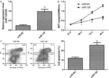
miR-126 is down-regulated in EOC tissues and cell lines
To determine whether miR-126 was involved in ovarian tumorigenesis, we evaluated the ex- pression levels of miR-126 in EOC tissues and cell lines. qRT-PCR assay showed that expres -sion of miR-126 was significantly decreased in EOC tissues compared with adjacent normal tissue, as well as was differentially decreased in four ovarian cancer cell lines compared with human ovarian surface epithelial cell line (HOSEpiC) (Figure 1A and 1B). SKOV3 cells exhibited the lowest expression of miR-126, and were selected for next studies (Figure 1B). To investigate the clinical relevance of miR-126 in EOC, the median (0.426) of all 40 patients was chosen as the cutoff point to divide two groups of all cases: low-miR-126 (<0.426, 23 cases) group and high-miR-126 expressing group (>0.426, 17 cases). It was found that miR-126 expression were negatively correlated with FIGO stage, histological grading and lymph node metastasis(all P<0.01), which are all indi-cators of poor prognosis (Table 1). Meanwhile, we did not find any correlation of miR-126 expression and age and tumor size. These stud -ies suggest that miR-126 is down-regulated in
miR-126 inhibits EOC cell proliferation and induces cell apoptosis
To investigate the function of miR-126, we restored it expression by transfected miR-126 mimic in SKOV3 (Figure 2A). Proliferation of SKOV3 cells was determined by MTT assay at the indicated time (1-4 days). With time longer, difference in the proliferation rate became more significant in SKOV3 cells with miR-126 restoration compared with miR-NC group (Figure 2B), which suggested that restoration of miR-126 inhibits SKVO3 cell proliferation. In addition, we also investigate effect miR-126 on cell apoptosis. Flow cytometer assay showed that restoration of miR-126 induced cells apop-tosis relative to miR-NC group (P<0.05, Figure 2C).
miR-126 inhibits EOC cell migration and inva-sion
We also investigate whether miR-126 effect on EOC migration and invasion in SKOV3 cells transfected with miR-126 mimic or miR-NC by wound heal and transwell chamber assay, respectively. Our results demonstrated that restoration of miR-126 expression could signifi -cantly suppress SKVO3 cell migratory (Figure 3A) and invasive (Figure 3B) capabilities.
IRS2 is a direct target of miR-126
[image:4.612.93.521.72.230.2]To investigate the underlying mechanism of
Figure 1. miR-126 expression is downregulated in epithelial ovarian cancer (EOC) tissues and cell lines. A. miR-126
expression in 40 EOC tissues and their corresponding adjacent normal tissues were detected by quantitative RT-PCR (qRT-RT-PCR) *P<0.05; **P<0.01 versus normal tissue. B. miR-126 expression in four ovarian cancer cell lines (SKOV3, A2780, OVCAR and HO-8910) and human ovarian surface epithelial cell line (HOSEpiC) were detected by
looked for its potential downstream targets using two publicly available algorithms (Target- scan6.2 and miRanda) to help identify miR-126 targets in EOC cells. Based on the predicted binding region of miR-126 in the 3’-UTR of IRS2, we propose that IRS2 is a direct target of miR-126 (Figure 4A). To further confirm this pro -pose, dual-luciferase reporter assay were per-formed, and found that restoration of miR-126 significantly decreased the luciferase activity of the IRS 3’-UTR-WT in SKVO3 cells (Figure 4B), while had no inhibition effect on the mutant IRS2-3’UTR reporter activity in SKVO3 cells (Figure 4B), indicting the direct regulation of miR-126 in the 3’UTR of IRS2 mRNA. In addi -tion, qRT-PCR and western blotting showed that restoration of miR-126 in SKOV3 cells sig-nificantly decrease IRS2 expression on mRNA level (Figure 4C) and protein level (Figure 4D). These results suggest that IRS2 is a direct tar-get of miR-126.
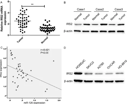
IRS2 expression was upregulated and inverse-ly correlated with miR-126 expression in EOC tissues
[image:5.612.93.522.73.383.2]Since it has been confirmed that IRS2 is a direct target of miR-126, we determined the expression of IRS2 expression in EOC tissues and corresponding normal tissue. We found that IRS2 expression on mRNA levels (Figure 5A) and protein levels (Figure 5B) were upregu-lated compared with matched normal tissue. Using Spearman’s correlation analysis, we found that IRS2 mRNA expression was inverse-ly correlated with miR-126 expression in EOC tissues (Figure 5C; r=-0.421, P<0.05). Mean- while, we also detected the IRS2 expression in four ovarian cancer cell lines (SKOV3, A2780, OVCAR and HO-8910) and human ovarian sur-face epithelial cell line (HOSEpiC). Western blot assay showed that IRS2 protein expression was obviously upregulated in four ovarian cancer Figure 2. miR-126 inhibits EOC cell proliferation and induced cell apoptosis. A. miR-126 expression was restored in SKOV3 cells after transfected with miR-126 mimic, assessed by qRT-PCR. B. Cell proliferation was determined by
cell lines compared with human ovarian sur-face epithelial cell line (HOSEpiC) (Figure 5D). Discussion
Accumulating evidence had shown that microR-NAs (miRmicroR-NAs), a class of small non-coding RNAs, are important regulators involved in cell proliferation, cycle, apoptosis, invasion and migration of multiple types of human cancers, including ovarian cancer [19, 20]. A larger num-ber of miRNAs has been identified to play a role in control cell proliferation, metastasis, and cell cycle in ovarian cancer [19, 20]. For example, Wen et al [21] found that that miR-338-3p func-tions as a tumor suppressor and suppresses tumor growth of EOC in vitro and in vivo through PI3K/AKT signaling pathways by targeting Runx2. Zhu et al [22] reported that miR-661 functions as tumor promoter by targeting the INPP5J gene, and then promoting cell prolifera-tion of ovarian cancer. Lan et al [23] showed
[image:6.612.93.519.75.392.2]proliferation and induces apoptosis partially by repression of PDGFRA. In the present study, our results first showed that miR-126 expres -sion was obviously decreased in EOC tissue and cell lines, and its expression was negative associated with FIGEO stage, histological grad -ing and lymph node metastasis. Our results also showed that restoration of miR-126 signifi -cantly inhibited EOC cell proliferation, migration and invasion, and induced cell apoptosis. These results suggested that miR-126 might play crucial roles in the carcinogenesis of EOC. The sequence encoding miR-126, located at intron 7 of the EGFL7 gene, had been showed that it expression could be epigenetically regu-lated with the EGFL7 gene [24]. Downregulation of miR-126 expression has been found in hepa-tocellular carcinoma [10], renal cell carcinoma [11], osteosarcoma [12], gastric cancer [13], cervix cancer [14], non-small cell lung cancer [15], colorectal cancer [16], and colon cancer Figure 3. miR-126 inhibits EOC cell migration and invasion. A. Cell migration was determined by wound healing as-say in SKVO3 cells transfected with miR-126 or miR-NC. B. Cell invasion was determined by invasion chamber asas-say
showed that miR-126 plays a potential role as a tumor suppressor in many kinds of cancers [10-17]. For ovarian cancer, only a study showed that miR-126 could inhibit serine/threonine p21-activated kinase 4 (PAK4) protein expres-sions in ovarian cancer cells [25]. However, the expression and function of miR-126 in ovarian cancer remains unclear. In this study, our results showed that miR-126 expression level was decreased in EOC tissue and cell lines, and that restoration of miR-126 significantly inhib -ited EOC cell proliferation, migration and inva-sion. These results suggested that miR-126 functions as tumor suppressor in epithelial ovarian cancer.
IRS2, located in the 13q34 region, is important member of the insulin receptor substrates (IRSs) family [26]. IRSs are cytoplasmic
[image:7.612.99.522.69.416.2]scaf-fold proteins that act as signaling intermedi-ates through which downstream intracellular signals are generated, which in turn allows ini-tiation of intracellular signaling cascades, such as the Wnt/ß-catenin pathway [27]. Overex- pression of IRS2 has been found in many types of cancers, including ovarian cancer [28]. It has been showed that IRS2 play an oncogenic role in various cancers, including ovarian cancer [29]. In this study, we confirmed IRS2 was a direct target of miR-126 by luciferase reporter assay. Restoration of miR-126 expression in SKVO3 cells inhibited IRS expression on mRNA level and protein level. Of note, our results showed that IRS2 expression was upregulated in EOC tissue and cell lines and its expression inversely correlated with miR-126 expression in EOC tissues. These results might suggest that Figure 4. IRS2 is a direct target of miR-126. A.The putative miR-126-binding sites and mutant (Mut) 3’-UTR IRS2
sites was shown. B. Luciferase assay were measured in SKVO3 cells cotransfected with miR-126 or miR-NC and
IRS2 3’UTR (Wt) or a mutant (Mut) reported plamid. Wt: Wide type; Mut: Mutant type.C. IRS2 mRNA expression was
miR-126 exerted tumor suppressor in epithelial ovarian cancer by repression of IRS2.
In summary, the results presented here first demonstrate that miR-126 expression level was decreased in EOC tissue and cell lines, and its expression was negatively correlated with advanced FIGEO stage, high histological grad -ing and lymph node metastasis. Our find-ing also demonstrated that miR-126 inhibited EOC cell proliferation, migration and invasion, as well as induced cell apoptosis via directly tar-geting IRS2, suggesting that miR-126 might be developed as a new therapeutic target in epi-thelial ovarian cancer.
Disclosure of conflict of interest None.
Address correspondence to: Xiaomeng Zhang, De- partment of Ophthalmology, The Second Hospital of
Jilin University, Changchun 130041, China. E-mail: zhangxm15106@sina.com
References
[1] Siegel R, Naishadham D, Jemal A. Cancer sta-tistics, 2013. CA Cancer J Clin 2013; 63: 11-30.
[image:8.612.93.519.72.420.2][2] Permuth-Wey J and Sellers TA. Epidemiology of ovarian cancer. Methods Mol Biol 2009; 472: 413-437.
Figure 5. IRS2 was up-regulated and inversely correlated with miR-126 expression in EOC tissues.A. IRS2 mRNA
expression in human EOC tissues and their corresponding normal tissues was determined by qRT-PCR. β-actin was
used as an internal control. *P<0.05; **P<0.01 versus normal tissue. B. IRS2 protein expression in human EOC
tissues and their corresponding normal tissues was determined by Weston bolt. β-actin was used as an internal
[3] Coleman RL, Monk BJ, Sood AK and Herzog TJ.
Latest research and treatment of advanced-stage epithelial ovarian cancer. Nat Rev Clin Oncol 2013; 10: 211-224.
[4] Legge F, Ferrandina G, Salutari V and Scambia G. Biological characterization of ovarian can -cer: prognostic and therapeutic implications. Ann Oncol 2005; 16 Suppl 4: iv95-101. [5] Valinezhad Orang A, Safaralizadeh R and
Kazemzadeh-Bavili M. Mechanisms of miRNA-Mediated Gene Regulation from Common Downregulation to mRNA-Specific Upre-gulation. Int J Genomics 2014; 2014: 970607.
[6] Bushati N and Cohen SM. microRNA functions. Annu Rev Cell Dev Biol 2007; 23: 175-205. [7] Hwang HW and Mendell JT. MicroRNAs in cell
proliferation, cell death, and tumorigenesis. Br J Cancer 2006; 94: 776-780.
[8] Calin GA and Croce CM. MicroRNA signatures
in human cancers. Nat Rev Cancer 2006; 6: 857-866.
[9] Volinia S, Calin GA, Liu CG, Ambs S, Cimmino A,
Petrocca F, Visone R, Iorio M, Roldo C, Ferracin
M, Prueitt RL, Yanaihara N, Lanza G, Scarpa A,
Vecchione A, Negrini M, Harris CC and Croce CM. A microRNA expression signature of
hu-man solid tumors defines cancer gene targets. Proc Natl Acad Sci U S A 2006; 103:
2257-2261.
[10] Zhao C, Li Y, Zhang M, Yang Y and Chang L. miR-126 inhibits cell proliferation and induces cell apoptosis of hepatocellular carcinoma cells partially by targeting Sox2. Hum Cell 2015; 28: 91-9.
[11] Khella HW, Scorilas A, Mozes R, Mirham L,
Lianidou E, Krylov SN, Lee JY, Ordon M, Stewart
R, Jewett MA and Yousef GM. Low Expression
of miR-126 Is a Prognostic Marker for Metastatic Clear Cell Renal Cell Carcinoma. Am J Pathol 2015; 185: 693-703.
[12] Jiang L, Tao C, He A and He X. Overexpression
of miR-126 sensitizes osteosarcoma cells to
apoptosis induced by epigallocatechin-3-gal-late. World J Surg Oncol 2014; 12: 383. [13] Chen H, Li L, Wang S, Lei Y, Ge Q, Lv N, Zhou X
and Chen C. Reduced miR-126 expression fa-cilitates angiogenesis of gastric cancer through
its regulation on VEGF-A. Oncotarget 2014; 5:
11873-11885.
[14] Wang X, Tang S, Le SY, Lu R, Rader JS, Meyers C and Zheng ZM. Aberrant expression of onco-genic and tumor-suppressive microRNAs in
cervical cancer is required for cancer cell
growth. PLoS One 2008; 3: e2557.
[15] Kim MK, Jung SB, Kim JS, Roh MS, Lee JH, Lee EH and Lee HW. Expression of microRNA miR-126 and miR-200c is associated with progno-sis in patients with non-small cell lung cancer. Virchows Arch 2014; 465: 463-471.
[16] Yamaguchi T, Iijima T, Wakaume R, Takahashi K, Matsumoto H, Nakano D, Nakayama Y, Mori
T, Horiguchi S and Miyaki M. Underexpression
of miR-126 and miR-20b in hereditary and nonhereditary colorectal tumors. Oncology 2014; 87: 58-66.
[17] Li N, Li X, Huang S, Shen S and Wang X. [miR-126 inhibits colon cancer proliferation and in-vasion through targeting IRS1, SLC7A5 and TOM1 gene]. Zhong Nan Da Xue Xue Bao Yi Xue Ban 2013; 38: 809-817.
[18] Zhang Q, Tang Q, Qin D, Yu L, Huang R, Lv G,
Zou Z, Jiang XC, Zou C, Liu W, Luo J, Zhao Z,
Muhammad S, Wang G, Chen YG and Wang X.
Role of microRNA 30a targeting insulin recep-tor substrate 2 in colorectal tumorigenesis. Mol Cell Biol 2015; 35: 988-1000.
[19] Zhang S, Lu Z, Unruh AK, Ivan C, Baggerly KA, Calin GA, Li Z, Bast RC Jr and Le XF. Clinically
relevant microRNAs in ovarian cancer. Mol Cancer Res 2015; 13: 393-401.
[20] Kinose Y, Sawada K, Nakamura K and Kimura T. The role of microRNAs in ovarian cancer. Biomed Res Int 2014; 2014: 249393.
[21] Wen C, Liu X, Ma H, Zhang W and Li H. miR3383p suppresses tumor growth of ovari-an epithelial carcinoma by targeting Runx2. Int J Oncol 2015; 46: 2277-2285.
[22] Zhu T, Yuan J, Wang Y, Gong C, Xie Y and Li H.
MiR-661 contributed to cell proliferation of hu-man ovarian cancer cells by repressing INPP5J expression. Biomed Pharmacother 2015; 75: 123-8.
[23] Lan H, Chen W, He G and Yang S. miR-140-5p
inhibits ovarian cancer growth partially by
re-pression of PDGFRA. Biomed Pharmacother
2015; 75: 117-22.
[24] Saito Y, Friedman JM, Chihara Y, Egger G, Chuang JC and Liang G. Epigenetic therapy up -regulates the tumor suppressor microRNA-126
and its host gene EGFL7 in human cancer
cells. Biochem Biophys Res Commun 2009; 379: 726-731.
[25] Luo P, Fei J, Zhou J and Zhang W. microR-NA-126 suppresses PAK4 expression in ovari-an covari-ancer SKOV3 cells. Oncol Lett 2015; 9: 2225-2229.
[26] Janssen JA and Varewijck AJ. Insulin analogs and cancer: a note of caution. Front Endocrinol (Lausanne) 2014; 5: 79.
[27] Longato L, de la Monte S, Kuzushita N,
Horimoto M, Rogers AB, Slagle BL and Wands JR. Overexpression of insulin receptor sub-strate-1 and hepatitis Bx genes causes prema-lignant alterations in the liver. Hepatology 2009; 49: 1935-1943.
[29] Tan Y, Cheung M, Pei J, Menges CW, Godwin AK and Testa JR. Upregulation of DLX5 promotes
ovarian cancer cell proliferation by enhancing