Review Article
Efficacy and safety of mesenchymal stromal cells on
left ventricular function after acute myocardial
infarction: a meta-analysis of randomized
controlled trials
Haidi Wu1, Dayong Deng2, Hongyan Cao1, Zikai Song1, Ling Qin1
1Department of Cardiology, First Hospital, Jilin University, Jilin Province, China; 2Department of Radiology, Jilin
Provincial Cancer Hospital, Changchun, Jilin Province, China
Received January 9, 2017; Accepted February 3, 2017; Epub April 15, 2017; Published April 30, 2017
Abstract: Mesenchymal stromal cells (MSCs) transfer has emerged as a new therapeutic modality for acute myo-cardial infarction (AMI), but the benefits and safety profile still remain controversial. We performed a meta-analysis to assess the efficacy and safety of MSCs transplantation in patients with AMI based on published randomized controlled trials (RCTs). A systematic literature search of Pub Med, EMBASE, and the Cochrane library from 1985 to 2016 was conducted. We identified RCTs involving subjects with AMI receiving MSCs therapy and following up for at least 3 months for inclusion. Pooled analyses were conducted using random effects models. The defined end points were left ventricular ejection fraction (LVEF), left ventricular end-diastolic volumes (LVEDV) and left ventricu-lar end-systolic volumes (LVESV), and major adverse cardiac event rates (MACEs). Seven trials with a total of 435 participants were included. Overall, MSCs therapy improved LVEF by 4.79% (95% confidence interval [CI] 2.12-7.46, P=0.0004), compared with the controls. There were trends toward reduced LVEDV and LVESV, but the differences were not significant (P=0.22 and P=0.09). Meta-analysis of the RCTs did not detect an association between MSCs and MACEs, all-cause death, heart failure, in-stent thrombosis, recurrent myocardial infarction, arrhythmia or re-hospitalization. In addition, Subgroup analysis also revealed greater increase in LVEF in favor of MSCs regarding duration of <6 months, 6 months and 12 months (P<0.00001, P=0.0003, P=0.0005, respectively) but not for >12 months (P=0.17) no matter what measurement was used. Based on the current clinical trials, transplantation of MSCs for patients with AMI induces a significant increase in LVEF and is safe in short-term follow-up.
Keywords: Mesenchymal stromal cells, acute myocardial infarction, left ventricular function
Introduction
Acute myocardial infarction (AMI) remains the leading cause of death worldwide despite remarkable progress in treatment and health care. Myocardial injury begins after 15-20 min-utes of coronary artery occlusion and the
pro-cedure is irreversible [1]. The final result is
necrosis and permanent loss of cardiomyo-cytes and the formation of scar tissue, which limit the ability to regenerate the lost cells. Although development of thrombolytic agents, percutaneous coronary intervention (PCI), and coronary artery bypass grafting (CABG) can relieve the cause of the infarction, how to recu-perate injured myocytes or regenerate new
ones can’t be solved. Therapy with stem cells with potential to regenerate damaged myocar-dium during AMI has emerged as a novel alter-native option.
Mesenchymal stromal cells (mesenchymal stem cells; MSCs), a new cell source for regen-erative therapy, are a heterogeneous group of cells that can be isolated from many adult tis-sue (e.g. bone marrow, adipose tistis-sue,
umbili-cal cord) and culture expanded [2]. They have
self-renewal and multi-lineage differentiation
capabilities [3], regeneration of all cell types in the tissue where they are located [4]. Thus, the
have the potential to either transform into car-diomyocytes, hence replacing lost tissue, or repair the injured vascular and cardiac cells
through paracrine effects [5-8].
Recently, there is a growing body of
meta-anal-ysis demonstrating the efficacy and safety of
bone morrow stem cells (BMSCs) in patients with AMI and CAD, the results showed an improvement of cardiac function and a good
security [9-11]. Meta-analysis regarding bone
marrow-derived mononuclear cell (BMMNC) for the treatment for AMI indicated that intracoro-nary infusion of BMMNC is safe, but does not enhance cardiac function, nor does it improve
clinical outcome [12]. A systematic review and
meta-analysis of clinical trials in participants with clinical conditions of ischemic stroke, Crohn’s disease, cardiomyopathy, myocardial infarction, graft versus host disease, and healthy volunteers revealed that MSC therapy
appears safe [13]. However, the efficacy and
safety of MSCs in patients with AMI are not known very well. Therefore, we conducted a
meta-analysis to accurately evaluate the effi -cacy and safety of MSCs for patients with AMI on the basis of collective data from published
randomized controlled trials (RCTs).
Materials and methods
Search strategy
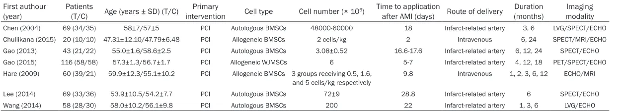
We conducted literature searches form data-base of Pub Med (1985-2016), Emdata-base (2003-2016), and the Cochrane library 2003-2016). The following search strategy was applied: mesenchymal stromal cells, mesenchymal stem cells, mesenchymal progenitor cells, bone marrow stromal cells, bone marrow mesenchy-mal stem cells, coronary artery disease, myo-cardial infarction, acute myomyo-cardial infarction. In addition, we manually searched the refer-ence lists of all original articles and previous systematic reviews. Only articles published in English were included (Table 1).
Eligibility criteria
Studies were included based on the following criteria: (1) published RCTs, (2) conducted in patients were clinically diagnosed with AMI<1 month, (3) participants in control group received
standard revascularization therapy or standard revascularization therapy with saline rather
than MSCs, (4) MSCs were derived from bone marrow, or umbilical cord, (5) the transplanted
MSCs were purified autologous or allogeneic
source, (6) MSCs were administered by intra-coronary or intravenous injection, (7) no restric-tions of MSCs dose. The exclusion criteria for studies were as follows: (1) patients were diag-nosed with chronic ischemic heart disease, angina, old myocardial infarction (OMI), or coro-nary chronic total occlusion (CTO), (2) trans-planted cells were other stem cell type other
than MSCs, (3) circulating MSCs were mobilized
from bone marrow with granulocyte colony stimulating factor (G-CSF), (4) no LVEF data were available, (5) less than 3 months of follow-up were recorded, (6) data were presented as median and range, or (7) publications were in languages other than English.
Data extraction
Two reviewers independently screened all titles, abstracts or article to identify studies that met the inclusion criteria. Discrepancies were resolved by consensus. Relevant data regard-ing study information, baseline characteristics, follow-up period, change in mean left ventricu-lar ejection fraction (LVEF), left ventricuventricu-lar end-diastolic volume (LVEDV) and left ventricular end-systolic volume (LVESV), and major adverse cardiac events (MACEs) were extracted from individual studies. Clinical trials with multiple publications, sequential follow-up durations or different outcome indicators were considered as a single study. Give the multiple modalities used for their assessment, magnetic reso-nance imaging (MRI) and single-photon emis-sion computed tomography (SPECT) data were preferred over echocardiographic data for pri-mary analysis if available.
Study outcomes
The primary end point was mean LVEF changes from baseline to follow-up. Changes in mean LVEDV, LVESV, and the incidence of MACEs were considered as the secondary endpoints. Statistical analysis
To assess risk of bias, assessment of the
qual-ity of studies was made in randomized trials
out-Table 1. Characteristics of studies included in the meta-analysis First authour
(year) Patients (T/C) Age (years ± SD) (T/C) interventionPrimary Cell type Cell number (× 106) Time to application after AMI (days) Route of delivery (months)Duration modalityImaging Chen (2004) 69 (34/35) 58±7/57±5 PCI Autologous BMSCs 48000-60000 18 Infarct-related artery 3, 6 LVG/SPECT/ECHO Chullikana (2015) 20 (10/10) 47.31±12.10/47.79±6.48 PCI Allogeneic BMSCs 2 cells/kg 2 Intravenous 6, 24 SPECT/MRI/ECHO Gao (2013) 43 (21/22) 55.0±1.6/58.6±2.5 PCI Autologous BMSCs 3.08±0.52 16.6-17.6 Infarct-related artery 6, 12, 24 SPECT/ECHO Gao (2015) 116 (58/58) 57.3±1.3/56.7±1.7 PCI Allogeneic WJMSCs 6 5-7 Infarct-related artery 4, 12, 18 PET/SPECT/ECHO Hare (2009) 60 (39/21) 59.9±12.3/55.1±10.2 PCI Allogeneic BMSCs 3 groups receiving 0.5, 1.6,
and 5 cells/kg respectively
9.8 Intravenous 1, 2, 3, 6, 12 ECHO/MRI
Lee (2014) 69 (33/36) 53.9±10.5/54.2±7.7 PCI Autologous BMSCs 72±9 28.8 Infarct-related artery 6 SPECT/ECHO Wang (2014) 58 (28/30) 58.0±10.2/56.1±9.8 PCI Autologous BMSCs 200 22 Infarct-related artery 1, 3, 6 LVG/ECHO
[image:3.792.89.718.85.197.2]come data, selective reporting and other bias (Table 2).
Statistical analyses were done in accordance
with the PRISMA statement [14]. Continuous
outcomes were expressed as weighted mean
differences with 95% confidence intervals (CI).
For most studies reporting mean ± SD at base-line and follow-up, but not the actual change (from baseline to follow-up) as mean ± SD, changes in SD were estimated according to a
previously used standardized formula [15]. In the presence of significant heterogeneity, a
random effects model was used to pool the
data; otherwise, a fixed effects model was used. Data were analyzed for heterogeneity by
I2 statistic, which was defined as low
(25%-50%), intermediate (50%-75%), or high (>75%). Potential reasons for observed heterogeneity
Additional subgroup analyses were performed within the RCTs based on the duration of follow-up in an attempt to gain more insight into pos-sible discriminating parameters or conditions that might improve outcome in future trials. The small number of RCTs in each meta-analysis precluded the conduct of planned sensitivity analyses.
Results
Search results
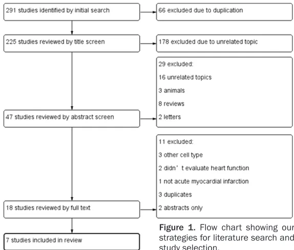
The initial search identified 291 citations using
the search strategies met the inclusion criteria (Figure 1); the numbers were reduced to 225 after removal of duplications, 207 were exclud-ed basexclud-ed on the title and abstract screen, the remaining 18 studies were assessed for eligi-Table 2. Quality assessment of study methodologies
RCT Random sequence generation concealmentAllocation Blinding of participants and personnel outcome assessorsBlinding of outcome dataIncomplete Selective reporting
Chen (2004) U U U U L L
Chullikana (2015) L L L L L L
Gao (2013) L L L L H L
Gao (2015) L L H L L L
Hare (2009) L L L L H L
Lee (2014) U U H L H L
Wang (2014) U U U L L L
L = low risk of bias, H = high risk of bias, U = unclear risk of bias.
Figure 1. Flow chart showing our strategies for literature search and study selection.
were explored with subgroup analyses based on different duration of follow-up. Odds ratios (ORs) were calculated for clinical outcomes of MA- CEs, all-cause death, heart failure, in-stent thrombosis,
recurrent MI, revasculariza -tion, arrhythmia and
rehospi-talization. Funnel plots were
constructed to explore possi-ble publication bias. A P value <5% was considered as
sta-tistically significant.
Metho-dological quality and all analy-ses of outcome data were conducted using Cochrane Review Manager 5.3.
[image:4.612.91.525.85.194.2] [image:4.612.89.376.226.470.2]bility. Among these, 3 studies using stem cells not derived from MSCs, 2 studies with non-rel-evant outcomes of heart function, 1 study involving old myocardial infarction, 3 trials duplicated after reading full text carefully and 2 only reported abstract because of ongoing
were excluded. Finally, a total of 7 RCTs [16-22]
were included in this meta-analysis. Characteristics of included studies
These 7 RCTs [16-22] included 435 patients of
AMI treated with PCI + MSCs in treatment group and PCI + placebo or PCI only in control group. All the studies reported the actual LVEF, LVEDV and LVESV from baseline to follow-up, but only
five studies assess the mean changes. We
studied the difference in mean changes in LVEF, LVEDV, and LVESV between patients receiving MSCs (n=185) and control treatment (n=172). However, 6 RCTs with patients received MSCs (n=189) and control treatment (n=177) performed safety analysis. All trials applied one of the two types of MSCs, including WJMSCs and BMSCs. 3.08 × 10⁶ to 6 × 10⁹ MSCs were
transplanted via the infarct-related artery in 5 studies and via the intravenous route in 2
stud-ies. The sample size in every study was rela -tively small, ranging from 10 to 58 participants.
Measurements of study outcome included MRI, echocardiography, LV angiography, and SPECT. The follow-up period ranged from 3 to 24 months, four groups included <6 months, 6 months, 12 months and >12 months follow-up
data were utilized for the subgroup analysis.
Remarkable heterogeneity was observed in each meta-analysis in this study.
Methodological quality assessment of included studies
Overall, the methodological quality of the included trials was good. All pooled studies were RCTs, among which four reported details
of the randomization process (Table 2),
includ-ing block randomization [17], sequential num
-ber method [18, 19] and sealed envelopes [20]. All four trials using adequate methods
concealed treatment allocation. As for blinding of participants and personnel, only three are
double blinding trials [17, 18, 20], 2 showed single blind [19] and open-label method [21], the other 2 was not unclear [16, 21]. Among the
trials, two did not show details of blinding
out-come assessors to treatment allocation [16, 21]. Moreover, at least 89% (ranging from 89 to 100%) of randomized patients were analyzed
[image:5.612.91.522.74.174.2]for the primary outcome in all studies.
Figure 2. Forest plot of the mean difference (MD, with 95% confidence interval [CI]) in left ventricular ejection frac -tion (LVEF).
[image:5.612.91.523.231.321.2]Mean differences in cardiac parameters
Overall, in 5 studies, MSCs therapy led to a
sig-nificant increase in LVEF by 4.79% (95% CI,
2.12-7.46%, P=0.0004, Figure 2). LVEDV decreased by 5.35 ml (95% CI, -13.97-3.26%, P=0.22, Figure 3) and LVESV decreased by 6.18 ml (95% CI -13.31 to 0.95, P=0.09, Figure 4) were recorded in patients treated with MSCs
compared with controls, but both differences
were not statistically significant. However, a
considerable degree of heterogeneity was observed in the LVEF comparisons (I2=98%).
Planned subgroup analysis regarding duration of follow-up was conducted to further explore possible statistical heterogeneity. We didn’t perform sensitivity analysis because of little
[image:6.612.88.526.74.164.2]sample size in each meta-analysis.
Figure 4. Forest plot of mean difference (MD, with 95% confidence interval [CI]) in left ventricular end-systolic vol -ume (LVESV).
[image:6.612.91.519.214.538.2]Subgroup analysis
The significance of MSCs follow-up period in
LVEF was examined. Subgroup analysis reve- aled that at <6 months of follow-up (n=3 RCTs), LVEF increased by + 2.53% (95% CI, 1.45-3.62;
P<0.00001). This beneficial effect was sus -tained and increased to a more pronounced effect of + 3.99% (95% CI, 1.82-6.16; P= 0.0003) at 6 months (n=5) and + 2.60% (95% CI, 1.13 to 4.06; P=0.0005) at 12 months (n=3) of follow-up, when compared with control. However, this treatment effect disappeared to the + 2.90 (95% CI, -1.21 to 7.02; P=0.17) at long-term follow-up of >12 months, (Figure 5). Additionally, different dose of MSCs, type of MSCs, the delivery method, modality of
assess-ment were also identified as possible causes of
heterogeneity. We didn’t conduct subgroup
analysis based on above influence factors because of poor sample size. Sensitivity analy -sis wasn’t performed either because of the same reason.
Relative risks of clinical outcomes
6 studies reported full details on clinical out-comes including MACEs, all-cause death, heart
failure, in-stent thrombosis, recurrent
myocar-dial infarction, revascularization, arrhythmia and rehospitalization. MACEs were defined as
all-cause death, heart failure, and recurrent myocardial infarction. Overall, MSCs transplan-tation resulted in a reduction of MACEs (OR 0.64, 95% CI 0.30 to 1.34, I2=0, P=0.24)
(Figure 6), compared with controls. There was also a trend toward reduced incidences of all-cause death (OR 0.76, 95% CI 0.17 to 3.50, I2=0, P=0.73), heart failure (OR 0.99, 95% CI
0.14 to 7.22, I2=0, P=0.99), in-stent
thrombo-sis (OR 0.30, 95% CI 0.01 to 8.33, I2 not
appli-cable, P=0.48), arrhythmia (OR 0.35, 95% CI 0.07 to 1.75, I2=40%, P=0.20) and
rehospotal-ization (OR 0.61, 95% CI 0.24 to 1.54, I2=0,
P=0.29) (Table 3), although without statistical
significance. A higher incidence of recurrent MI and revascularization was observed (OR 1.00,
3.00, respectively) in MSC-treated patients,
albeit not to statistically significant extents. The
[image:7.612.97.519.72.198.2]95% CI was wide (0.13 to 7.48, 0.12 to 75.19, respectively) that may be primarily due to the limited study population. These results mani-fested that MSCs therapy is safe for patients with AMI.
Figure 6. Forest plot of odds ratio (OR, with 95% confidence interval [CI]) in major adverse cardiac events (MACEs).
Table 3. Summary of clinical outcomes
Outcome Studies reporting Events/MSCs Events/Control OR 95% CI P-value
MACEs 6 15/180 18/165 0.64 0.30 1.34 0.24
All-cause death 6 2/181 3/165 0.76 0.17 3.50 0.73
Heart failure 2 1/79 1/78 0.99 0.14 7.22 0.99
Instent thrombosis 4 0/119 1/88 0.3 0.01 8.33 0.48
Recurrent MI 4 1/119 1/116 1.00 0.13 7.48 1.00
Rrevascularization 3 1/109 0/106 3.00 0.12 75.19 0.50
Arrhythmia 5 9/153 14/135 0.35 0.07 1.75 0.20
Rehospitalization 4 11/140 11/125 0.61 0.24 1.54 0.29
MSCs, mesenchymal stromal cells; OR, odds ratio; CI, confidence interval; MACEs, major adverse cardiovascular events; MI,
[image:7.612.92.523.255.374.2]Publication bias
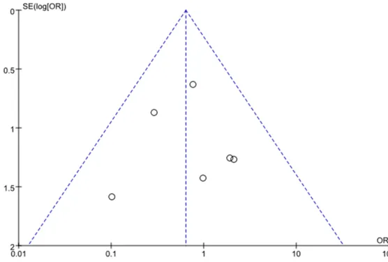
Funnel plots of LVEF and MACEs showed that studies were equally distributed around the overall estimate, suggesting that there was no evidence for publication bias (Figures 7, 8). Discussion
This meta-analysis summarized the efficacy
and the safety of MSCs administration in patients with AMI after PCI. The results revealed
that MSCs therapy may beneficial in improving
cardiac function, supporting the utility of this method as a feasible strategy for AMI. Inter-
ies demonstrated discordant results with a decrease in LVEF with treatment or a more robust increase in LVEF in the control group
[28-30]. Our meta-analysis got the similar
results with most studies. The modest improve-ment of LVEF was mainly due to a sustained LVEDV and LVESV. The results of subgroup analysis based on follow-up duration showed
that MSCs’ beneficial impact on LVEF disap -peared when >12 months. It was similar to
Wollert et al [31] research in which LVEF
increased by 6.7% at 6- month in the treatment group but the effect was lost at 18 month. Chen
[image:8.612.91.375.73.264.2]et al [32] also showed initial benefit at 3, 6 and
Figure 7. Funnel plot for left ventricular ejection fraction outcome data of the included studies.
Figure 8. Funnel plot for safety outcome data of the included studies.
estingly, when subgroup anal-ysis was performed by
follow-up duration, this beneficial
effect of MSCs therapy on car-diac function disappeared when >12 months. As for sa- fety evaluation, our analysis was unable to detect associa-tions between MSCs treat-ment and all of the adverse events in patients subjected to MSCs therapy compared with controls. Although a high-er incidence of myocardial
re-infarction and revasculariza -tion was observed, the small
sample size included may be
a contributory factor. It was a remarkable fact that the CI was wide in our study, which may be explained by relatively small studies and low number of events in all outcomes. Therefore, clinical trials of larger scale and longer-term follow-up should be
conduct-ed to fully assess the efficacy
and possible adverse effects of MSCs in patients with AMI. Different stem cells have be- en used to access the associ-ation with coronary artery dis-ease (CAD) in recent years, although the results are con-troversial. Most studies in patients with CAD have shown a greater increase in LVEF with stem cells than control
[image:8.612.90.368.315.503.2]-9 moths of LVEF but that was subsequently lost at 12 month. This may be explained by the dynamic nature of LVEF raising the issue of
possible time-limited benefit or discrepancy in
the measurements of LVEF when multiple modalities were used for the same patient or the lower number of studies in >12 months group.
We think that our meta-analysis shows strong indications that MSCs therapy is effective in improving LV function for short-term clinical outcome in patients with AMI. Although the number of patients treated with MSCs is still too small, this new cell therapy might prove to be more effective. It is thought that mesenchy-mal cell populations or cardiac-derived stem cells exhibit more cardioprotective and regen-erative potential. As a novel technique, cell-based therapy for CAD has the potential to improve myocardial function and act as adjunc-tive therapy to medical and reperfusion strate-gies. In studies of pathological mechanism, administration of BMCs has been shown to regenerate areas of infracted myocardium and coronary capillaries through differentiation into new cardiomyocytes and vascular endothelial
cells [33-36]. However, data suggest that the
main effect of BMCs may be mediated through their paracrine effects via secretion of
cyto-kines, fibroblast growth factors, and vascular
endothelial growth factors that are involved in angiogenesis, inhibition of cardiomyocyte
apoptosis, and cell-cell transaction [5, 6, 37, 38]. The result is the replacement of lost myo -cardial tissue and saving of ischemic and hiber-nating cardiomyocytes and improvement of LV function.
Cell-base therapy could cause immediate adverse events such as acute infusion toxicity and fever and organ system related adverse events such as cardiovascular, gastrointestinal, renal, pulmonary, neurological, hematological, infection related adverse events, death and malignancy. Our analysis indicated that MSCs were safe in respect of cardiovascular adverse events and death when used in patients with AMI. We didn’t perform meta-analysis of other
adverse events because of the small size
included. We should note that immunomodula-tory activities of MSCs could induce acute infu-sion toxicity, fever and malignancy. Large-scale prospective trials in patients with AMI required to further evaluate the safety for MSCs administration.
Formal testing of publication bias was per-formed using funnel plots. It was not possible to fully exclude publication bias, although the
results showed no statistical significance. There was significant heterogeneity in the effi -cacy of MSCs administration in our meta-analy-sis. After subgroup analysis based on duration, heterogeneity remained in every subgroup. This heterogeneity may be linked to multiple factors including cell type, number of injected cells, method of preparation and injection, imaging modality, and patient selection. As we known, sensitivity analysis will access the stability of the combined effect. However, we didn’t per-form sensitivity analysis because of the small
size of RCTs in every subgroup.
Our meta-analysis has several limitations. Firstly, there are two studies published in
abstract form only that may influence results
despite of our comprehensive search strategy. Secondly, the numbers of patients included are relatively small, which may provide an
inaccu-rate indication of the efficacy and safety of
MSCs and precluded the performance of sensi-tivity analysis. Finally, subgroup analysis did not include factors such as cell therapy timing, cell numbers, cell types, delivery method, or imag-ing modality due to the limited number of included trials.
Conclusion
This is the first meta-analysis accessing the efficacy and safety of MSCs on cardiac repair
in patients with AMI. MSCs transplantation appears to improve left ventricular function and be safe in patients with AMI in short-term. Large
randomized double-blinded prospective stud -ies with long-term follow-up are needed to improve the accuracy of the results.
Disclosure of conflict of interest None.
Address correspondence to: Ling Qin,Department of Cardiology, First Hospital, Jilin University, No.71 Xinmin Street, Changchun, Jilin Province, China. E-mail: [email protected]
References
[2] Haack-Sorensen M, Friis T, Bindslev L, Mortensen S, Johnsen HE and Kastrup J. Com-parison of different culture conditions for hu-man mesenchymal stromal cells for clinical stem cell therapy. Scand J Clin Lab Invest 2008; 68: 192-203.
[3] Bernardo ME, Pagliara D and Locatelli F. Mes-enchymal stromal cell therapy: a revolution in regenerative medicine? Bone Marrow Trans-plant 2012; 47: 164-171.
[4] Volarevic V, Bojic S, Nurkovic J, Volarevic A, Lju-jic B, Arsenijevic N, Lako M and Stojkovic M. Stem cells as new agents for the treatment of infertility: current and future perspectives and challenges. Biomed Res Int 2014; 2014: 507234.
[5] Giannotti G, Doerries C, Mocharla PS, Mueller MF, Bahlmann FH, Horvath T, Jiang H, Sorren-tino SA, Steenken N, Manes C, Marzilli M, Ru -dolph KL, Luscher TF, Drexler H and Land-messer U. Impaired endothelial repair capacity of early endothelial progenitor cells in prehy-pertension: relation to endothelial dysfunction. Hypertension 2010; 55: 1389-1397.
[6] Sieveking DP, Buckle A, Celermajer DS and Ng MK. Strikingly different angiogenic properties of endothelial progenitor cell subpopulations: insights from a novel human angiogenesis as-say. J Am Coll Cardiol 2008; 51: 660-668. [7] Gnecchi M, Zhang Z, Ni A and Dzau VJ. Para
-crine mechanisms in adult stem cell signaling and therapy. Circ Res 2008; 103: 1204-1219. [8] Fazel S, Cimini M, Chen L, Li S, Angoulvant D,
Fedak P, Verma S, Weisel RD, Keating A and Li RK. Cardioprotective c-kit+ cells are from the bone marrow and regulate the myocardial bal-ance of angiogenic cytokines. J Clin Invest 2006; 116: 1865-1877.
[9] Liu B, Duan CY, Luo CF, Ou CW, Sun K, Wu ZY, Huang H, Cheng CF, Li YP and Chen MS. Effec-tiveness and safety of selected bone marrow stem cells on left ventricular function in pa-tients with acute myocardial infarction: a meta-analysis of randomized controlled trials. Int J Cardiol 2014; 177: 764-770.
[10] Sadat K, Ather S, Aljaroudi W, Heo J, Iskandrian AE and Hage FG. The effect of bone marrow mononuclear stem cell therapy on left ventric-ular function and myocardial perfusion. J Nucl Cardiol 2014; 21: 351-367.
[11] Mathiasen AB, Jorgensen E, Qayyum AA, Haack-Sorensen M, Ekblond A and Kastrup J. Rationale and design of the first randomized, double-blind, placebo-controlled trial of intra-myocardial injection of autologous bone-mar-row derived mesenchymal stromal cells in chronic ischemic heart failure (MSC-HF Trial). Am Heart J 2012; 164: 285-291.
[12] de Jong R, Houtgraaf JH, Samiei S, Boersma E and Duckers HJ. Intracoronary stem cell infu-sion after acute myocardial infarction: a meta-analysis and update on clinical trials. Circ Car-diovasc Interv 2014; 7: 156-167.
[13] Lalu MM, McIntyre L, Pugliese C, Fergusson D, Winston BW, Marshall JC, Granton J, Stewart DJ; Canadian Critical Care Trials Group. Safety of cell therapy with mesenchymal stromal cells (SafeCell): a systematic review and meta-anal-ysis of clinical trials. PLoS One 2012; 7: e47559.
[14] Moher D, Liberati A, Tetzlaff J, Altman DG; PRISMA Group. Preferred reporting items for systematic reviews and meta-analyses: the PRISMA statement. PLoS Med 2009; 6: e1000097.
[15] Hristov M, Heussen N, Schober A and Weber C. Intracoronary infusion of autologous bone marrow cells and left ventricular function after acute myocardial infarction: a meta-analysis. J Cell Mol Med 2006; 10: 727-733.
[16] Chen SL, Fang WW, Ye F, Liu YH, Qian J, Shan SJ, Zhang JJ, Chunhua RZ, Liao LM, Lin S and Sun JP. Effect on left ventricular function of in-tracoronary transplantation of autologous bone marrow mesenchymal stem cell in pa-tients with acute myocardial infarction. Am J Cardiol 2004; 94: 92-95.
[17] Chullikana A, Majumdar AS, Gottipamula S, Krishnamurthy S, Kumar AS, Prakash VS and Gupta PK. Randomized, double-blind, phase I/ II study of intravenous allogeneic mesenchy-mal stromesenchy-mal cells in acute myocardial infarc-tion. Cytotherapy 2015; 17: 250-261.
[18] Gao LR, Pei XT, Ding QA, Chen Y, Zhang NK, Chen HY, Wang ZG, Wang YF, Zhu ZM, Li TC, Liu HL, Tong ZC, Yang Y, Nan X, Guo F, Shen JL, Shen YH, Zhang JJ, Fei YX, Xu HT, Wang LH, Tian HT, Liu DQ and Yang Y. A critical challenge: dosage-related efficacy and acute complica -tion intracoronary injec-tion of autologous bone marrow mesenchymal stem cells in acute myo-cardial infarction. Int J Cardiol 2013; 168: 3191-3199.
[19] Gao LR, Chen Y, Zhang NK, Yang XL, Liu HL, Wang ZG, Yan XY, Wang Y, Zhu ZM, Li TC, Wang LH, Chen HY, Chen YD, Huang CL, Qu P, Yao C, Wang B, Chen GH, Wang ZM, Xu ZY, Bai J, Lu D, Shen YH, Guo F, Liu MY, Yang Y, Ding YC, Yang Y, Tian HT, Ding QA, Li LN, Yang XC and Hu X. Intracoronary infusion of Wharton’s jelly-de-rived mesenchymal stem cells in acute myo-cardial infarction: double-blind, randomized controlled trial. BMC Med 2015; 13: 162. [20] Hare JM, Fishman JE, Gerstenblith G, DiFede
Men-dizabal AM, Lowery MH, Rouy D, Altman P, Wong Po Foo C, Ruiz P, Amador A, Da Silva J, McNiece IK, Heldman AW, George R and Lardo A. Comparison of allogeneic vs autologous bone marrow-derived mesenchymal stem cells delivered by transendocardial injection in pa-tients with ischemic cardiomyopathy: the POSEIDON randomized trial. JAMA 2012; 308: 2369-2379.
[21] Lee JW, Lee SH, Youn YJ, Ahn MS, Kim JY, Yoo BS, Yoon J, Kwon W, Hong IS, Lee K, Kwan J, Park KS, Choi D, Jang YS and Hong MK. A ran-domized, open-label, multicenter trial for the safety and efficacy of adult mesenchymal stem cells after acute myocardial infarction. J Kore-an Med Sci 2014; 29: 23-31.
[22] Wang X, Xi WC and Wang F. The beneficial ef -fects of intracoronary autologous bone marrow stem cell transfer as an adjunct to percutane-ous coronary intervention in patients with acute myocardial infarction. Biotechnol Lett 2014; 36: 2163-2168.
[23] Wohrle J, von Scheidt F, Schauwecker P, Wi-esneth M, Markovic S, Schrezenmeier H, Hom -bach V, Rottbauer W and Bernhardt P. Impact of cell number and microvascular obstruction in patients with bone-marrow derived cell ther-apy: final results from the randomized, double-blind, placebo controlled intracoronary Stem Cell therapy in patients with Acute Myocardial Infarction (SCAMI) trial. Clin Res Cardiol 2013; 102: 765-770.
[24] Grajek S, Popiel M, Gil L, Breborowicz P, Lesiak M, Czepczynski R, Sawinski K, Straburzynska-Migaj E, Araszkiewicz A, Czyz A, Kozlowska-Skrzypczak M and Komarnicki M. Influence of bone marrow stem cells on left ventricle perfu-sion and ejection fraction in patients with acute myocardial infarction of anterior wall: randomized clinical trial: Impact of bone mar -row stem cell intracoronary infusion on im-provement of microcirculation. Eur Heart J 2010; 31: 691-702.
[25] Rodrigo SF, van Ramshorst J, Hoogslag GE, Boden H, Velders MA, Cannegieter SC, Roelofs H, Al Younis I, Dibbets-Schneider P, Fibbe WE, Zwaginga JJ, Bax JJ, Schalij MJ, Beeres SL and Atsma DE. Intramyocardial injection of autolo-gous bone marrow-derived ex vivo expanded mesenchymal stem cells in acute myocardial infarction patients is feasible and safe up to 5 years of follow-up. J Cardiovasc Transl Res 2013; 6: 816-825.
[26] Assmus B, Rolf A, Erbs S, Elsasser A, Haber-bosch W, Hambrecht R, Tillmanns H, Yu J, Corti R, Mathey DG, Hamm CW, Suselbeck T, Tonn T, Dimmeler S, Dill T, Zeiher AM, Schachinger V; REPAIR-AMI Investigators. Clinical outcome 2 years after intracoronary administration of
bone marrow-derived progenitor cells in acute myocardial infarction. Circ Heart Fail 2010; 3: 89-96.
[27] Can A, Ulus AT, Cinar O, Topal Celikkan F, Sim-sek E, Akyol M, Canpolat U, Erturk M, Kara F and Ilhan O. Human umbilical cord mesenchy-mal stromesenchy-mal cell transplantation in myocardial ischemia (HUC-HEART Trial). A study protocol of a phase 1/2, controlled and randomized trial in combination with coronary artery by-pass grafting. Stem Cell Rev 2015; 11: 752-760.
[28] Hopp E, Lunde K, Solheim S, Aakhus S, Arne-sen H, Forfang K, EdvardArne-sen T and Smith HJ. Regional myocardial function after intracoro-nary bone marrow cell injection in reperfused anterior wall infarction-a cardiovascular mag-netic resonance tagging study. J Cardiovasc Magn Reson 2011; 13: 22.
[29] Roncalli J, Mouquet F, Piot C, Trochu JN, Le Cor-voisier P, Neuder Y, Le Tourneau T, Agostini D, Gaxotte V, Sportouch C, Galinier M, Crochet D, Teiger E, Richard MJ, Polge AS, Beregi JP, Man-rique A, Carrie D, Susen S, Klein B, Parini A, Lamirault G, Croisille P, Rouard H, Bourin P, Nguyen JM, Delasalle B, Vanzetto G, Van Belle E and Lemarchand P. Intracoronary autologous mononucleated bone marrow cell infusion for acute myocardial infarction: results of the ran-domized multicenter BONAMI trial. Eur Heart J 2011; 32: 1748-1757.
[30] Nair V, Madan H, Sofat S, Ganguli P, Jacob MJ, Datta R, Bharadwaj P, Sarkar RS, Pandit AJ, Ni-tyanand S, Goel PK, Garg N, Gambhir S, George PV, Chandy S, Mathews V, George OK, Talwar KK, Bahl A, Marwah N, Bhatacharya A, Bhar-gava B, Airan B, Mohanty S, Patel CD, Sharma A, Bhatnagar S, Mondal A, Jose J, Srivastava A and Trial MI. Efficacy of stem cell in improve -ment of left ventricular function in acute myo-cardial infarction--MI3 Trial. Indian J Med Res 2015; 142: 165-174.
[31] Wohrle J, Merkle N, Mailander V, Nusser T, Schauwecker P, von Scheidt F, Schwarz K, Bommer M, Wiesneth M, Schrezenmeier H and Hombach V. Results of intracoronary stem cell therapy after acute myocardial infarction. Am J Cardiol 2010; 105: 804-812.
[32] Chen S, Liu Z, Tian N, Zhang J, Yei F, Duan B, Zhu Z, Lin S and Kwan TW. Intracoronary trans-plantation of autologous bone marrow mesen-chymal stem cells for ischemic cardiomyopa-thy due to isolated chronic occluded left anterior descending artery. J Invasive Cardiol 2006; 18: 552-556.
clini-cal evidence. Antioxid Redox Signal 2009; 11: 1865-1882.
[34] Orlic D, Kajstura J, Chimenti S, Bodine DM, Leri A and Anversa P. Bone marrow stem cells re-generate infarcted myocardium. Pediatr Trans-plant 2003; 7 Suppl 3: 86-88.
[35] Rota M, Kajstura J, Hosoda T, Bearzi C, Vitale S, Esposito G, Iaffaldano G, Padin-Iruegas ME, Gonzalez A, Rizzi R, Small N, Muraski J, Alvarez R, Chen X, Urbanek K, Bolli R, Houser SR, Leri A, Sussman MA and Anversa P. Bone marrow cells adopt the cardiomyogenic fate in vivo. Proc Natl Acad Sci U S A 2007; 104: 17783-17788.
[36] Hao L, Hao J, Fang W, Han C, Zhang K and Wang X. Dual isotope simultaneous imaging to evaluate the effects of intracoronary bone marrow-derived mesenchymal stem cells on perfusion and metabolism in canines with acute myocardial infarction. Biomed Rep 2015; 3: 447-452.
[37] Nygren JM, Jovinge S, Breitbach M, Sawen P, Roll W, Hescheler J, Taneera J, Fleischmann BK and Jacobsen SE. Bone marrow-derived he-matopoietic cells generate cardiomyocytes at a low frequency through cell fusion, but not transdifferentiation. Nat Med 2004; 10: 494-501.
![Figure 2. Forest plot of the mean difference (MD, with 95% confidence interval [CI]) in left ventricular ejection frac-tion (LVEF).](https://thumb-us.123doks.com/thumbv2/123dok_us/1398824.674733/5.612.91.523.231.321/figure-forest-difference-confidence-interval-ventricular-ejection-lvef.webp)
![Figure 4. Forest plot of mean difference (MD, with 95% confidence interval [CI]) in left ventricular end-systolic vol-ume (LVESV).](https://thumb-us.123doks.com/thumbv2/123dok_us/1398824.674733/6.612.91.519.214.538/figure-forest-difference-confidence-interval-ventricular-systolic-lvesv.webp)
![Figure 6. Forest plot of odds ratio (OR, with 95% confidence interval [CI]) in major adverse cardiac events (MACEs).](https://thumb-us.123doks.com/thumbv2/123dok_us/1398824.674733/7.612.92.523.255.374/figure-forest-confidence-interval-adverse-cardiac-events-maces.webp)