Original Article
Propofol ameliorates ischemia/reperfusion
induced cerebral injury by upregulation
of microRNA-206 expression
Yong Wang1, Rui Zhang2, Yanli Huang1
1Intensive Care Unit of Ninth Hospital, Xi’an 710054, Shaanxi, P. R. China; 2Neurosurgery of Ninth Hospital, Xi’an 710054, Shaanxi, P. R. China
Received December 17, 2015; Accepted March 19, 2016; Epub June 15, 2016; Published June 30, 2016
Abstract: Aims: The aim of this study is to investigate the role of miR-206 in propofol induced protective effects in ce-rebral ischemia/reperfusion injury and to explore its possible mechanism. Methods: Hypoxia/reoxygenation (H/R) model of mouse motoneuron-like cell line NSC-43 was established, and the expression miR-206 upon propofol treatment was analyzed by Realtime PCR. Cells were transfected with miR-206 mimic (miR-206) or scramble control (NC) and subjected to H/R, followed by propofol treatment or not. Cell viability and apoptosis was detected by CCK8
assay, flow cytometry and Western blot analysis of apoptosis-related proteins. Target of miR-206 was predicted by bioinformatics analysis and verified by Dual-luciferase reporter gene assay and Western blot. An in vivo model of rat cerebral ischemia/reperfusion injury was used to verify the target of miR-206. Before or after propofol treatment, hippocampi were taken for TUNEL assay and Western analysis of Orthodenticle Homeobox 2 (OTX2) expression.
Results: Expression of miR-206 was significantly upregulated after propofol treatment in rat hippocampi. MiR-206 transfection significantly increased cell viability and decreased apoptosis rate, which was consistent with the effect
of propofol treatment. Both miR-206 transfection and propofol treatment after H/R inhibited the expression of Bax but upregulated Bcl-2 expression. In addition, 3’untranslated region (3’UTR) of OTX2 has a putative binding site for miR-206. MiR-206 could directly bind to the OTX2 3’UTR and negatively regulate the expression of OTX2 protein. Conclusions: Our results indicate that miR-206 was upregulated after propofol treatment in H/R model to decrease
the expression of OTX2 protein, reduce the apoptosis of neural cells and finally suppress cerebral ischemia/reperfu -sion injury.
Keywords: Propofol, miR-206, hypoxia-reoxygenation, hippocampal neurons
Introduction
Cerebrovascular disease is one of the common clinical diseases with high morbidity and mor-tality, which greatly endanger human health. Ischemic stroke, with the pathological features of hypoxia-induced brain damage and cerebral ischemia, accounts for 80-85% of the cerebro-vascular diseases [1, 2]. Promptly restore blood supply in ischemic area of patients is the ma- jor strategy for treatment clinically, but severe brain ischemia-reperfusion injury may occur as a result [3]. Currently, there is no effective clini-cal treatment against the brain tissue damage caused by hypoxia and ischemia-reperfusion (I/R). In addition, the molecular mechanisms for the development of such damage are not yet clear.
brain tissue and further investigations are needed.
MiRNAs are a class of 18-22 nucleotide (nt), highly conserved, non-coding RNAs that pre-dominantly serve as translational repressor by binding to complementary sequences in the 3’ untranslated region (UTR) of their target mRNAs [11]. It is reported that propofol affected the miRNA profile in multiple tissues and cells to regulate cell activity. For example, propofol pro-moted the apoptosis of ovarian epithelial cells through upregulating let-7i [12], and propofol could inhibit the metastasis of esophageal squamous cell carcinoma by suppressing the expression of miR-143 [13]. It remained to be clarified whether miRNAs are involved during propofol treatment of I/R. Therefore, in this study we attempt to find out whether miR-206 is involved in the cerebral protection of propofol after I/R and to find molecular mechanisms to support the development of propofol as a ther-apeutic drug.
Material and methods
Cerebral infraction and the reperfusion rat model
A total of 60 adult male SD rats weighing 230-250 g were used. They were allowed standard chow pellets and drinking water ad libitum. The experimental protocol was ethically approved by the Animal Care Committee of Xi’an Ninth Hospital. Rats were randomly divided into four groups (15 animals each). Rats of the sham-operated control (sham) and ischemia/reperfu-sion (I/R) groups were intravenously (i.v.) admin-istered saline solution. Rats of the lipid micro-sphere +I/R group (LM+I/R) were i.v. adminis-trated 1 ml/kg lipid microsphere, and propofol +I/R group were received propofol preparation (50 mg/kg) dissolved in lipid microsphere 24 hr before I/R. All rats except those of the sham control group were exposed to 90 min of cere-bral ischemia followed by 24 hr of reperfusion, and another 24 hr of reperfusion was per-formed after the rats were fully awake. After I/R or sham operation, all rats were sacrificed by decapitation. The brains were carefully dissect-ed out on ice for following experiments. Cerebral I/R was performed as described previ-ously [14]. Briefly, rats were anesthetized at the time of the operation, the right and left
com-mon carotid arteries were exposed by a midline ventral incision in the neck. The bilateral carot-id artery was separated from the adjacent tis-sues and vagus nerve. Ischemia was induced by bilateral clamping of the common carotid arteries for 90 min. Following cerebral isch-emia, the arteries were declamped to restore circulation. The skin was sutured for reperfu-sion. Rats of the sham control group were exposed to the same procedure except for carotid occlusion.
Construction of H/R model in NSC-43
Mouse motoneuron-like NSC-43 cells were cul-tured in DMEM (Gibco, Thermo Fisher Scientific Inc, Vilnius, Lithuania, USA) medium supple-mented with 10% heat inactivated fetal bovine serum (FBS) and 1% penicillin-streptomycin at 37°C, 5% CO2 Cells were seeded into 24-well plates at 1 × 10⁵ cells/well. For hypoxia, the
cul-ture media was replaced by synthetic isch-aemia solution and the NSC-43 s were then placed in hypoxic conditions with 5% CO2 and 95% N2 at 37°C for 24 h. After hypoxia, the medium was washed off, and the NSC-43 s were returned to reperfusion solution and cul-tured with 5% CO2 and 95% O2 for 6 h. At the same time, prepared propofol was added to the medium at the concentration of 150 μM.
RNA extraction and real-time PCR assay
The total RNA from each treatment was extract-ed using TRIzol® isolation reagent (Thermo Fisher Scientific, Vilnius, Lithuania) according to the manufacture’s construction. Following gel electrophoresis verification of RNA integrity and quantification using UV spectrophotome-ter, 0.5 ug of total RNA was reverse transcribed into cDNA using a miRNA cDNA Kit (TAKARA, Dalian, China) with specific primers. The expres-sion of small nuclear U6 was used as internal control. Quantitative Real-time PCR was per-formed using KAPA SYBR FAST qPCR Kits (Kapa biosystems, Boston, Massachusetts, USA). Forward primer used for miR-206 amplification was 5-AATGTAAGGAAGTGTG-3, and the reverse primer is provided by kit. Primers used for amplification of U6 are as follows, forward: 5’-CTCGCTTCGGCAGCACA-3’ and reverse: 5’-A- ACGCTTCACGAATTTGCGT-3’. The relative ex- pression levels were evaluated using the 2-ΔΔCt
MiRNA transfection of NSC-43
NSC-43 cells were seeded at 24-well plate and incubated to a contingency of 70%-90% prior to transfection with 31.25 uM of miR-206 mimics or negative control (NC) (Ribobio, Guangzhou, China) using Lipofectamine 2000 (Thermo Fisher Scientific, Vilnius, Lithuania) according to the manufacturer’s instructions. Cells were collected at 48 h post-transfection for further experiments or subjected to H/R treatment.
CCK-8 assay
After hypoxia culture, NSC-43 cells were re- turned to reperfusion solution and cultured with 5% CO2 and 95% O2 for 6 h. After washing twice with PBS, cells were suspended in fresh DMEM containing 10% of Cell Counting Kit-8 (Biyuntian, Beijing, China) and incubated for 1 h at 37°C. The absorbance values at 450 nm in each well were measured with ELx800 (BioTek, Winooski, Vermont, USA).
Annexin V-FITC staining and flow cytometry analysis
NSC-43 cells were harvested after trypsin digestion, washed with PBS twice and stained by Annexin V-FITC Apoptosis Detection Kit I (BD bioscience, San Jose, CA, USA) according to the manufacturer’s instruction. Cells were immedi-ately analyzed by flow cytometry. Cells undergo-ing apoptosis were Annexin V positive and PI negative, and cells that were necrosis were Annexin V negative and PI positive. Cells observed to be Annexin V and PI positive were in end stage apoptosis or already dead.
Western blot analysis
Cells were collected and re-suspended in RIPA lysis buffer with 1% PMSF to extract the total protein. Each sample was centrifuged at 12,000 × g for 10 min at 4°C for collecting the supernatant. Protein assay kit (Biyuntian, Beijing, China) using bovine serum albumin (BSA) as the standard was used to measure the total protein. Protein was separated by sodium dodecyl sulfate-polyacrylamide gel electropho-resis (SDS-PAGE) and expression of BAX, Bcl-2, OTX2 and GAPDH was detected with the follow-ing antibodies: rabbit anti-rat OTX2 (ab11413; 1:1000), rabbit anti-rat BAX (ab7977; 1:1000), mouse anti-rat monoclonal anti-Bcl-2 (AB11-
7115; 1:1000) and rabbit anti-rat monoclonal GAPDH (ab181602; 1:5000). Goat anti-rabbit (ab181603; 1:5000) or goat anti-mouse (ab 1018; 1:2000) antibodies conjugated to HRP were used as secondary antibodies. All antibodies were purchased from Abcam (Cam- bridge, MA, USA). Signal detection was per-formed using chemiluminescence reaction (ECL) (Beyotime Institute of Biotechology, Bei- jing, China).
Dual-luciferase reporter gene assay
One of the miR-206 binding sites on the 3’UTR of Otx2 gene with lowest P value was chosen. Luciferase reporter plasmids were generated by insertion of wild type or mutant Otx2 3’UTR fragment containing the miR-206 binding site into the multiple cloning site (Spe-1 and Hind III) downstream of the luciferase reporter gene in the pMIR-REPORT™ Luciferase (Thermo Fisher Scientific, Vilnius, Lithuania). HEK293T cells were seeded at a concentration of 5 × 10⁴
cells per well in 24-well plates and transfected with 1 ug constructed luciferase reporters and 10 ng pMIR-REPORT™ β-gal Control Plasmid as an internal control for transfection efficiency. And, 100 nmol miRNA mimics or negative con-trol RNA were also transfected. Luminescence was measured at 24 h after transfection using the Dual-Luciferase® Reporter Assay System (Sigma Chemical Co., St. Louis, MO, USA) according to the manufacturer’s instructions. Measurements of luminescence were per-formed on the luminometer (Glomax 20/20, Promega, Madison, WI, USA).
Immunocytochemistry
transparency, sections were mounted with neu-tral gum.
Cells with yellow or brown granules in the cyto-plasm or membrane of hippocampal cells were positive cells under light microscopy. Five fields at high-magnification were randomly taken and positive cells were counted. Positive rate was the ratio of the number of positive cells to the number of total cells. Cells without positive staining or with a positive rate less than 1% were scored as 1. Cells with a positive rate more than 1% and less than 25% were scored as 2. Cells with a positive rate between 25% and 50% were scored as 3. Cells with a positive
rate over 50% were scored as 4. According to the strength of staining, cells with no staining were scored as 0; cells with yellow staining were scored as 1; cells with medium brown staining were scored as 2; cells with dark brown staining were scored as 3. The product of posi-tive rate score and staining strength score was accepted as the final score. Score 0-1 was neg-ative, 2-3 was weakly positive, 4-6 was medium positive, and >6 was strong positive.
TUNEL staining
Brains of rats were dissected and fixed in 10% formaldehyde and embedded in paraffin. Paraffin sections were cut at 4 μm and sections were dewaxed and rehydrated in graded alco-hols. Subsequently, the sections were incubat-ed for 20 min at 20-37°C in proteinase K (with-out DNase). Then sections were incubated with 0.3% hydrogen peroxide to inactivate endoge-nous peroxidase activity, followed by PBS wash-ing for 3 times. TUNEL stainwash-ing was performed according to the instructions of the In situ cell death detection kit-POD (Roche, Basel, Swit- zerland). The incorporated fluorescein-dUTP was detected with 50 ul of biotinylated anti-body for 60 min at 37°C, followed by PBS or HBSS washing once. The reaction was stopped by 2 × SSC for 10 min at room temperature. After PBS washing, 50 ul Streptavidin-HRP were added to slides and the labeled antibody was visualized DAB chromogenic reagent. The frequency of TUNEL-positive cells that with yel-low or brown staining were evaluated under a 400-fold microscope magnification. Five fields at high-magnification were randomly taken and positive cells were counted. The average of positive cells was calculated.
Statistical analysis
Data were expressed as mean ± S.D. Statistical significance was determined with paired t-tests using SPSS 16.0 (SPSS Statistics/IBM Corp, Chicago, IL, USA). P-values < 0.05 were consid-ered statistically significant.
Results
MiR-206 is upregulated after propofol treat-ment
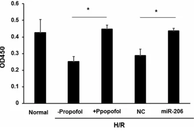
[image:4.612.89.289.72.225.2]We first evaluated the expression of miR-206 in rat ischemia/reperfusion model with or without propofol treatment by Real-time PCR. MiR-206 Figure 1. Expression of miR-206 in NSC-34 cells.
Normal NSC-34 cells orNSC-34 cells administrated with H/R were treated with propofol (H/R+ propofol) or left untreated (H/R), and miR-206 expression was analyzed by Real time PCR. *P < 0.05, **P < 0.01, t test.
[image:4.612.92.287.316.448.2]expression was significantly downregulated in NSC-34 cells subjected to H/R compared with untreated cells (P < 0.05), and its expression was significantly upregulated after propofol treatment (Figure 1) (P < 0.05). These results indicate that miR-206 may play a role in the protection of neural cell injury induced by H/R.
Overexpression of mir-206 increases the vi-ability of NSC-34 cells
To investigate the involvement of miR-206 in propofol induced protection, viability of H/R treated cells or normal control cells was ana-lyzed by CCK-8 assay. As expected, propofol treatment significantly increased cell viability (P < 0.05). Transfection with miR-206 mimics also upregulated of cell viability compared with the NC group (P < 0.05) (Figure 2). These results suggest that the protection effects of propofol in neural cell injury may be dependent on the upregulation miR-206 expression.
Overexpression of miR-206 inhibits apoptosis after exposure to H/R
We next investigate the role of miR-206 in regu-lating cell apoptosis after exposure to H/R.
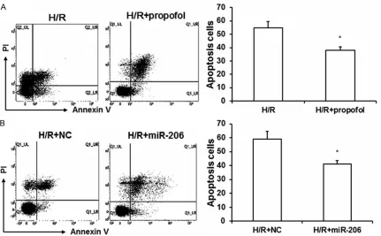
NSC-34 cells were stained with Annexin V-FITC and propidium iodide, and then analyzed by flow cytometry. There was an obvious reduction in the number of apoptotic cells when propofol was administrated compared with the untreat-ed H/R group (P < 0.05) (Figure 3A). A similar reduction in the number of apoptotic cells was observed when cells were transfected with miR-206 compared with that transfected with NC (Figure 3B). To confirm this finding, apopto-sis-related proteins were analyzed by Western blot after exposure to H/R. Apoptotic protein Bax was significantly downregulated while anti-apoptotic protein Bcl-2 was upregulated upon propofol treatment compared with untreated cells (Figure 4A), and miR-206 transfection resulted in the same expression tendency of these proteins compared with cells transfected with NC (Figure 4B). Overall, overexpression of miR-206 induced apoptosis of NSC-34 cells, which may contribute to the protection effect of propofol.
OTX2 is a target of miR-206
[image:5.612.93.524.71.338.2]To figure out the molecular mechanisms in which the miR-206 are involved, its target Figure 3. Transfection of miR-206 mimic inhibited NSC-34 cell apoptosis. A. NSC-34 cells administrated with H/R
were treated with propofol (H/R+ propofol) or left untreated (H/R). Cell apoptosis was analyzed by flow cytometry
and the apoptosis rate was summarized. B. NSC-34 cells were transfected with miR-206 mimic or NC, and
genes were predicted by TargetScan (www.tar-getscan.org/), and we found that the 3’UTR of
Otx2 contains potential binding sites for miR-206. As it is reported that OTX2 participates in cell apoptosis, miR-206 may inhibit NSC-34 cell apoptosis through targeting OTX2. To validate whether miR-206 could directly target OTX2, the 3’UTR fragment of Otx2 containing the miR-206 binding site was cloned into the
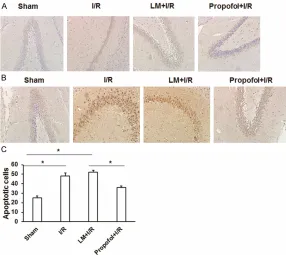
[image:6.612.90.374.75.265.2]pMIR-pus was detected by TUNEL staining. As shown in Figure 6A and 6C, a visible elevation in the number of TUNEL-positive cells was observed in hippocampus of I/R group as compared to sham control. Pre-administration of propofol to cerebral I/R-exposed rats caused a reduction of TUNEL-positive cell number as compared to I/R and lipid microsphere +I/R (LM+I/R) groups. The number of OTX2 protein expressed cells Figure 4. Western blot analysis of apoptosis-related proteins. A. NSC-34 cells
administrated with H/R were treated with propofol (H/R+ propofol) or left untreated (H/R). Cell lysates were prepared and analyzed by Western blot for BAX, Bcl-2 and GAPDH protein. B. NSC-34 cells were transfected with miR-206 mimic or NC, and underwent H/R. Cell lysates were prepared and analyzed by Western blot for BAX, Bcl-2 and GAPDH protein. *P < 0.05, t test.
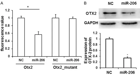
Figure 5.Otx2 is a direct target of miR-206. A. The wildtype Otx2 3’UTR lu-ciferase reporter construct (Otx2) or mutant Otx2 3’UTR luciferase reporter construct (Otx2_mutant) was co-transfected with miR-206 mimic or NC, and luciferase activities were assayed 24 h post transfection. *P < 0.05 com-pared with NC, t test. B. NSC-34 cells were transfected with miR-206mim-ic or scramble (NC). Cell lysates were prepared and analyzed by Western blot for OTX2 and GAPDH protein. Representative Western blot results were shown. *P < 0.05, t test.
REPOR Luciferase control vec-tor, pMIRREPORT β-gal was used as internal control. In addition, we generated a mu- tated reporter construct in which the miR-206 seed ma- tch sequence in Otx2 3’UTR was mutated (Otx2_mutant). Co-transfection of the Otx2
3’UTR construct and miR-206in HEK293T cells yielded a significantly reduced lucifer-ase activity (Figure 5A, P < 0.05). In contrast, no signifi-cant difference in luciferase activity was observed bet- ween miR-206 mimic and NC when co-transfected with Otx2_mutant constructs (Fi- gure 5A).
To determine whether miR- 206 could regulate Otx2 ex- pression, we performed West- ern blot and found that expre- ssion of OTX2 protein is signi- ficantly downregulated upon miR-206 mimic transfection compared with NC (Figure 5B, P<0.05). These data demon-strate a direct interaction between Otx2 mRNA and miR- 206.
Expression of OTX2 is down-regulated upon propofol treatment
[image:6.612.91.374.354.506.2]hippocam-was upregulated in I/R group compared to sham control, and decreased upon pre-admin-istration of propofol when compared to I/R and LM+I/R groups (Figure 6B). These results indi-cated that OTX2 protein may be involved in propofol-induced protection.
Discussion
Ischemia-reperfusion (I/R) injury that caused by ischemia and perfusion is a common patho-physiological process during operations, which has been paid more and more attention. MiR-206 is a recently identified miRNA that play important roles in the differentiation of myo-cytes as well as tumor metastasis and apopto-sis. Dey et al. reported that miR-206 promoted myogenesis and accelerated myogenic differ-entiation by inhibiting Pax7 expression [15]. Studies of Fu et al. demonstrated that miR-206 could repress tumor proliferation and invasion in breast cancer by targeting Cx43 [16]. In
addi-rching we found that Otx2 is a potential target of miR-206. The transcription factor OTX2, a member of the OTX family that is characterized by the presence of a conserved 60-amino acid residue homeodomain, binds to the promoter sequence of corresponding DNA sequence to promote gene transcription [18]. In pre-streak embryos, Otx2 is expressed in the entire murine epiblast, but during gastrulation, its expression becomes progressively restricted to the rostral part of the embryo that corresponds to the pre-sumptive fore- and mid-brain [19]. Actually, OTX2 is important for brain morphogenesis and some sensory organ development [20]. Here we confirmed the direct binding of miR-206 to the 3’UTR of Otx2 mRNA, and proved that OTX2 protein was significantly downregulated upon miR-206 overexpression.
[image:7.612.90.376.71.326.2]We also constructed a rat model of cerebral I/R injury to study the function of propofol and OTX2 in vivo. Propofol treatment ameliorated Figure 6. TUNEL-based assay for apoptosis of brain in vivo. A. Rats were
divid-ed into four groups according to different i.v. drug treatments: sham-operat-ed control (sham) group, ischemia/reperfusion (I/R) group, lipid microsphere +I/R group (LM+I/R) and propofol +I/R group. Brains of each group were
harvested 24 h after the final injection for TUNEL staining. Representative
images of the hippocampal CA1 region, stained for TUNEL-positive apoptotic nuclei (brown) in the four treatment groups. B. Brains of each group were
harvested 24 h after the final injection for immunohistochemical staining of
OTX2 protein. Representative images of the hippocampal CA1 region were shown. C. Statistical results of TUNEL staining. *P < 0.05.
the damage in hippocampus caused by I/R and in turn, upregulated OTX2 protein expression, demonstrating the protective effect of propofol and the involvement of OTX2 protein.
In summary, propofol ameliorates cerebral da- mage after I/R, and one possible mechanism is the upregulation of OTX2 expression mediated by downregulation of 206. Therefore, miR-206 could be applied as a therapeutic target for the treatment of cerebral I/R.
Acknowledgements
We thank Xijing Zhang (Chief physician, Anes- thetic ICU of Xi’an Xijing Hospital) for help in experiments.
Disclosure of conflict of interest
None.
Address correspondence to: Yong Wang, Intensive Care Unit of Ninth Hospital, No. 151 2nd South Ring Road, Xi’an 710054, Shaanxi, P. R. China. Tel: 86- 29-82322755; E-mail: cvo555@163.com
References
[1] Nurmi ME and Jehkonen M. [Recognition and rehabilitation of impaired awareness of illness, i.e. anosognosia in a patient with cerebrovas-cular disease]. Duodecim 2015; 131: 228-234.
[2] Khatri R, McKinney AM, Swenson B and Janardhan V. Blood-brain barrier, reperfusion injury, and hemorrhagic transformation in acute ischemic stroke. Neurology 2012; 79: S52-57.
[3] Yun Q, Jiang M, Wang J, Cao X, Liu X, Li S and Li B. Overexpression Bax interacting factor-1 pro-tects cortical neurons against cerebral isch-emia-reperfusion injury through regulation of ERK1/2 pathway. J Neurol Sci 2015; 357: 183-191.
[4] Schartner M, Seth A, Noirhomme Q, Boly M, Bruno MA, Laureys S and Barrett A. Complexity of multi-dimensional spontaneous eeg de-creases during propofol induced general an-aesthesia. PLoS One 2015; 10: e0133532. [5] Liu Y, Shi L, Liu C, Zhu G, Li H, Zhao H and Li S.
Effect of combination therapy of propofol and
sevoflurane on MAP2K3 level and myocardial
apoptosis induced by ischemia-reperfusion in rats. Int J Clin Exp Med 2015; 8: 6427-6435. [6] Gan X, Xing D, Su G, Li S, Luo C, Irwin MG, Xia
Z, Li H and Hei Z. Propofol attenuates small in-testinal ischemia reperfusion injury through
inhibiting NADPH oxidase mediated mast cell activation. Oxid Med Cell Longev 2015; 2015: 167014.
[7] Hu Q, Zhou D, Li X, Yang N, Guo P, Xu D and Li X. Renoprotective effects of propofol on the ex-pression of iNOS protein in rats with ischemia reperfusion injury. Int J Clin Exp Med 2015; 8: 776-780.
[8] Spahr-Schopfer I, Vutskits L, Toni N, Buchs P, Parisi L and Muller D. Differential neurotoxic effects of propofol on dissociated cortical cells and organotypic hippocampal cultures. Anes- thesiology 2000; 92: 1408-1417.
[9] Zhan RZ, Qi S, Wu C, Fujihara H, Taga K and Shimoji K. Intravenous anesthetics differen-tially reduce neurotransmission damage ca- used by oxygen-glucose deprivation in rat hip-pocampal slices in correlation with N-methyl-D-aspartate receptor inhibition. Crit Care Med 2001; 29: 808-813.
[10] Shibuta S, Sriranganathan V, Inoue T, Shimizu T, Tomi K and Mashimo T. The effects of propo-fol on NMDA- or nitric oxide-mediated neuro-toxicity in vitro. Neuroreport 2001; 12: 295-298.
[11] Jiang C, Chen X, Alattar M, Wei J and Liu H. MicroRNAs in tumorigenesis, metastasis, diag-nosis and progdiag-nosis of gastric cancer. Cancer Gene Therapy 2015; 22: 291-301.
[12] Su Z, Hou X and Wen Q. Propofol induces apop-tosis of epithelial ovarian cancer cells by up-regulation of microRNA let-7i expression. Eur J Gynaecol Oncol 2014; 35: 688-691.
[13] Zhang Y, Li J, Liu Y, Chen L and Yuan J. Propofol inhibits proliferation and invasion of osteosar-coma cells by regulation of microRNA-143 ex-pression. Oncol Res Featur Preclin Clin Cancer Therapeut 2014; 21: 201-207.
[14] Huang XP, Ding H, Lu JD, Tang YH, Deng BX and Deng CQ. Effects of the combination of the main active components of astragalus and
panax notoginseng on inflammation and apop -tosis of nerve cell after cerebral ischemia-re-perfusion. Am J Chin Med 2015; 43: 1419-1438.
[15] Dey BK, Gagan J and Dutta A. MiR-206 and -486 induce myoblast differentiation by down-regulating Pax7. Mol Cell Biol 2011; 31: 203-214.
[16] Fu Y, Shao ZM, He QZ, Jiang BQ, Wu Y and Zhuang ZG. Hsa-miR-206 represses the prolif-eration and invasion of breast cancer cells by targeting Cx43. Eur Rev Med Pharmacol Sci 2015; 19: 2091-2104.
[17] Chen QY, Jiao DM, Yan L, Wu YQ, Hu HZ, Song J, Yan J, Wu LJ, Xu LQ and Shi JG. Comprehen-
sive gene and microRNA expression profiling
[18] Hoch RV, Lindtner S, Price JD and Rubenstein JL. OTX2 transcription factor controls regional patterning within the medial ganglionic emi-nence and regional identity of the septum. Cell Rep 2015; 12: 482-494.
[19] Mallamaci A, Di Blas E, Briata P, Boncinelli E and Corte G. OTX2 homeoprotein in the devel-oping central nervous system and migratory cells of the olfactory area. Mech Dev 1996; 58: 165-178.