Original Article
Preventive effect of traditional Chinese medicine (TCM)
in the rats with irradiation induced pulmonary fibrosis
Hai-Ming Jiang1*, Ye-Jia Hu2*, Yu-Zhu Li3, Xiao-Feng Du3, Xin-Lei Hao3, Wei-Qiang Zhang3, Li-Hua Yang3
1Department of Emergency, Affiliated Hospital of Binzhou Medical University, Yantai 264000, China; 2
Depart-ment of Physiology, School of Medical Science, Binzhou Medical University, Yantai 264000, China; 3Department
of Critical Care Medicine, Yantai Affiliated Hospital of Binzhou Medical University, Yantai 264000, China. *Equal
contributors.
Received September 13, 2015; Accepted March 25, 2016; Epub July 15, 2016; Published July 30, 2016
Abstract: In this study the effect of traditional Chinese medicine (TCM) on pulmonary fibrosis caused in rats on ex-posure to radiations was demonstrated. The radiation source for inducing pulmonary fibrosis in rats was Cobalt-60 irradiator emitting radiations at a dose of 22 Gy. TCM or dexamethasone was administered to the rats at a dose of 12 and 5 mg/kg, respectively. The treatment was continued for a period of 21 days followed by measurement of rate of mortality and lung index values. Results showed that the rate of mortality in the animals treated with TCM was reduced to a marked level compared to the DEX treated group. TCM also led to inhibitory effect on the tissue damage in the pulmonary tissues compared to the untreated rats. In addition the level of MDA was decreased, ac-tivity of SOD increased and the alveolar epithelial type II (AE2) cells protected on treatment of rats with TCM. It also improved the expression of transformation factor β1 (TGF-β1), interleukin (IL)-6, IL-10, and tumor necrosis factor-α (TNF-α) along with the activation of Nrf-2. Therefore, TCM exhibits preventive effect on the pulmonary fibrosis in rats caused on exposure to radiations.
Keywords: Pulmonary fibrosis, necrosis factor, preventive, activation, damage
Introduction
Pulmonary injuries induced on exposure to radi-ations like pneumonitis and lung fibrosis, are associated with the decrease in therapeutic efficiency of tumor treatment methods and affect the quality of life very badly during sur-vival periods [1]. Currently, various malignant diseases in thoracic cavity including lung, esophageal and breast cancers, malignant lym-phoma and thymoma are treated using radia-tion therapy. In humans the complicaradia-tions associated with pulmonary fibrosis start to develop after 6 months of the exposure to the radiations [2]. The rateof pulmonary injury inci-dence in the cancer patients undergoing radio-therapy has been found to be 20.3-36.9% [3-6]. The pulmonary fibrosis begins with prolif-eration of the fibroblasts and accumulation of collagen leading to the disruption of pulmonary tissues [7]. Exposure to radiations induces gen-eration of reactive oxygen species (ROS) which lead to harmful effects on DNA structure,
mem-brane lipid peroxidation, signal transduction pathway and transcription factor activation [8]. The stress induced by ROS following exposure to radiations continues during the pulmonary fibrosis through the oxidant-producing enzyme activation, enhanced mitochondrial membrane permeability, and respiratory burst activation in the phagocytic cells [9]. At present the drugs including steroids and other anti-inflammatory candidates are being used for the treatment of pulmonary fibrosis [2]. However, these drugs produce several side effects and also the miti-gation of fibrosis is inefficient.
Animals and treatment strategy
Themale Sprague-Dawley rats weighing 200 ± 10 g were obtained from the Experimental Animal Center of Shandong Engineering Rese- arch Center for Natural Drugs (Yantai, China). The animal experiments were performed ac- cording to the guidelines for the Care and Use of Laboratory Animals of Yantai University. The animals were provided with free access to water and food on a 12 h light and dark cycle. For the surgery of the animal’s sodium pento-barbital anesthesia was used.
The pulmonary fibrosis rat model was prepared by exposing the thorax cavity of the animals to gamma-rays emitted by 60Co irradiator [Reviss
Services (UK), Ltd., Buckinghamshire, UK]. The rats were randomly divided into 4 groups of 10 each; TCM, dexamethasone (DEX) negative con- trol (irradiated and untreated) group and posi-tive control (no irradiation no treatment) group. The animals in the TCM and DEX groups rece- ived TCM and DEX at the dose of 20 and 5 mg/ kg, respectively. The animals in the positive and negative control groups received normal saline.
Processing of lung samples for histopathologi-cal examination
Two animals from each group were sacrificed on the day 20, 40, 80 and 160 after the treat-ment was started to extract the lungs. Right lung after paraformaldehyde fixing and gradient ethyl alcohol dehydration was paraffin embed-ded. The samples were cut into 3-μm thin sec-tions and stained with hematoxylin and eosin (H&E), Masson’s trichrome (Masson) and Sirius red. The left lung was immediately stored under liquid nitrogen atmosphere for further analy-ses. From the aortic artery of each animal blood
The left lung was lysed in the lysis buffer for 30 min at 110°C. The supernatant isolated was subjected to measurement of absorbance at 565 nm. For determination of the collagen con-tent in the lung samples hydroxyproline (Hyp) assay as per the manufacturer’s protocol (Nan- jing Jiancheng Bioengineering Institute, Nan- jing, China) was used.
Serum SOD activity
For determination of the SOD activity suppres-sion of ferricytochromec reduction caused by the sample treatment was measured using xan-thine/xanthine oxidase. The method involves suppression in the reduction of nitrobluetetra-zolium (NBT). For this purpose MDA content kit available commercially (Nanjing Jiancheng Bio- engineering Institute) was used as per the man-ual protocol.
Serum malondialdehyde (MDA) content
The serum content of MDA was determined according to the manual protocol using the SOD activity kit (Nanjing Jiancheng Bioengin- eering Institute). For determination of the MDA content production of chromogen in the reac-tion between MDA and 2-thiobarbituric acid was measured.
Immunohistochemical analyses
anti-mouse/rabbit IgG for 45 min using the Polymer-HRP Detection System (Santa Cruz Biotechnology, Inc., Santa Cruz, CA, USA). Dia- minobenzidine (DAB; Dako, Glostrup, Denmark) visualization of the slides using Mayer’s hema-toxylin counterstaining and ethyl alcohol dehy-dration was performed.
Level of cytokine in serum
TGF-β1 ELISA kit (Boster Biological Technology) was used to measure the content of TGF-β1 as per the manual protocol. Optical density was measured at 455 nm and value was calculated from the plot. For the purpose of measuring IL-6, IL-10, and TNF-α content flow cytometry bead assay (BD™ CBA Flex Set; BD, Sparks, MD, USA) was used.
Western blot analysis
Left lung tissues were lysed in radio immuno-precipitation assay (RIPA) lysis buffer and the
tissue lysate was centrifuged at 15000 rpm and 4°C for 10 min for 15 min. BCA protein assay kit (Beyotime Institute of Biotechnology, Jiangsu, China) was employed to measure the content of proteinsin the supernatant. The pro-teinsafter electrophoresis on 10-12% SDS-PAGE gel in reducing conditions wereelectrob-lotted onto nitrocellulose membrane (Millipore Corp., Billerica, MA, USA). The non-fat milk blo- cked membranes were incubated overnight with primary antibodies including NQO-1 (Milli- pore) Nrf-2, HO-1, or β-actin (Cell Signaling Technology, Inc., Danvers, MA, USA). The mem-brane was washed with PBS and then incubat-ed with secondary antibody for 45 min. Visua- lization of the protein bands was performed with enhanced chemiluminescence bearing Su- per Signal detection kit (Boster Biological Te- chnology).
Statistical analysis
All the data are presented as the means ± stan-dard deviations (SD). The differences between the groups were examined using a One-way ANOVA followed by Dunnet’s t-test. P<0.05 was takes as statistically significant difference. Results
Effect of TCMon mortality and lung morphol-ogy
[image:3.612.92.522.74.222.2]The rate of mortality in the positive control, negative control, DEX-treated and TCM treated groups was 0, 40, 30 and 5%, respectively. In the rats treated with TCM, examination of the lungs showed normal morphology without any lesion and bleeding spaces. However, the lungs in the negative control group showed presence
Figure 1. Effect of TCM and DEX on the morphology of the lungs in rats at day 120 after irradiation.
[image:3.612.91.287.268.419.2]of excessive lesions and bleeding spaces on the outer surface. DEX treatment on day could only prevent the appearance of lesions on the days 20 and 40 whereas on the days 80 and 160 lung morphology was similar to those of negative control group (Figure 1).
TCM reduces the lung index score
Calculation of lung index revealed that in TCM treated rats it was significantly lower compared to negative control group on the day 20, 40, 80 and 120 after radiation exposure. However, in the positive control group lung index was lower and similar to that of the TCM treated rats (Figure 2).
TCM modulates the serum redox state
Rats treated with TCM were found to possess markedly reduced content of MDA compared to negative control group on the days 20, 40, 80 and 160 after irradiation. In DEX treated rats MDA content was reduced on day 20 and 40 only but was similar to negative control groups on the day 80 and 160 (Figure 3A). In TCM treated rats SOD activity was increased com-pared to negative control group on the days 20, 40, 80 and 160 after irradiation. DEX treat-ment increased the SOD activity on the day 20 and 40 after irradiation and the activity was similar to negative control group on the rest of the tested days (Figure 3B).
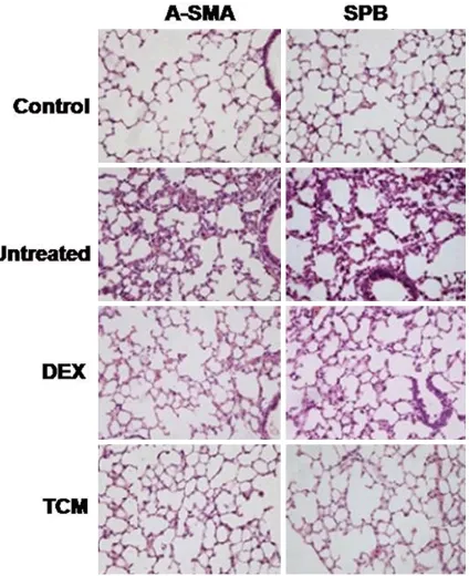
Effect of TCM on α-SMA and SPB
Results from immunohistochemistry showed inhibitory effect of TCM on the expression of α-SMAa (myo) fibroblast marker which was
increased on radiation exposure (Figure 4). However, TCM treatment increased the expres-sion of SPB which was reduced in the rats after exposure to radiations (Figure 4).
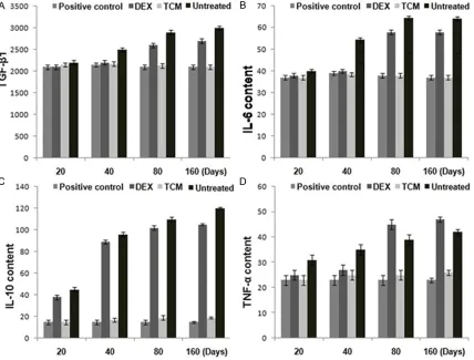
Effect of TCM on level of serum cytokine
TCM treated rats showed reduced expression of TGF-β1 on day 20, 40, 80 and 160 after irra-diation. In DEX treated rats the level of TGF-β1 was reduced on days 20 and 40 and was simi-lar to those of negative control group on the day 80 and 160 (Figure 5A). Therefore, TCM treat-ment reversed the promoting effect of radia-tions on TGF-β1 until day 160.
Rats treated with TCM also showed reduced IL-6, IL-10 and TNF-α expression on the day 20, 40, 80 and 160 after irradiation. Comparison of the expression of these three proteins in DEX treated and negative control groups showed similar results on day 80 and 160. In positive control and TCM treated groups the expression of the proteins was almost similar (Figure 5B-D).
Inactivation of Nrf-2 by TCM activates Nrf-2
Western blot analysis showed activation of pro-teins including HO-1, Nrf-2, and NQO-1 in the rats treated with TCM markedly higher com-pared to DEX treated rats (Figure 6). The TCM treated animals showed higher expression of the above proteins on all the tested days (20, 40, 80 and 160) after irradiation. Comparison of the expression of these proteins in DEX treat-ed and negative control groups revealtreat-ed that DEX only exhibited enhancing effect until day
40 there after the expression was similar with those of negative control group.
Discussion
Reactive oxygen species (ROS) produced in the cells on exposure to radiations leads to harmful
effects on DNA structure, membrane lipid per-oxidation, signal transduction pathway and transcription factor activation [8]. The stress induced by ROS following exposure to radia-tions continues during the pulmonary fibrosis through the oxidant-producing enzyme activa-tion, enhanced mitochondrial membrane
[image:5.612.101.525.72.592.2]meability, and respiratory burst activation in the phagocytic cells [9]. The results from the present study revealed that TCM treatment reverses the pulmonary fibrosis induced on exposure to radiations in the rats. The rate of mortality and the lung index score in TCM treat-ed rats was rtreat-eductreat-ed marktreat-ed compartreat-ed to the untreated and even DEX treated rats.
of MDA along with the inflammatory cytokines and increased the SOD activity in the rats. Therefore, TCM induced decrease in the oxida-tive stress can partly be due to enhanced expression of antioxidant proteins. Pulmonary fibrosis is induced by the proliferation and mat-uration of the fibroblasts which in turn is caused by cytokine, TGF-β1 [16]. The degree of
[image:6.612.94.522.74.399.2]expres-Figure 5. The effect of TCM on the expression of transforming growth factor (A) β1 (TGF-β1), (B) interleukin (IL)-6, (C) IL-10, and (D) tumor necrosis factor α (TNF-α) after 20, 40, 80 and 160 of irradiation.
Figure 6. Effect of TCM and DEX on the activation of nuclear transcription factor NF-E2-related factor 2 (Nrf-2), heme oxygenase-1 (HO-1) and NAD(P) H: quinone oxidoreductase-1 (NQO-1) protein expression after 120 days of irradiation.
[image:6.612.91.368.454.558.2]sion of TGF-β1 has been found to correlate with the rate of incidence of pulmonary fibrosis [17]. Other factors found to be associated with the fibroblast proliferation and the induction of pul-monary fibrosis is the TNF-α [18] as well as pro-inflammatory cytokines like IL-1 and IL-6 devel-op which also develdevel-op the fibrous connective tissue. The expression of these cytokines was found to be significantly reduced in the TCM treated rats.
Nrf2 plays a critical role in the regulation of the major antioxidant enzymes HO-1 and NQO-1. Our western blot analysis results revealed that TCM significantly enhanced the expression lev-els of Nrf-2, HO-1, and NQO-1 in the rat lung tissues compared with radiation only and DEX-treated rats.
In summary the present study demonstrates that TCM induces marked decrease in the rate of morbidity and reverses the radiation induced pulmonary fibrosis in the rats.
Disclosure of conflict of interest
None.
Address correspondence to: Dr. Li-Hua Yang, De- partment of Critical Care Medicine, Yantai Affiliated Hospital of Binzhou Medical University, 717 Jinbu Street, Muping District, Yantai 264000, Shandong, China. Tel: 2123456; Fax: 0086-535-2123456; E-mail: yanglh3001@gmail.com
References
[1] Stone HB, Coleman CN, Anscher MS and Mc-Bride WH. Effects of radiation on normal tis-sue: consequences and mechanisms. Lancet Oncol 2003; 4: 529-536.
[2] Epperly MW, Guo H, Gretton JE and Greenberg-er JS. Bone marrow origin of myofibroblasts in irradiation pulmonary fibrosis. Am J Respir Cell Mol Biol 2003; 29: 213-224.
[3] Matsuo Y, Shibuya K, Nakamura M, Nara-bayashi M, Sakanaka K, Ueki N, Miyagi K, Nori-hisa Y, Mizowaki T, Nagata Y, Hiraoka M. Dose-volume metrics associated with radiation pneumonitis after stereotactic body radiation therapy for lung cancer. Int J Radiat Oncol Biol Phys 2012; 83: e545-e549.
[4] Minor GI, Yashar CM, Spanos WJ Jr, Jose BO, Silverman CL, Carrascosa LA, Farmer M, Paris KJ. The relationship of radiation pneumonitis to treated lung volume in breast conservation therapy. Breast J 2006; 12: 48-52.
[5] Rosenzweig KE, Zauderer MG, Laser B, Krug LM, Yorke E, Sima CS, Rimner A, Flores R, Rusch V. Pleural intensity-modulated radio-therapy for malignant pleural mesothelioma. Int J Radiat Oncol Biol Phys 2012; 83: 1278-1283.
[6] Liu HW, Seftel MD, Rubinger M, Szwajcer D, Demers A, Nugent Z, Schroeder G, Butler JB, Cooke A. Total body irradiation compared with BEAM: long-term outcomes of peripheral blood autologous stem cell transplantation for non-Hodgkin’s lymphoma. Int J Radiat Oncol Biol Phys 2010; 78: 513-520.
[7] Tsoutsou PG, Koukourakis MI. Radiation pneu-monitis and fibrosis: mechanisms underlying its pathogenesis and implications for future research. Int J Radiat Oncol Biol Phys 2006; 66: 1281-1293.
[8] Zhao W, Robbins ME. Inflammation and chron-ic oxidative stress in radiation-induced late normal tissue injury: therapeutic implications. Curr Med Chem 2009; 16: 130-143.
[9] Zhang Y, Zhang X, Rabbani ZN, Jackson IL, Vu-jaskovic Z. Oxidative stress mediates radiation lung injury by inducing apoptosis. Int J Radiat Oncol Biol Phys 2012; 83: 740-748.
[10] deVries HE, Witte M, Hondius D, Rozemuller AJ, Drukarch B, Hoozemans J, van Horssen J. Nrf2-induced antioxidant protection: a promis-ing target to counteract ROS-mediated dam-age in neurodegenerative disease? Free Radic Biol Med 2008; 45: 1375-1383.
[11] Yang G, Li X, Li X, Wang L, Li J, Song X, Chen J, Guo Y, Sun X, Wang S, Zhang Z, Zhou X, Liu J. Traditional Chinese medicine in cancer care: a review of case series published in the Chinese literature. Evid Based Complement Alternat Med 2012; 7510-7546.
[12] Liu J, Li X, Liu J, Ma L, Li X, Fønnebø V. Tradi-tional Chinese medicine in cancer care: a re-view of case reports published in Chinese lit-erature. Forsch Komplementarmed 2011; 18: 257-263.
[13] Li X, Yang G, Li X, Zhang Y, Yang J, Chang J, Sun X, Zhou X, Guo Y, Xu Y, Liu J, Bensoussan A. Traditional Chinese medicine in cancer care: a review of controlled clinical studies published in Chinese. PLoS One 2013; 8: e60338. [14] Zhang L, Dong XW, Wang JN, Tang JM, Yang JY,
Guo LY, Zheng F, Kong X, Huang YZ, Chen SY. PEP-1-CAT-transduced mesenchymal stem cells acquire an enhanced viability and pro-mote ischemia-induced angiogenesis. PLoS One 2012; 7: e52537.