Original Article
Repair of minor tissue defect in fingers by transfer of
free arterialized venous flaps
Ruihua Li, Shilian Kan, Ketong Gong
Department of Hand and Microsurgery, Tianjin Hospital, Tianjin 300211, China
Received November 18, 2015; Accepted January 25, 2016; Epub July 15, 2016; Published July 30, 2016
Abstract: Objective: Reconstruction of skin and soft tissue defects in the fingers is a huge challenge in recent years. We present a retrospective analysis of nine patients with arterialized venous flaps used to cover defects in the fingers. Methods: Nine patients with various finger tissue defects were treated using arterial venous flap from the hand. Seven patients were men and two were women with the mean age 28.3 years, of which eight had injury by machine crush, one by chain saw. Fracture and dislocation in five cases and with tendon rupture in five cases. The defects ranged from 2.5 cm × 2.5 cm to 4.5 cm × 4.5 cm. All flaps were transferred as free flaps. Results: Among the 9 patients, flaps survived in all patients with one experiencing partial epidermal necrosis. After flap transplan-tation, mild swelling occurred in the flaps, but began to decrease on the second week. All the patients reported a normal use of their finger activities in daily living and work. Except the scar, no donor-site morbidity was reported. Conclusion: The arterialized venous flap has good function with low donor site morbidity. It is an excellent flap for reconstruction for finger defects with better survival rate.
Keywords: Arterialized venous flaps, finger, skin reconstruction
Introduction
Reconstruction of skin and soft tissue defects in the fingers remains one of the biggest chal-lenges [1]. Use of skin flaps such as palmar advancement flap, cross-finger flap, distally based homodigital island flaps, and pedicled perforator finger flaps proves to be excellent treatment but creates large scars, poor sensa-tion, with chance of neuroma in the pedicle [2-6]. Use of free flaps in the reconstructive sur-gery as an alternative source is gaining moment in recent years, though it requires highly skilled surgeon. Among the free flaps commonly used in the finger reconstruction is superficial pal-mar branch of the radial artery flap, ulnar artery perforator free flap, posterior interosseous per-forator flap, arterialized venous flap, and free serratus anterior fascia flap [1, 7-9].
Nakayama et al introduced arterialized venous flap (AVF) from the experimental study [10]. Yoshimura et al were the first to use AVFs clini-cally for repairing skin defects of the fingers [11]. They used thirteen arterialized venous
satisfac-tory results were reported in several other case reports with AVFs [16-18].
We present our results in nine patients with soft-tissue and skin injuries in the fingers treat-ed with the arterializtreat-ed venous flap. We designed arterialized venous flap based on the shape of the wound and the flaps had ante-grade perfusion fashion. We treated nine patients from October 2005 to November 2011 in our department by AVF and achieved satis-factory results.
Materials and methods
[image:2.612.89.526.84.211.2]From October 2005 to November 2011, over a period of 6 year and 2 months 9 patients with various finger tissue defects were treated using arterial venous flap from the hand. Seven patients were men and two were women with the age ranged from 17 to 48 years (mean 28.3 years). Fingers were associated with bone and soft tissue defect having tendons, blood ves-sels and nerves exposed with no free skin for grafting. Eight cases had injury by machine crush, one by chain saw. Fracture and disloca-tion in five cases and tendon rupture in five cases All patients underwent emergency sur-gery, with a time delay after injury of 2 to 8 hours (mean 4.8 hours). The defects ranged from 2.5 cm × 2.5 cm to 4.5 cm × 4.5 cm. All flaps were transferred as free flaps. All of the cases were performed by the senior hand sur-geon. The details of patients are presented in Table 1.
Surgical procedures
Healthy skin and soft tissue were retained in the site, and debris, inactive skin were removed. Before the flap placement, fracture dislocation
is fixed by Kirschner wire, tendons were repaired by surgery and nerve rupture underwent nerve anastomosis. Arteriovenous anastomosis was performed to repair the arterial defects. The flap was designed according to the size of the defect. In the donor skin from ipsilateral fore-arm palm, two longitudinal superficial veins region was selected and marked by methylene blue. The flap should be slightly larger than the range of defects range. The cut was made at the proximal and distal region to free the vein in the flap. The length of the vein was based according to the subject area. The flap when cut doesn’t carry the deep fascia. Cut flaps ranged from 3.0 cm × 3.0 cm to 5.0 cm × 5.0 cm. The afferent vessels were anastamosed to the digital arteries and the efferent veins were anastamosed to dorsal veins. After the surgery, anti-inflammatory, anti coagulant, and anti spasm drugs were administered to the patients. Close monitoring of color, temperature, and blood flow through the flap was observed. Results
After the first day of flap transplantation, mild swelling occurred in the flaps, with the increas-ing in swellincreas-ing in the second day havincreas-ing small blisters in the peripheral region. On the fifth day swelling in the flap began to decrease with the slow disappearance of blisters. On the ninth day, complete disappearance of the swelling with the flap skin color being normal. Among the 9 patients, all flaps survived with one expe-riencing partial epidermal necrosis in the flap. According to the Chinese Medical Society of Hand Surgery functional evaluation, fingers in four patients were excellent and in five patients were good. The patients reported a normal use of their finger activities of daily living and work. No donor-site morbidity was reported.
Table 1. Patient characteristics
Case Sex Age (yrs) Affected Finger Pattern of defect Defect size (mm) Flap type
1 F 23 Right middle finger Severe damage in the dorsal skin 4 cm × 4.5 cm Flap from the ipsilateral forearm palm
2 M 17 Left little finger Severe damage in the dorsal skin 2.5 cm × 2.5 cm Flap from the ipsilateral forearm palm
3 M 48 Left middle finger Severe damage in the volar skin 3.5 cm × 2.5 cm Flap from the ipsilateral forearm palm
4 M 29 Left index finger Severe damage in the dorsal skin 3.0 cm × 2.5 cm Flap from the ipsilateral forearm palm
5 M 33 Right ring finger Severe damage in the radial volar
and radial dorsal skin
4.5 cm × 4.0 cm Flap from the ipsilateral forearm palm
6 M 42 Right index finger Severe damage in the volar skin 3.0 cm × 2.5 cm Flap from the ipsilateral forearm palm
7 F 22 Left ring finger Severe damage in the volar skin 4.0 cm × 2.5 cm Flap from the ipsilateral forearm palm
8 M 20 Left middle finger Severe damage in the dorsal skin 4.0 cm × 3.5 cm Flap from the ipsilateral forearm palm
Typical case reports
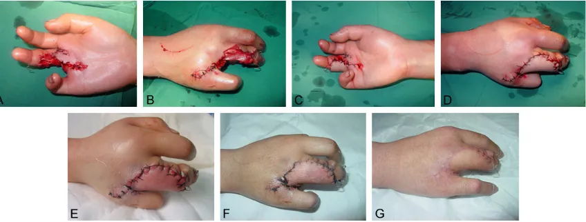
A 23 years old woman suffered a mechanical injury with severe damage in the dorsal skin of right middle finger and complete loss in the ring finger while working (Figure 1). The patient underwent emergency surgery on January 17, 2007. After shortening ring finger by debride-ment surgery in proximal base plane, middle finger fracture fixed by cross section pinning. Near the middle finger, skin defect in the dorsal metacarpal region was 4 cm × 4.5 cm. A 5 cm × 5 cm venous flap was designed from the ipsi-lateral forearm palm. The donor site was closed primarily. Flap was grafted over the whole area of wound. The afferent vessels were anasta-mosed to the digital arteries and the efferent veins were anastamosed to dorsal veins. The nerve was sutured to the radial digital nerve. There was mild swelling with the skin tempera-ture higher (ranging 0.6-1.20°C) than the nor-mal, postoperatively. On the 2nd 105 day tiny blisters appeared at the periphery of the flap, but gradually disappeared on the 4th day. On the 9th 106 day, the swelling disappeared. After 14 days stitches, the flaps survived with the skin having soft texture and color close to the normal skin of the fingers. After 4 months follow up of the flap, texture and color of the skin was closer to the normal dorsal skin. Discussions
Arterialized venous flaps have been widely used for the closure of smaller defects in fin-gers. These flaps are considered as a potential
[image:3.612.97.521.73.234.2]reconstructive option for large dorsal digital defects with exposed bone, joint, and/or exten-sor tendons when local flaps are inadequate or unusable [8]. Designed arterialized venous flap from the venous network of the forearm applied in five patients with various defects ranging in size from 6 × 8 cm to 10 × 12 cm had full sur-vival in 4 patients with 30% partial necrosis in one patient [19]. In another study, six patients with multiple soft tissue finger defects were covered by syndactylizing arterialized venous flaps, with palmar forearm was donor site and none of the patients suffered flap loss [20]. Large skin defects of the hand with AVFs rang-ing in size from 6 cm × 3 cm to 14 cm × 9 cm in 12 patients showed remarkable edema post-operatively, and partial necrosis of the flap only developed in three cases [21]. However, in our study only one case suffered partial necrosis. In another study, reconstruction of severe and extensive contractures of the palm in four patients using large AVFs showed complete survival with uneventful clinical courses [22]. Majority of AVFs were applied in antegrade per-fusion fashion. However, an experiment study indicated that flaps from human cadavers indi-cated that retrograde arterialization could increase blood circulation in the periphery of arterialized venous flaps [23]. Studies by Koch et al applying the retrograde arterialized venous flaps in 13 patients to resurface the skin and soft-tissue defects proved effective suggesting that retrograde perfusion enhances blood flow in the periphery of arterialized venous flaps giv-ing better results in terms of flap survival [24].
However, in our studies all the flaps were applied in antegrade perfusion fashion, with 8 flaps survived out of 9 flaps, with one having partial necrosis. This is similar to the studies by Woo et al who applied antegrade approach in hand with a 98% (151/154) success rate with 5.2% partial loss rate [17]. Hyza et al also indi-cated with high survival rate with their experi-ence of 13 venous free flaps in 12 patients with large dorsal digital defects [25].
Partial necrosis occurs in the flaps due to con-gestion and edema in the flaps bringing compli-cations post operatively which requires close monitoring of color, temperature, and blood flow through the flap [26, 27]. Flap swelling was found to be most pronounced during the first post operative week, but was slowly resolving in the second week in our study. This was simi-lar to earlier studies [17, 25]. In another report-ed study on ten patients, AVFT flap congestion and edema was seen in all cases. In seven cases, a single vein through the flap served as both the inflow and outflow conduits in seven cases and a separate efferent vein within the same flap was utilized as the outflow vessel in three. Congestion seen was transient due to the relatively small flap sizes (2.5 cm × 2 cm to 4.5 cm × 3 cm) [24]. Partial or complete necro-sis occurs commonly when operated under less than ideal circumstances, such as a chronically infected or open wounds, or largely avascular recipient sites [27].
In our opinion, the arterialized venous flap is an attractive option to cover the finger defects with low donor-site morbidity.
Disclosure of conflict of interest
None.
Address correspondence to: Ruihua Li, Department of Hand and Microsurgery, Tianjin Hospital, 406 Jiefang South Road, Hexi District, Tianjin 300211, China. Tel: +8622-28332917; Fax: +8622-2833- 2918; E-mail: ruihli66@163.com
References
[1] Liu Y, Jiao H, Ji X, Liu C, Zhong X, Zhang H, Ding X and Cao X. A comparative study of four types of free flaps from the ipsilateral extremity for finger reconstruction. PLoS One 2014; 9: e104014.
[2] Foucher G, Delaere O, Citron N and Molderez A. Long-term outcome of neurovascular pal-mar advancement flaps for distal thumb inju-ries. Br J Plast Surg 1999; 52: 64-68.
[3] Kappel DA and Burech JG. The cross-finger flap. An established reconstructive procedure. Hand Clin 1985; 1: 677-683.
[4] Li YF and Cui SS. Innervated reverse island flap based on the end dorsal branch of the digital artery: surgical technique. J Hand Surg Am 2005; 30: 1305-1309.
[5] Moschella F and Cordova A. Reverse homodigi-tal dorsal radial flap of the thumb. Plast Reconstr Surg 2006; 117: 920-926.
[6] Toia F, Marchese M, Boniforti B, Tos P and Delcroix L. The little finger ulnar palmar digital artery perforator flap: anatomical basis. Surg Radiol Anat 2013; 35: 737-740.
[7] Iglesias M, Fonseca-Lazcano JA, Moran MA, Butron P, Díaz-Morales M. Revascularization of arterialized venous flaps through a total retro-grade reverse blood flow: Randomized experi-mental trial of viability. Plast Reconstr Surg Glob Open 2013; 1: e34.
[8] Yan H, Fan C, Zhang F, Gao W, Li Z and Zhang X. Reconstruction of large dorsal digital de-fects with arterialized venous flaps: our experi-ence and comprehensive review of literature. Ann Plast Surg 2013; 70: 666-671.
[9] Ulrich D, Fuchs P, Bozkurt A and Pallua N. Free serratus anterior fascia flap for reconstruction of hand and finger defects. Arch Orthop Trauma Surg 2010; 130: 217-222.
[10] Nakayama Y, Soeda S, Kasai Y. Flaps nour-ished by arterial inflow through the venous system: an experimental investigation. Plast Reconstr Surg 1981; 67: 328-334.
[11] Yoshimura M, Shimada T, Imura S. The venous skin graft method for repairing skin defects of the fingers. Plast Reconstr Surg 1987; 79: 243-250.
[12] Inoue G, Maeda N and Suzuki K. Resurfacing of skin defects of the hand using the arteri-alised venous flap. Br J Plast Surg 1990; 43: 135-139.
[13] Fukui A, Inada Y, Maeda M, Mizumoto S, Yajima H and Tamai S. Venous flap-its classification and clinical applications. Microsurgery 1994; 15: 571-578.
[14] Nishi G, Shibata Y, Kumabe Y, Hattori S, Okuda T. Arterialized venous skin flaps for the injured finger. J Reconstr Microsurg 1989; 5: 357-365. [15] Chen HC, Tang YB, Noordhoff MS. Four types of
venous flaps for wound coverage: a clinical ap-praisal. J Trauma 1991; 31: 1286-1293. [16] Koch H, Moshammer H, Spendel S, Pierer G
[17] Woo SH, Kim KC, Lee GJ, Ha SH, Kim KH, Dhawan V and Lee KS. A retrospective analysis of 154 arterialized venous flaps for hand re-construction: an 11-year experience. Plast Reconstr Surg 2007; 119: 1823-1838.
[18] Nakazawa H, Kikuchi Y, Honda T, Isago T, Morioka K and Itoh H. Use of an arterialised venous skin flap in the replantation of an am-putated thumb. Scand J Plast Reconstr Surg Hand Surg 2004; 38: 187-191.
[19] Yilmaz M, Menderes A, Karataş O, Karaca C, Barutçu A. Free arterialised venous forearm flaps for limb reconstruction. Br J Plast Surg 1996; 49: 396-400.
[20] Kayalar M, Levent K, Sugun TS, Gurbuz Y, Savran A, Kaplan I. Syndactylizing arterialized venous flaps for multiple finger injuries. Microsurgery 2014; 34: 527-534.
[21] Woo SH, Jeong JH and Seul JH. Resurfacing relatively large skin defects of the hand using arterialized venous flaps. J Hand Surg 1996; 21: 222-229.
[22] Nakazawa H, Nozaki M, Kikuchi Y, Honda T, Isago T. Successful correction of severe con-tracture of the palm using arterialized venous flaps. J Reconstr Microsurg 2004; 20: 527-531.
[23] Moshammer HE, Schwarzl FX, Haas FM, Maechler H, Pierer G, Wiltgen M, Koch H. Retrograde arterialized venous flap: An experi-mental study. Microsurgery 2003; 23: 130-134.
[24] Koch H, Scharnagl E, Schwarzl FX, Haas FM, Hubmer M, Moshammer HE. Clinical applica-tion of the retrograde arterialized venous flap. Microsurgery 2004; 24: 118-124.
[25] Hyza P, Vesely J, Novak P, Stupka I, Sekac J, Choudry U. Arterialized venous free flaps-a re-constructive alternative for large dorsal digital defects. Acta Chir Plast 2008; 50: 43-50. [26] Kong BS, Kim YJ, Suh YS, Jawa A, Nazzal A, Lee
SG. Finger soft tissue reconstruction using ar-terialized venous free flaps having 2 parallel veins. J Hand Surg 2008; 33: 1802-1806. [27] Jared W, Garlick JW, Goodwin IA, Wolter K AJ.