Original Article
PD-L1-expressing neutrophils as a novel indicator to
assess disease activity of rheumatoid arthritis
Qing Luo1*, Lulu Zeng2*, Hanying Mei3, Zikun Huang1, Zhongqin Luo1, Jianqing Ye2, Xue Li2, Yang Guo2, Junming Li1
1Department of Clinical Laboratory, The First Affiliated Hospital of Nanchang University, Nanchang 330006,
Jiangxi, China; 2Department of Medical College, Nanchang University, Nanchang 330006, Jiangxi, China; 3 Depart-ment of Rheumatology, Jiujiang First Peoples Hospital, Jiujiang 332000, Jiangxi, China. *Equal contributors. Received November 21, 2016; Accepted December 25, 2016; Epub May 15, 2017; Published May 30, 2017
Abstract: Background: It is well-known that increased frequency of neutrophils was found in patients with Rheumatoid arthritis (RA), and neutrophils are the most abundant immune cells in Synovial fluid (SF) of RA patients. Costimulatory molecules plays an important role in determining the activation status and function of immune cells. However, the immunomodulatory roles and mechanisms of costimulatory molecules on neutrophils in RA are poorly understood. Objective: To determine the frequency of PD-L1-expressing neutrophils in patients with RA and tested the hypothesis that their frequency correlated with the activity of RA. Method: The expression of costimulatory mol-ecules including PD-1, PD-L1, Tim-3, CD40, TIGIT, CD80 and CD86 on neutrophils were determined by flow cytom-etry. The frequencies of PD-L1-expressing neutrophils in patients with RA were further analyzed for their correlation with markers of autoimmune response, inflammation, disease activity and severity of RA. Results: Compared with healthy controls (HC), the frequency of PD-L1-expressing neutrophils in peripheral blood (PB) and synovial fluid (SF) were significantly elevated in RA patients (P = 0.0001) (P < 0.0001). And, the frequency of PD-L1-expressing neu-trophils in SF of RA patients was significantly increased than that in autologous PB (P < 0.0001). Furthermore, we found that the frequency of PD-L1-expressing neutrophils in patients with RA was increased significantly in subjects with high RF titre, high levels of inflammatory markers and high Disease activity score 28 (DAS28). Conclusion: The frequency of PD-L1-expressing neutrophils is elevates in patients with RA and correlates with the disease activity of RA.
Keywords: Rheumatoid arthritis, PD-L1, neutrophils, synovial fluid, disease activity score 28
Introduction
Rheumatoid arthritis (RA) is a chronic debilitat-ing systemic autoimmune disease character-ized by inflammation and destruction of the joints. About 1% of the population suffers from RA, and many patients develop long-term joint damage, severe illness and disability [1]. Ac- cumulating studies have shown that several factors are involved in the pathogenesis of RA, including genetic, infectious, environmental, and hormonal factors [2-4]. Unbalance of adap-tive and innate immune systems driving the excessive immune responses is observed in RA. Due to its heterogeneity and multiplicity, the etiology of RA still remains elusive [5]. Although the etiopathology of RA is not fully understood, it is known that neutrophils,
mac-rophages, synovial fibroblasts, T cells and B cells are involved in the mechanisms that drive the onset of RA [6]. Neutrophils, which are part of the innate immunity, are crucial for patho-genic defense. They are the first cell type to arrive at sites of inflammation [7]. And, neutro-phils are by far the most abundant immune cell in SF of the joints of RA patients [8, 9]. Neutrophils have been reported to lead to the pathology of RA by releasing inflammatory mediators and regulating the functions of other immune cells including macrophages, NK cells, dendritic cell (DC), T cells and B cells [10]. Nevertheless, the roles of neutrophils in RA pathogenesis have not been well elucidated, especially, the regulative effect on immune cells.
which functions as an immunomodulatory mol-ecule. The engagement of PD-L1 with its recep-tor, programmed death 1 (PD-1), delivers inhibi-tory signals to target cells such as activated T-cell and B-cell, thus helps to maintain the bal-ance between effective immunity, tolerbal-ance and immunopathology [11]. PD-L1 is broadly expressed on a variety of immune cells, includ-ing T cells, B cells, dendritic cells, and mono-cytes. Recent evidence indicates that PD-L1 is also expressed on neutrophils and associated with the development of numerous diseases, including the infection of human immunodefi-ciency virus [12], sepsis [13], Burkholderia pseudomallei-Infected disease [14], tuberculo-sis [15] and systemic lupus erythematosus [16]. However, the frequency and roles of PD-L1-expressing neutrophils in RA has not been established.
In the present study, we determined the fre-quency of PD-L1-expressing neutrophils in pa- tients with RA and tested the hypothesis that their frequency correlated with the activity of RA.
Materials and methods
Subjects
Fresh peripheral blood (PB) was harvested by venipuncture from 67 patients with RA which fulfilled the American College of Rheumatology criteria for RA [17], and 52 healthy controls (HC) that unrelated to the patients, without inflam-matory and autoimmune diseases. Synovial fluid (SF) was obtained from 27 cases of the 67 RA patients. RA disease activity was measured using the disease activity score 28 (DAS28) [18]. The study was approved by the Ethics Committee of the First Affiliated Hospital of Nanchang University (No. 019) and was carried out in compliance with the Helsinki Declaration. Informed consent was obtained from all the participants before they entered the study.
Flow cytometry analysis
The peripheral blood and synovial fluid were drawn and analyzed immediately for the molec-ular phenotypes of neutrophils by flow cytome-try. The following antibodies were used: ECD-conjugated anti-CD3, PC5-ECD-conjugated an- ti-CD15 (BD Biosciences, San Diego CA, USA), PE-conjugated anti-PD1, anti-Tim3, anti-TIGIT, anti-CD86, and anti-PDL1, FITC-con-jugated
anti-CD80 (MIH clones, e Bioscience, San Diego, CA, USA). The neutrophils were identified as CD15+CD3- populations [16] and the mem-branous markers were detected by flow cytom-etry with triple staining. Briefly, 50 microliters of fresh heparinized whole blood were incubated simultaneously with 5 μL ECD-conjugated anti-CD3, 5 μL PC5-conjugated anti-CD15 and 5 μL fluorescence-conjugated antibodies targeting other membranous molecules on ice in the dark for 30 minutes. Cells incubated with PE- and FITC-conjugated mouse IgG were used as isotype controls. All flow samples were analyzed with a CYTOMICS FC 500 flow cytom-eter (BECKMAN COULTER) and associated software programs (CXP).
ESR, CRP and autoantibodies measurement
Erythrocyte sedimentation rate (ESR) was de- termined according to the instructions de- scribed by the manufacturer. C-reactive Protein (CRP) and Rheumatoid factor (RF) were mea-sured by nephelometry. Anticitrullinated protein antibodies (ACPA) of IgG in serum were mea-sured by commercially available ELISA kits (kexin, Shanghai, China).
Statistical analysis
Statistical analysis and graphic presentation were carried out with GraphPad Prism version 5.0 (GraphPad Software, San Diego, CA). A t- test was used where the normality test passed; otherwise, the nonparametric Mann-Whitney test was used to analyze the data. Likewise, the Pearson method or the nonparametric Spearman method was used for correlation analysis. Value of P < 0.05 is considered as significant difference.
Results
Characteristics of study subjects
Characteristics of RA patients and healthy con-trols (HC) enrolled in this study are included in
therapy with disease modifying antirheumatic drugs (DMARDs).
The frequency of PD-L1-expressing neutrophils is elevated in patients with RA
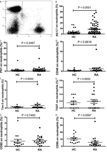
The neutrophils were identified in peripheral blood as CD15+CD3- populations and analyzed for the expression of costimulatory mo- lecules including PD-1, PD-L1, Tim-3, TIGIT, CD40, CD80 and CD86 by flow cytometry. Data showed that the frequency of PD-L1 positive neutrophils was significantly elevated in RA patients compared to HC (Figure 1B, P = 0.0001). No significant difference was ob- served in the frequency of PD1-, CD40-, Tim-3-, TIGIT-, CD80-, or CD86-expressing neutrophils between RA individuals and HC (Figure 1). Neutrophils are the most abundant immune cells in synovial fluid of the joints of RA patients [8, 9]. And, researchers have reported that the changes in synovial fluid may reflect the devel-opment and progression of RA much more directly and clearly [4, 19]. We therefore inves-tigated PD-L1 expression on neutrophils in the synovial fluid of RA patients. As shown in Figure 2, the frequency of PD-L1 positive neutrophils in synovial fluid was significantly increased than that in peripheral blood of HC (P < 0.0001) or that in peripheral blood of cases (P < 0.0001).
The frequency of PD-L1-expressing neutro-phils correlated with markers of autoimmune response
Rheumatoid arthritis (RA) is characterized by auto-antibodies overproduction such as RF and ACPA. Thus, the hallmark antibodies of RA including RF and ACPA were determined and analyzed for their correlation with the
frequen-cy of PD-L1-expressing neutrophils in this study. Data showed that 41 patients were positive for RF and 37 patients were positive for ACPA in all 49 patients who received auto-antibodies detection. As shown in
Figure 3, the frequency of PD-L1-expressing neutrophils was significantly increased in patients with positive RF. And, the frequency of PD-L1-expressing neutrophils trends to elevate in patients with positive ACPA, but a significant difference was not reached. Moreover, the correlations between the fre-quency of PD-L1-expressing neutrophils in the synovial fluid and the hallmark
antibod-Table 1. Baseline characteristics of rheumatoid arthritis patients
Categories RA patients (n = 67) trols (n = 52)Healthy
con-Females,% 79% 77%
Age (years, average ± SD) 55 ± 13 50 ± 11 DAS28 (average ± SD) 3.9 ± 1.8 -RF (IU/ml) (49 patients) 447.2 ± 701.1 -ACPA (RU/ml) (49 patients) 524.6 ± 967.4 -CRP (mg/L) (49 patients) 12.8 ± 17.3
-ESR (mm/h) 37.9 ± 31.3
-ies in serum of RA were investigated but no obvious correlation was found (data no shown). These results showed that the elevated fre-quency of PD-L1-expressing neutrophils is cor-related with the markers of autoimmune re- sponse, suggested that PD-L1-expressing neu-trophils may associated with the pathogenesis of RA.
The frequency of PD-L1-expressing neutrophils correlated with markers of inflammation
RA is characterized by synovial hyperplasia and inflammation. RA Patients are frequently ac- companied by the elevated levels of inflamma-tory markers. In order to investigate the correla-tion between the frequency of PD-L1-expressing neutrophils and inflammatory markers, the markers of inflammation including ESR and CRP were determined and analyzed for their correlation with the frequency of PD-L1-ex- pressing neutrophils in RA patients. As shown in Figure 4, positive correlations between the frequency of PD-L1-expressing neutrophils and ESR, CRP were found. Moreover, we investigat-ed the correlation between the frequency of PD-L1-expressing neutrophils in the synovial fluid and the inflammatory markers in serum of RA. No obvious correlation was found (data no shown). These results indicated that the fre-quency of PD-L1-expressing neutrophils asso- ciated with markers of inflammation.
The frequency of PD-L1-expressing neutrophils correlated with disease activity of RA
[image:3.612.92.319.97.215.2]mark-Figure 1. Expression of PD-L1, PD1, CD40, TIGIT, CD80 and CD86 on neutrophils in peripheral blood. A. Gates were then set on neutrophils based on CD15 and CD3, neutrophils were defined as CD15+CD3– FSC high, SSC high. B.
[image:4.612.93.525.71.682.2]Mann-Whit-Figure 2. Expression of PD-L1 on neutrophils in synovial fluid and peripheral blood. A. The frequency of PD-L1 posi-tive neutrophils in synovial fluid was significantly increased than that in peripheral blood of HC (P < 0.0001, Mann-Whitney test). B. The frequency of PD-L1 positive neutrophils in synovial fluid was significantly increased than that in peripheral blood of cases (P < 0.0001, Mann-Whitney test).
ney test). C. The frequency of PD1-expressing neutrophils was similar between HC and RA patients (P = 0.2467, Mann-Whitney test). D. The frequency of CD40-expressing neutrophils was similar between HC and RA patients (P
= 0.0616, Mann-Whitney test). E. The frequency of Tim-3-expressing neutrophils was similar between HC and RA patients (P = 0.8564, Mann-Whitney test). F. The frequency of TIGIT-expressing neutrophils was similar between HC and RA patients (P = 0.5650, t-test). G. The frequency of CD80-expressing neutrophils was similar between HC and RA patients (P = 0.7475, t-test). H. The frequency of CD86-expressing neutrophils was similar between HC and RA patients (P = 0.0587, t-test). HC: healthy control subject; RA: rheumatoid arthritis; PD-L1: programmed death ligand 1; PD-1: programmed death 1; TIGIT: T cell immunoreceptor with Ig and immunoreceptor tyrosine-based inhibitory domains; Tim-3: T cell immunoglobulin domain- and mucin domain containing molecule-3.
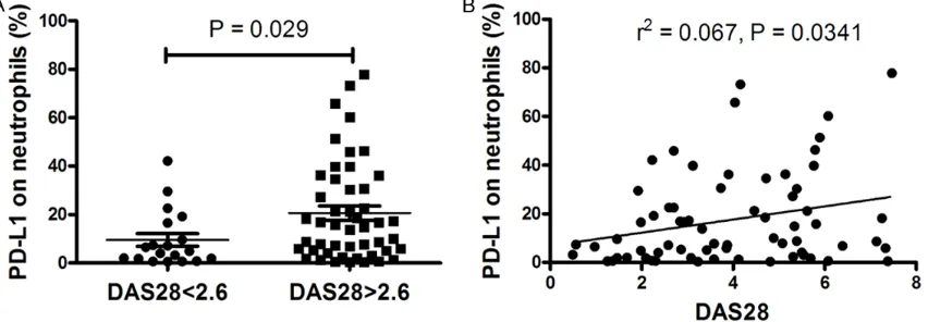
ers, such as RF and ACPA, were reported to cor-relate with disease activity and the severity of joint destruction of RA [10]. And, ESR and CRP are traditionally valuable for calculating DAS28, a scoring system for assessing the severity in patients with RA. Thus, patients with RA were further classified into active and remission patients according to the DAS28 and analyzed for their relation with the frequency of PD-L1-expressing neutrophils. Data showed that the frequency of PD-L1-expressing neutrophils in patients with active RA was significantly higher compared with patients with remission RA (P = 0.029) (Figure 5A). Furthermore, we found that there was a positive correlation between the frequency of PD-L1-expressing neutrophils and the DAS28 score (r2 = 0.067, P = 0.034) (Figure
5B), which demonstrated that the frequency of PD-L1-expressing neutrophils is correlated with disease activity of RA. We also investigated the correlation between the frequency of PD-L1-expressing neutrophils in the synovial fluid and DAS28. No obvious correlation was found (data no shown).
Subsequently, we compared the frequency of PD-L1-expressing neutrophils between
new-onset and late-new-onset patients of RA. Data showed that the frequency of PD-L1-expressing neutrophils trends to elevate in new-onset patients of RA, but a significant difference was not reached (data no shown).
Discussion
[image:5.612.94.516.187.333.2]Figure 3. Correlation of frequency of expressing neutrophils with autoantibody. A. The frequency of PD-L1-expressing neutrophils was significantly increased in RA patients with positive RF (P = 0.0105, Mann-Whitney test). B. The frequency of PD-L1-expressing neutrophils trends to elevate in patients with positive ACPA, but a significant difference was not reached (P = 0.197, t-test).
Figure 4. Correlation of frequency of PD-L1-expressing neutrophils with inflammatory markers. A. The frequency of PD-L1-expressing neutrophils in RA patients correlated significantly with ESR (r2 = 0.1105, P = 0.0068, Pearson
method). B. The frequency of PD-L1-expressing neutrophils in RA patients correlated significantly with CRP (r2 =
0.1183, P = 0.0155, Pearson method).
[image:6.612.97.523.522.670.2]TIGIT, have been reported to have abnormal expression on T cells, monocyte or NK cells of RA patients [22-25]. In this study, for the first time, we investigated the expression of CD80, CD86, PD1, PD-L1, Tim-3, CD40 and TIGIT on neutrophils from RA patients, and showed that the frequency of PD-L1-expressing neutrophils was significantly increased in RA patients com-pared with HC. Moreover, our research revealed that the frequency of PD-L1-expressing neutro-phils was associated with disease activity of RA.
RA is associated with the development of auto-antibodies, which are often detectable before clinical symptoms of the disease become ap- parent. In this study, the serous levels of RF and ACPA, the hallmark antibodies of RA were first determined and analyzed for their relation with the frequency of PD-L1-expressing neutro-phils. Data showed that the frequency of PD- L1-expressing neutrophils was significantly in- creased in patients with positive RF, suggested that PD-L1-expressing neutrophils may associ-ated with autoimmune responses of RA. How- ever, although tend to increase in patients with positive ACPA, the frequency of PD-L1-expressing neutrophils showed no statistic relation with ACPA titer. As ACPA titer often cor-relate positively with disease activity and the severity of joint destruction and would decrease following therapy with DMARDs, the poor cor-relation between the frequency of PD-L1-ex- pressing neutrophils and ACPA may be due to the fact that majority of RA patients had re- ceived therapy prior to participation in the study.
It is well-known that autoimmune response is a kind of chronic inflammation against self anti-gens. So the correlation between the frequency of PD-L1-expressing neutrophils and inflamma-tory markers was analyzed. Our results showed that the frequency of PD-L1-expressing neutro-phils was positively related with ESR and CRP. Considering the application of ESR and CRP for calculating DAS28, our research investigated the correlation between the frequency of PD- L1-expressing neutrophils and DAS28. Sub- sequent results resulted from the DAS28 of RA patients confirmed our speculations. We also compared the frequency of PD-L1-expressing neutrophils between new-onset and late-onset patients of RA. Data showed that the frequency of PD-L1-expressing neutrophils trends to
ele-vate in new-onset patients of RA, but a signifi-cant difference was not reached. Thus, we established the correlation between the fre-quency of PD-L1-expressing neutrophils and disease activity of RA.
In the report, we identified that the frequency of PD-L1-expressing neutrophils from peripheral blood and synovial fluid of RA were up-regu- lated. Also, the elevation of the frequency of PD-L1-expressing neutrophils from peripheral blood was correlated with RF titre, inflammato-ry markers and disease activity of RA. But the elevation of the frequency of PD-L1-expressing neutrophils from synovial fluid was not associ-ated with RF titre, inflammatory markers and disease activity of RA. This may be that periph-eral blood indicate systemic perspectives and synovial fluid indicate local perspectives in RA. PD-L1, an immunoregulatory molecule that be- longs to the B7 superfamily, was identified as ligands for PD-1, and engagement of PD-1 by PD-L1 induced negative signaling by the recruit-ment of phosphatases, such as SHP-2, and de- phosphorylation of effector molecules involved in downstream TCR signaling [27, 28]. By far, there are only a few papers which reported the expression of PD-L1 on neutrophils. Some re- searches found that the expression of PD-L1 was up-regulated in infectious diseases and considered that this up-regulation of PD-L1 on neutrophils is involved in immunosuppression [12-15]. It is worth mentioning that our recent researches suggested that the up-regulation of PD-L1 on neutrophils may serves as a negative feedback mechanism [16].
pressing neutrophils may serves as a negative feedback mechanism to prevent potential tis-sue damage caused by excessive autoimmune responses in patients with RA.
Conclusions
To our knowledge, this is the first report on the characteristics of PD-L1-expressing neutrophils in RA. Additionally, our research established a correlation between the frequency of PD-L1-expressing neutrophils and disease activity and severity of RA, which might improve our under-standing of neutrophil’s role in RA.
Acknowledgements
This study was supported by the National Natural Science Foundation of China (81- 360459) and Jiangxi Provincial Natural Science Foundation of China (20151BAB215031). We would like to acknowledge the help from Dr. Rui Wu at department of rheumatology, the First Affiliated Hospital of Nanchang University, Nanchang, Jiangxi, China.
Disclosure of conflict of interest
None.
Authors’ contribution
Conceived and designed the experiments: QL LZ JL. Performed the experiments: QL LZ HM ZH ZL JY XL YG. Analyzed the data: QL LZ JL. Wrote the paper: QL LZ JL. Contributed re- agents/materials/analysis tools: HM ZH ZL JY. All authors read and approved the manuscript.
Address correspondence to: Drs. Junming Li and Zikun Huang, Department of Clinical Laboratory, First Affiliated Hospital of Nanchang University, 17 Yongwai Zheng Jie, Nanchang 330006, Jiangxi, China. Tel: (+86) 88692794; Fax: (+86) 0791-88623153; E-mail: lisir361@163.com (JML); yfyhzk@163.com (ZKH)
References
[1] Goekoop-Ruiterman YP and Huizinga TW. Rheumatoid arthritis: can we achieve true drug-free remission in patients with RA? Nat Rev Rheumatol 2010; 6: 68-70.
[2] Dugowson CE, Koepsell TD, Voigt LF, Bley L, Nelson JL and Daling JR. Rheumatoid arthritis in women. Incidence rates in group health
co-operative, Seattle, Washington, 1987-1989. Arthritis Rheum 1991; 34: 1502-7.
[3] Li X, Yuan FL, Lu WG, Zhao YQ, Li CW, Li JP and Xu RS. The role of interleukin-17 in mediating joint destruction in rheumatoid arthritis. Bio-chem Biophys Res Commun 2010; 397: 131-5. [4] Yuan FL, Li X, Lu WG, Li CW, Xu RS and Dong J.
IL-33: a promising therapeutic target for rheu-matoid arthritis? Expert Opin Ther Targets 2011; 15: 529-34.
[5] Pratt AG, Isaacs JD and Mattey DL. Current concepts in the pathogenesis of early rheuma-toid arthritis. Best Pract Res Clin Rheumatol 2009; 23: 37-48.
[6] Cascão R, Rosário HS, Souto-Carneiro MM and Fonseca JE. Neutrophils in rheumatoid arthri-tis: more than simple final effectors autoimmu-nity reviews. Autoimmun Rev 2010; 9: 531-5. [7] Nathan C. Neutrophils and immunity:
challeng-es and opportunitichalleng-es. Nat Rev Immunol 2006; 6: 173-82.
[8] Mohr W, Westerhellweg H and Wessinghage D. Polymorphonuclear granulocytes in rheumatic tissue destruction. III. an electron microscopic study of PMNs at the pannus-cartilage junction in rheumatoid arthritis. Ann Rheum Dis 1981; 40: 396-9.
[9] Wittkowski H, Foell D, af Klint E, De Rycke L, De Keyser F, Frosch M, Ulfgren AK and Roth J. Ef- Ef-fects of intra-articular corticosteroids and anti-TNF therapy on neutrophil activation in rheu-matoid arthritis. Ann Rheum Dis 2007; 66: 1020-5.
[10] Wright HL, Moots RJ and Edwards SW. The mul-tifactorial role of neutrophils in rheumatoid ar-thritis. Nat Rev Rheumatol 2014; 10: 593-601. [11] Keir ME, Butte MJ, Freeman GJ and Sharpe AH.
PD-1 and its ligands in tolerance and immuni-ty. Annu Rev Immunol 2008; 26: 677-704. [12] Bowers NL, Helton ES, Huijbregts RP, Goepfert
PA, Heath SL and Hel Z. Immune suppression by neutrophils in HIV-1 infection: role of PD-L1/ PD-1 pathway. PLoS Pathog 2014; 10: e1003993.
[13] Wang JF, Li JB, Zhao YJ, Yi WJ, Bian JJ, Wan XJ, Zhu KM and Deng XM. Up-regulation of Pro-grammed cell death 1 ligand 1 on neutrophils may be involved in sepsis-induced immuno-suppression: an animal study and a prospec-tive case-control study. Anesthesiology 2015; 122: 852-63.
[14] Buddhisa S, Rinchai D, Ato M, Bancroft GJ and Lertmemongkolchai G. Programmed death li-gand 1 on Burkholderia pseudomallei-infected human polymorphonuclear neutrophils im-pairs T cell functions. J Immunol 2015; 194: 4413-21.
Banchereau J, Chaussabel D and O’Garra A. Programmed death ligand 1 is over-expressed by neutrophils in the blood of patients with ac-tive tuberculosis. Eur J Immunol 2011; 41: 1941-47.
[16] Luo Q, Huang Z, Ye J, Deng Y, Fang L, Li X, Guo Y, Jiang H, Ju B, Huang Q and Li J. PD-L1-ex-pressing neutrophils as a novel indicator to as-sess disease activity and severity of systemic lupus erythematosus. Arthritis Res Ther 2016; 18: 47.
[17] Arnett FC, Edworthy SM, Bloch DA, McShane DJ, Fries JF, Cooper NS, Healey LA, Kaplan SR, Liang MH and Luthra HS. The American Rheu-matism Association 1987 revised criteria for the classification of rheumatoid arthritis. Ar- Ar-thritis Rheum 1988; 31: 315-24.
[18] Prevoo ML, van’t Hof MA, Kuper HH, van Leeu-wen MA, van de Putte LB and van Riel PL. Mod- Mod-ified disease activity scores that include twen-ty-eight-joint counts. Development and va- lidation in a prospective longitudinal study of patients with rheumatoid arthritis. Arthritis Rheum 1995; 38: 44-8.
[19] Lajas C, Abasolo L, Bellajdel B, Hernández- García C, Carmona L, Vargas E, Lázaro P and Jover JA. Costs and predictors of costs in rheu-Costs and predictors of costs in rheu-matoid arthritis: a prevalence-based study. Ar- Ar-thritis Rheum 2003; 49: 64-70.
[20] Hock BD, Taylor KG, Cross NB, Kettle AJ, Hamp-ton MB and McKenzie JL. Effect of activated human polymorphonuclear leucocytes on T lymphocyte proliferation and viability. Immu- nology 2012; 137: 249-58.
[21] Pillay J, Kamp VM, van Hoffen E, Visser T, Tak T, Lammers JW, Ulfman LH, Leenen LP, Pickkers P and Koenderman L. A subset of neutrophils in human systemic inflammation inhibits T cell responses through Mac-1. J Clin Invest 2012; 122: 327-36.
[22] Raptopoulou AP, Bertsias G, Makrygiannakis D, Verginis P, Kritikos I, Tzardi M, Klareskog L, Catrina AI, Sidiropoulos P and Boumpas DT. The programmed death 1/programmed death ligand 1 inhibitory pathway is up-regulated in rheumatoid synovium and regulates peripheral T cell responses in human and murine arthri-tis. Arthritis Rheum 2010; 62: 1870-80. [23] Moret FM, van der Wurff-Jacobs KM, Bijlsma
JW, Lafeber FP and van Roon JA. Synovial T cell hyporesponsiveness to myeloid dendritic cells is reversed by preventing PD-1/PD-L1 interac-tions. Arthritis Res Ther 2014; 16: 497.
[24] Li S, Peng D, He Y, Zhang H, Sun H, Shan S, Song Y, Zhang S, Xiao H, Song H and Zhang M. Expression of TIM-3 on CD4+ and CD8+ T cells in the peripheral blood and synovial fluid of rheumatoid arthritis. APMIS 2014; 122: 899-904.
[25] Wang F, Hou H, Wu S, Tang Q, Liu W, Huang M, Yin B, Huang J, Mao L, Lu Y and Sun Z. TIGIT expression levels on human NK cells correlate with functional heterogeneity among healthy individuals. Eur J Immunol 2015; 45: 2886-97. [26] Wang J, Shan Y, Jiang Z, Feng J, Li C, Ma L and
Jiang Y. High frequencies of activated B cells and T follicular helper cells are correlated with disease activity in patients with new-onset rheumatoid arthritis. Clin Exp Immunol 2013; 174: 212-20.
[27] Nishimura H, Nose M, Hiai H, Minato N and Honjo T. Development of lupus-like autoim-mune diseases by disruption of the PD-1 gene encoding an ITIM motif-carrying immunorecep-tor. Immunity 1999; 11: 141-51.
[28] Chemnitz JM, Parry RV, Nichols KE, June CH and Riley JL. SHP-1 and SHP-2 associate with immunoreceptor tyrosine-based switch motif of programmed death 1 upon primary human T cell stimulation, but only receptor ligation pre-vents T cell activation. J Immunol 2004; 173: 945-54.
[29] Terrazas LI, Montero D, Terrazas CA, Reyes JL and Rodríguez-Sosa M. Role of the program- med Death-1 pathway in the suppressive activ-ity of alternatively activated macrophages in experimental cysticercosis. Int J Parasitol 2005; 35: 1349-58.
[30] Kuang DM, Zhao Q, Peng C, Xu J, Zhang JP, Wu C and Zheng L. Activated monocytes in peritu-moral stroma of hepatocellular carcinoma fos-ter immune privilege and disease progression through PD-L1. J Exp Med 2009; 206: 1327-37.